• Presents with airway compression, dysphagia, or infection, or may be asymptomatic
• Complete surgical excision is treatment of choice
• Excellent long-term clinical prognosis
Imaging
• Best study: Contrast-enhanced MR T1WI or T2WI to show well-defined, solitary, smooth-bordered mass
Macroscopic
• Grossly tubular, altered by infection
• Cut sections show clear serous to mucoid material
Microscopic
• Cyst lined by respiratory-type epithelium
• Cyst wall with mucoserous glands, hyaline cartilage, and smooth muscle
Top Differential Diagnoses
• Teratoma
• Dermoid cyst
• Branchial cleft cyst
• Thyroglossal duct cyst
• Cystic hygroma (lymphangioma)
Cyst Wall With Cartilage and Glands This hematoxylin and eosin shows all the major features of a bronchogenic cyst: A cyst space lined by respiratory-type epithelium with a fibrous connective tissue wall that contains mucoserous glands , hyaline cartilage and smooth muscle . Multiple step sections may be needed to identify the different components.
Ciliated Respiratory Epithelium High-power image shows ciliated respiratory-type epithelium overlying a band of smooth muscle closely associated with cartilage .
Wall Contents Hematoxylin and eosin shows the fibrous wall of a bronchogenic cyst. Seromucinous glands are found throughout the wall and may contribute to the mucoid material found within the cyst.
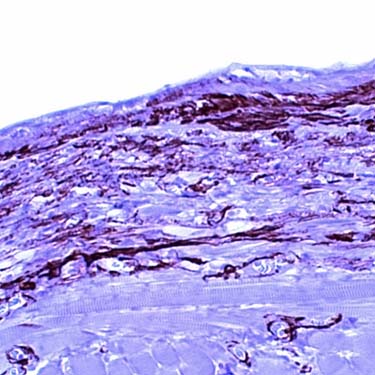
Highlighting of Smooth Muscle Smooth muscle, highlighted with smooth muscle actin, is characteristic within the wall of a bronchogenic cyst. Other cysts in the differential diagnosis, such as branchial cleft cysts and thyroglossal duct cysts, lack smooth muscle.
TERMINOLOGY
Synonyms
• Bronchial cyst
Definitions
• Rare congenital malformation of ventral foregut
Enteric cyst, neurenteric cysts are part of same family
ETIOLOGY/PATHOGENESIS
Embryogenesis
• Derived from small buds of diverticula that separate from foregut during formation of tracheobronchial tree
• Usually between 26th-40th day of gestation
CLINICAL ISSUES
Epidemiology
• Incidence
Rare
• Age
Pediatric population; rare in adults
Only gold members can continue reading. Log In or Register to continue
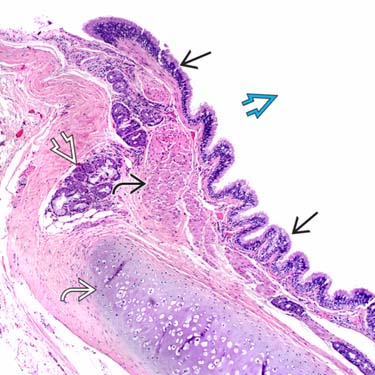
 lined by respiratory-type epithelium
  with a fibrous connective tissue wall that contains mucoserous glands
  , hyaline cartilage
  and smooth muscle
  . Multiple step sections may be needed to identify the different components.
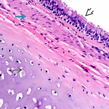
 overlying a band of smooth muscle
  closely associated with cartilage
  .
.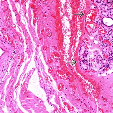
 are found throughout the wall and may contribute to the mucoid material found within the cyst.