Patient Story
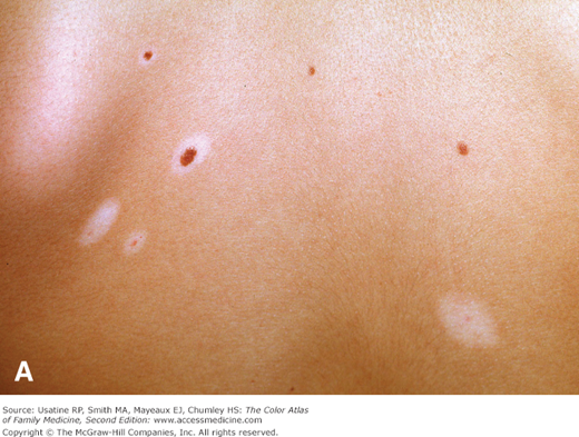
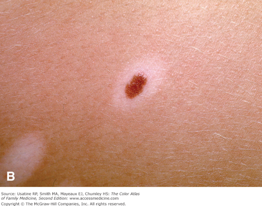
A young woman comes to the office because her husband has noted that the moles on her back are changing (Figure 162-1). A few have white halos around the brown pigmentation and some have lost their pigment completely, with a light area remaining. She has no symptoms but wants to make sure these are not skin cancers. Halo nevi are an uncommon variation of common nevi. These appear benign and the patient is reassured.
Introduction
Most nevi are benign tumors caused by the aggregation of melanocytic cells in the skin. However, nevi can occur on the conjunctiva, sclera and other structures of the eye. There are also nonmelanocytic nevi that are produced by other cells as seen in Becker nevi and comedonal nevi. Although most nevi are acquired, many nevi are present at birth.
Epidemiology
- Acquired nevi are common lesions, forming during early childhood; few adults have none.
- Prevalence appears to be lower in dark-skinned individuals.
- Present in 1% of neonates increasing through childhood and peaking at puberty; new ones may continue to appear in adulthood. In a population study of children (N = 180, ages 1 to 15 years) in Barcelona, the mean number of nevi was 17.5.1
- Adults typically have 10 to 40 nevi scattered over the body. In a population study in Germany, 60.3% of 2823 adults (mean age: 49 years; 50% women) exhibited 11 to 50 common nevi and 5.2% had at least 1 atypical nevus.2
- The peak incidence of melanocytic nevi (MN) is in the fourth to fifth decades of life; the incidence decreases with each successive decade.3
Etiology and Pathophysiology
- Benign tumors composed of nevus cells derived from melanocytes, pigment-producing cells that colonize the epidermis.
- MN represent proliferations of melanocytes that are in contact with each other, forming small collections of cells known as nests. Genetic mutations present in common nevi as well as in melanomas include BRAF, NRAS, and c-kit.4
- Sun (UV) exposure, skin-blistering events (e.g., sunburn), and genetics play a role in the formation of new nevi.3
- Nevi commonly darken and/or enlarge during pregnancy. Melanocytes have receptors for estrogens and androgens and melanogenesis is responsive to these hormones.3
- Three broad categories of MN are based on location of nevus cells2:
- Junctional nevi—Composed of nevus cells located in the dermoepidermal junction; may change into compound nevi after childhood (except when located on the palms, soles, or genitalia) (Figure 162-2).
- Compound nevi—A nevus in which a portion of nevus cells have migrated into the dermis (Figure 162-3).
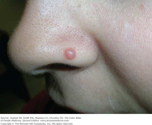
- Dermal nevi—Composed of nevus cells located within the dermis (usually found only in adults). These are usually raised and have little to no visible hyperpigmentation (Figures 162-4 and 162-5).
- Junctional nevi—Composed of nevus cells located in the dermoepidermal junction; may change into compound nevi after childhood (except when located on the palms, soles, or genitalia) (Figure 162-2).
- Special categories of nevi:
- Halo nevus—Compound or dermal nevus that develops a symmetric, sharply demarcated, depigmented border (Figure 162-1). Most commonly occurs on the trunk and develops during adolescence. Repigmentation may occur.
- Blue nevus—A dermal nevus that contains large amounts of pigment so that the brown pigment absorbs the longer wavelengths of light and scatters blue light (Tyndall effect) (Figure 162-6). Blue nevi are not always blue and color varies from tan to blue, black, and gray. Types of blue nevi include amelanotic, desmoplastic, atypical, and malignant variants; genetic mutations seen in blue nevi are often different than those seen in common nevi and include the Gαq class of G-protein α subunits, Gnaq, and Gna11 proteins.4 The nodules are firm because of associated stromal sclerosis. Usually appears in childhood on the extremities, dorsum of the hands and face. A rare variant, the cellular blue nevus is large (>1 cm), frequently located on the buttocks, and may undergo malignant degeneration.
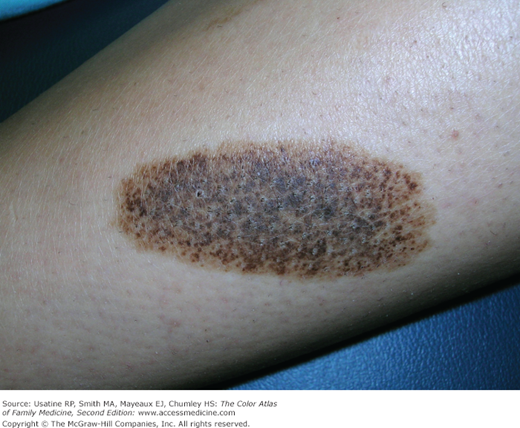
- Nevus spilus—Hairless, oval, or irregularly shaped brown lesion with darker brown to black dots containing nevus cells (Figure 162-7). May appear at any age or be present at birth; unrelated to sun exposure.
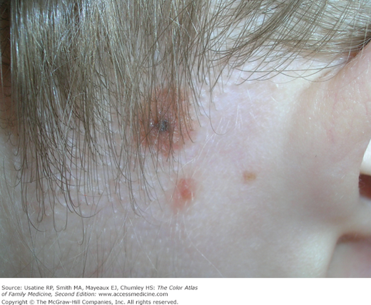
- Spitz nevus (formerly called benign juvenile melanoma because of its clinical and histologic similarity to melanoma)—Hairless, red, or reddish brown dome-shaped papules generally appearing suddenly in children, sometimes following trauma (Figures 162-8 and 162-9). The pink color is caused by increased vascularity. Most importantly, these should be fully excised with clear margins.
- Nevus of Ota—Dark brown nevus that occurs most commonly around the eye and can involve the sclera (Figure 162-10).
- Halo nevus—Compound or dermal nevus that develops a symmetric, sharply demarcated, depigmented border (Figure 162-1). Most commonly occurs on the trunk and develops during adolescence. Repigmentation may occur.
- Both acquired and congenital MN hold some risk for the development of melanoma; the number of MN, especially more than 100, is an important independent risk factor for cutaneous melanoma.5
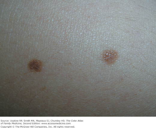
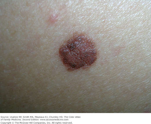
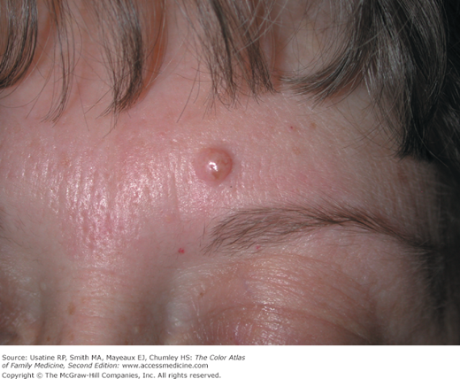
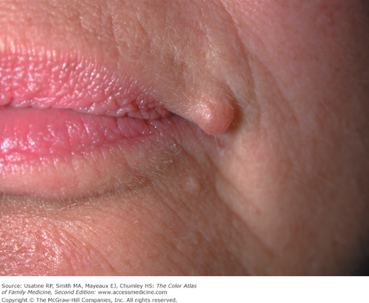
Figure 162-6
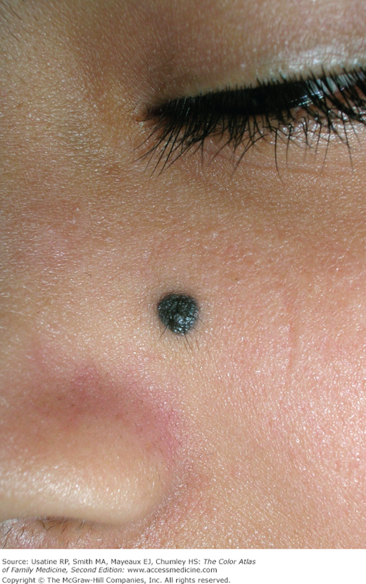
Blue nevus on the left cheek that could resemble a melanoma with its dark color. In this case it was fully excised with a 5-mm punch with a good cosmetic result. Blue nevi are benign and do not need to be excised unless there are suspicious changes. (Courtesy of Richard P. Usatine, MD.)