Patient Story
Five years ago, a young woman awoke with the inability to move the left side of her face. She was pregnant at that time. On examination it was found that she had absent brow furrowing, weak eye closure, and dropping of her mouth angle (Figure 233-1). She was diagnosed with Bell’s palsy and was provided eye lubricants and guidance on keeping her left eye moist. Her physician discussed the available evidence about treatment with steroids. She chose not to take the steroids because of her pregnancy.
Figure 233-1
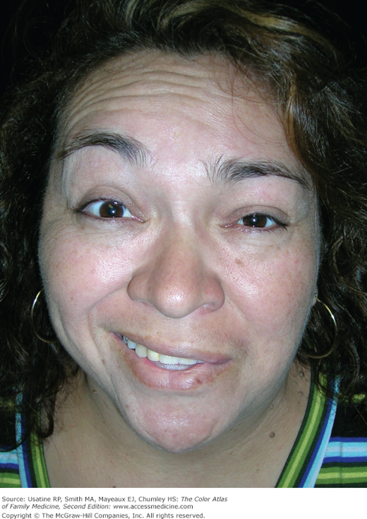
Bell’s palsy with loss of brow furrowing and dropped angle of the mouth on the affected left side of her face demonstrated during a request to smile and raise her eyebrows. The Bell’s palsy has been present for 5 years and the patient is being evaluated by ear, nose, and throat (ENT) for surgery to restore facial movement. (Courtesy of Richard P. Usatine, MD.)
Introduction
Epidemiology
- In a Canadian study, incidence was 13.1 to 15.2/100,000 adults.1
- In a study of United States military members, the incidence was 42.77/100,000, with higher incidence in females, blacks, and Hispanics; arid climate and cold months were independent predictors of risk with adjusted relative risk ratios of 1.34 and 1.31, respectively.2
- Women who develop Bell’s palsy in pregnancy have a 5-fold increased risk over national average of preeclampsia or gestational hypertension.3
- Seventy percent of cases of acute peripheral facial nerve palsy are idiopathic (Bell’s palsy); 30% have known etiologic factors such as trauma, diabetes mellitus, polyneuritis, tumors, or infections such as herpes zoster, leprosy (Figure 233-2) or Borrelia.4

Etiology and Pathophysiology
- Etiology of Bell’s palsy is currently unknown and under debate; the prevailing theory suggests a viral etiology from the herpes family.
- The facial nerve becomes inflamed, resulting in nerve compression.
- Compression of the facial nerve compromises muscles of facial expression, taste fibers to the anterior tongue, pain fibers, and secretory fibers to the salivary and lacrimal glands.
- This is a lower motor neuron lesion; the upper and lower portions of the face are affected (Figure 233-1). In upper motor neuron lesions (e.g., cortical stroke), the upper third of the face is spared, while the lower two-thirds are affected as a result of the bilateral innervation of the orbicularis, frontalis, and corrugator muscles, which allows sparing of upper face movement.


