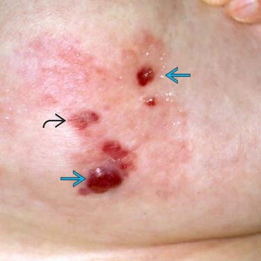
Clinical Appearance as Small Papules and Patches An atypical vascular lesion (AVL) typically presents as small papules and patches in radiated skin at a median time of 3 years post radiotherapy. The breast or chest wall are most common sites. The lesions can be solitary or multiple.
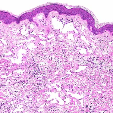
Atypical Vascular Lesion in Superficial Dermis Low-magnification view shows a superficially located, lymphangioma-like vascular lesion composed of dilated vascular structures lined by cytologically bland endothelial cells.
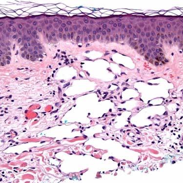
Higher Magnification of Superficial Atypical Vascular Lesion Higher magnification of this superficial dermal AVL shows a proliferation of dilated vascular channels lined by small, relatively bland-appearing endothelial cells.
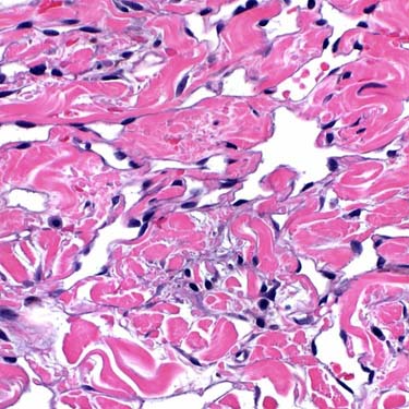
Dissecting Growth Pattern Is Typically Present AVLs often have a dissecting growth pattern consisting of jagged, anastomosing channels that infiltrate dermal collagen; a pattern that mimics well-differentiated angiosarcoma. Unlike angiosarcoma, however, there is no significant nuclear atypia, no multilayering of cells, and rarely any mitotic activity.
TERMINOLOGY
Abbreviations
• Atypical vascular lesion (AVL)
Synonyms
• Atypical vascular proliferation
Definitions
• Benign cutaneous vascular lesion presenting as small papule or patch in radiated skin, composed of thin-walled lymphatic vessels, usually limited to dermis
ETIOLOGY/PATHOGENESIS
Environmental Exposure
• Radiation (40-60 Gy)
• Median latency: 3 years post radiotherapy
CLINICAL ISSUES
Epidemiology
• Incidence
Exact incidence unknown
• Age
Wide age range; median in late 50s
Usually 1 decade earlier than radiation-associated cutaneous angiosarcoma
Site
• Skin of breast or chest wall most common site, following radiotherapy for breast cancer
• Less frequent in radiation fields associated with gynecological and other malignancies
Presentation
• Small, flesh-colored papule or erythematous patch
• Solitary or multiple
• Rarely presents as large patch
Treatment
• Complete excision of all lesions recommended
Only gold members can continue reading. Log In or Register to continue


 and patches
  in radiated skin at a median time of 3 years post radiotherapy. The breast or chest wall are most common sites. The lesions can be solitary or multiple.