Fig. 4.1
Benign urothelial cells. The top left corner shows benign superficial urothelial cells (umbrella cells) and the bottom right corner has benign intermediate/basal type urothelial cells. Although the non-superficial urothelial cells have a high N/C ratio, they have a smooth nuclear contour and do not show nuclear enlargement, placing them in the “negative” category. The follow-up diagnosis was benign (Bladder washing, TP, high mag.)
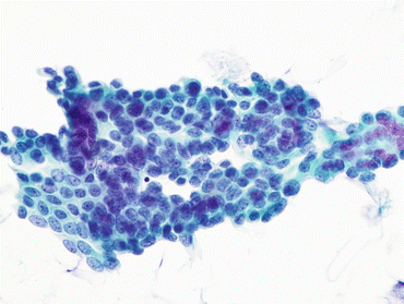
Fig. 4.2
Reactive urothelial cells. Superficial urothelial cells with slight nuclear enlargement and prominent chromocenters. There is no nuclear hyperchromasia, clumped chromatin, or nuclear contour irregularity. These changes are consistent with the “negative” category (Bladder washing, TP, high mag.)
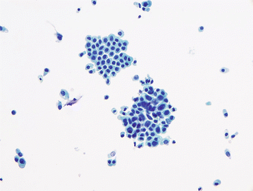
Fig. 4.3
Atypical urothelial cells (AUC). Two groups of urothelial cells are shown. The group at the top is composed of intermediate type urothelial cells with smooth nuclear contours, and no features of atypia. The urothelial cells in the group on the bottom have high N/C ratios, and nuclear contour irregularity. Nuclear chromasia is similar in both groups. Due to the cytologic atypia seen in the group on the bottom this case should be categorized as AUC (bladder washing, TP, intermediate mag.)
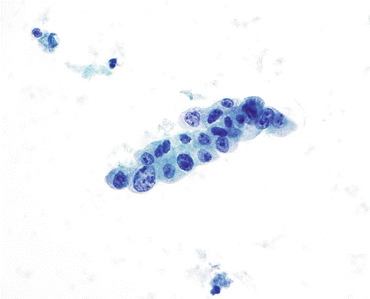
Fig. 4.4
Atypical urothelial cells (AUC). Atypical urothelial cells with high N/C ratios and nuclear contour irregularity. The absence of hyperchromasia and the presence of degenerated clumped chromatin preclude a diagnosis of SHGUC (Bladder washing, TP, high mag.)
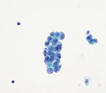
Fig. 4.5
Atypical urothelial cells (AUC). Atypical urothelial cells with high N/C ratio, enlarged nuclei, prominent chromocenters and mild nuclear contour irregularity. The chromatin is unevenly distributed yet hypochromatic. Concurrent bladder biopsy showed acute cystitis with extensive reactive epithelial changes. (Bladder Washing, TP, high mag.)
Fig. 4.6
Atypical urothelial cells (AUC). Atypical urothelial cells with high N/C ratios. (a) The image shows enlarged nuclei (compared to the neighboring benign urothelial cells) and mild nuclear contour irregularities. The chromatin is uniform and hypochromatic, precluding a diagnosis of SHGUC (Bladder washing, TP, high mag.). (b) Abnormal nuclear contours are present in a degenerated aggregate of urothelial cells. The cellular changes are worrisome, but the degree of degeneration precludes a definitive diagnosis. Degenerative cytoplasmic vacuolization is demonstrated (Bladder washing, TP, intermediate mag.)
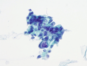
Fig. 4.7
Atypical urothelial cells (AUC). Urothelial cells with high N/C ratios display enlarged nuclei and conspicuous nuclear contour irregularity. The chromatin is clumped yet hypochromatic, precluding a diagnosis of SHGUC. (Bladder Barbotage, ThinPrep, high mag.)
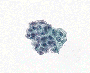
Fig. 4.8
Atypical urothelial cells (AUC). Urothelial cells with high N/C ratio, and nuclear hyperchromasia. Due to the degeneration, it is difficult to ascertain further chromatinic detail, therefore precluding a diagnosis of SHGUC. (Voided urine, TP, high mag.)
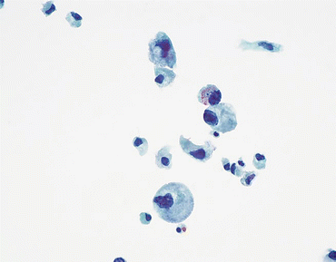
Fig. 4.9
Atypical urothelial cells (AUC). Urothelial cells with high N/C ratio (up to 50 %), and nuclear hyperchromasia are shown. The chromatin is coarse and the nuclear membranes are irregular. While the features are worrisome for high grade urothelial carcinoma, due to extensive degeneration and N/C ratio being less than 70 %, AUC diagnosis may be more appropriate. Follow up showed high grade urothelial carcinoma in the kidney; the urinary bladder had no pathology. (Bladder washing, TP, high mag.)
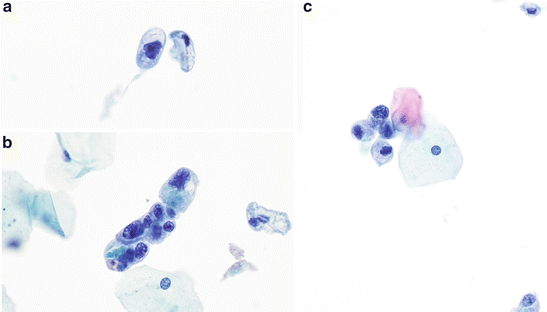
Fig. 4.10
Atypical urothelial cells (AUC). Urothelial cells with high N/C ratios, and nuclear hyperchromasia. (a) Atypical urothelial cell (AUC) (upper left). Urothelial cell displays irregular nuclear contours. (b)Group of urothelial cells with markedly irregular nuclear contours and variation in nuclear size (lower left). In comparison to the neighboring squamous cells there is mild nuclear hyperchromasia. There are degenerative cellular changes, such as partial loss of the cytoplasm and loss of crisp nuclear detail. (c) Small aggregate of atypical urothelial cells adjacent to squamous cells (right). The urothelial cell nuclei also show degeneration, but the one cell with the high N/C ratio is worrisome for carcinoma. The patient is a 36-year-old woman with recurrent urolithiasis, and no history of urothelial carcinoma. Her age and history are low-risk factors for bladder cancer. These three figures display the entire amount of atypical cells that are present in the specimen; therefore, these cytologic features warrant the diagnosis AUC (Voided, TP, high mag.)
Criteria
To standardize the criteria of the AUC category, a strict morphological definition based upon the characteristics of AUC is indispensable. The diagnosis AUC is defined as cellular changes that fulfill the major (required) criterion and only one minor criterion (the presence of two or more minor criteria, including hyperchromasia is diagnostic of SHGUC, unless there are marked degenerative changes):
Major criterion (required)
Non-superficial and non-degenerated urothelial cells with an increased nuclear cytoplasmic (N/C) ratio (>0.5) (Explanatory Note 1)
Minor criteria (one required):
Nuclear hyperchromasia (Explanatory Note 2)
Irregular nuclear membranes (chromatinic rim or nuclear contour) (Explanatory Note 3)
Irregular, coarse, clumped chromatin
Based on the presence of the one major criterion and one of the minor criteria noted above, a diagnosis of AUC may be rendered. Normal intermediate and basal urothelial cells, typically observed in instrumented urine specimens, should be identified and categorized as “normal” or NHGUC despite the fact that they have a high N/C ratio and may appear mildly hyperchromatic (Fig. 4.1). These cells frequently occur in groups, show uniform, round nuclei and inconspicuous nucleoli with finely dispersed, smooth chromatin (see Chap. 3).
Stay updated, free articles. Join our Telegram channel

Full access? Get Clinical Tree


