7 Appendix Diseases
Anatomy of the Appendix
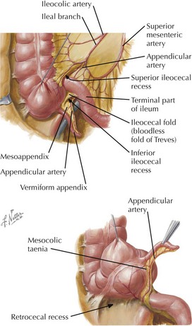
• Appendix develops as a diverticulum of the cecum (cecal bud) in embryonic week 8, as part of caudal midgut.
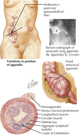
• Appendix is variable in length (2-20 cm) and may become inflamed and enlarged owing to fecal impaction and/or infection (appendicitis).
• Small mesentery (mesoappendix) connects with terminal ileum and contains appendiceal blood vessels and lymphatics.
• Tissue layers include mucosa, lamina propria, inner circular and outer longitudinal smooth muscle, and adventitia (peritoneum and mesentery).
• Taeniae coli (triple longitudinal muscle bands of the cecum) merge into a single, outer longitudinal muscle layer on appendix.
Location and Position of Appendix
• Typical locations: retrocecal-retrocolic, pelvic (descending), subcecal, ileocecal (anterior to ileum), ileocecal (posterior to cecum)


