May be indistinguishable histologically and immunohistochemically from apocrine carcinoma
• Primary cutaneous adenoid cystic carcinoma
Cribriform pattern within islands that are relatively uniform in size/shape and not interconnected
Deeply infiltrative, almost always with perineural invasion
• Tubular apocrine adenoma
Well circumscribed, noninfiltrative
Tubular islands with evidence of decapitation secretion
Islands not crowded together
Minimal cytologic atypia
Often myoepithelial cell layer present
• Hidradenocarcinoma
Larger solid and cystic nodules, infiltrative
Admixture of cell types: Clear, squamoid, poroid, mucinous
Hyalinized stroma
Apocrine Carcinoma at Scanning Magnification Low magnification shows a densely cellular dermal proliferation of packed glands and ductal structures, with many lumina easily identified.
Apocrine Carcinoma With Prominent Snouts and Secretions Higher magnification of the ductal/glandular structures shows enlarged, cuboidal to columnar-shaped cells with prominent apical snouts and secretions , typical of apocrine differentiation.
Apocrine Carcinoma With Perineural Invasion High-magnification view of apocrine carcinoma with perineural invasion shows glands lined by cuboidal cells surrounding a nerve. Focal snouts and secretions of cytoplasm are present within the lumina.
Immunohistochemistry for GCDFP-15 Apocrine carcinoma stains diffusely with GCDFP-15. The islands are crowded together, and lumina can be seen in many of the islands.
TERMINOLOGY
Synonyms
• Cribriform apocrine carcinoma
Definitions
• Malignant tumor with apocrine differentiation
CLINICAL ISSUES
Site
• Axilla and anogenital regions are most common sites, but other sites described
Presentation
• Dermal nodule, often asymptomatic
• May present within nevus sebaceus
• No history of breast carcinoma
Treatment
• Wide local excision; role of chemotherapy or radiotherapy unclear
Only gold members can continue reading. Log In or Register to continue


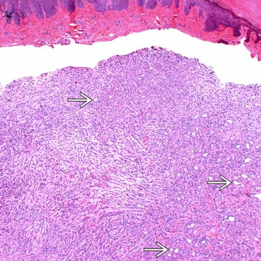
 easily identified.
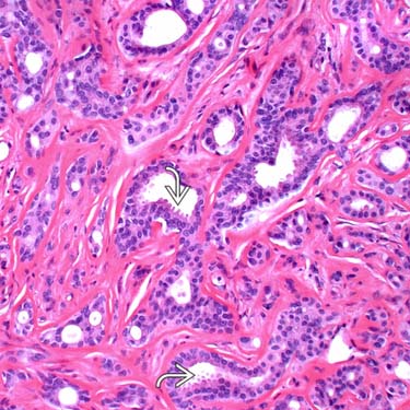
 , typical of apocrine differentiation.
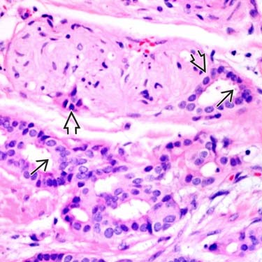
 shows glands lined by cuboidal cells surrounding a nerve. Focal snouts and secretions of cytoplasm
  are present within the lumina.
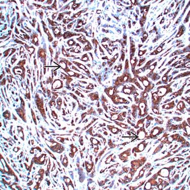
 can be seen in many of the islands.


