4 Aortic regurgitation
Salient features
History
• Asymptomatic (but may have normal or depressed left ventricular function)
• Dyspnoea and fatigue (from left ventricular impairment and low cardiac output initially on exertion)
• Symptoms of left ventricular failure in later stages
• Angina pectoris is less common than in aortic stenosis; usually indicates coronary artery disease.
Examination
Pulse
• Collapsing pulse (large volume, rapid fall with low diastolic pressure)
• Visible carotid pulsation in neck (dancing carotids or Corrigan’s sign)
• Capillary pulsation in fingernails (Quincke’s sign)
• A booming sound heard over femorals (‘pistol-shot’ femorals or Traube’s sign)
• To and fro systolic and diastolic murmur produced by compression of femorals by stethoscope (Duroziez’s sign or murmur).
Heart
• Heart sounds are usually normal.
• Apex beat will be displaced outwards and forceful may be seen and/or felt.
• Third heart sound is heard (in early systole with bicuspid aortic valve).
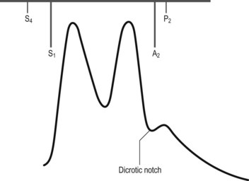
• Early diastolic, high-pitched murmur is heard at the left sternal edge with the diaphragm; if not readily apparent, it is important to sit the patient forward and auscultate with the patient’s breath held at the end of expiration (Fig. 4.1). When the ascending aorta is dilated and displaced to the right, the murmur may be heard along the right sternal border as well.
• An ejection systolic murmur may be heard at the base of the heart in severe aortic regurgitation (without aortic stenosis). This murmur may be as loud as grade 5 or 6, and underlying organic stenosis can be ruled out only by investigations.
• Ejection click suggests underlying bicuspid aortic valve.
• Mid-diastolic murmur of Austin–Flint may be heard at apex. It is typically low pitched, similar to the murmur of mitral stenosis but without preceding opening snap.
• Loud pulmonary component of second sound (suggests pulmonary hypertension).
General examination
• Head nodding in time with the heart beat (de Musset’s sign) may be present.
• Visible carotid pulsation may be obvious in the neck: dancing carotids or Corrigan’s sign.
• Blood pressure indicates wide pulse pressure.
• Look for systolic pulsations of the uvula (Muller’s sign).
• Check pupils for Argyll Robertson pupil of syphilis.
• Look for stigmata of Marfan syndrome: high-arched palate, arm span greater than height.
• Check joints for ankylosing spondylitis and rheumatoid arthritis.
Questions
Mention a few causes of chronic aortic regurgitation:
• Hypertension (accentuated tambour quality of second sound)
• Idiopathic dilatation of the aortic root and annulus
• Seronegative arthritis (ankylosing spondylitis, Reiter syndrome)
How would you investigate a patient with aortic regurgitation?
• Chest radiograph is usually normal in mild aortic regurgitation; possibly valvular calcification, cardiomegaly.
• ECG (Fig. 4.2) typically shows features of left ventricular hypertrophy and strain (increased QRS amplitude and ST/T wave changes in precordial leads) and left atrial hypertrophy (wide P wave in lead II and biphasic P in lead V1).
• Echocardiogram is indicated to confirm the diagnosis of aortic regurgitation, determine aetiology, assess valve morphology, acquire a semiquantitative estimate of severity of regurgitation, assess LV dimension, mass and systolic function, assess aortic size, in estimating the degree of pulmonary hypertension (when tricuspid regurgitation is present), and in determining whether there is rapid equilibration of aortic and LV diastolic pressure. Doppler is the best method for detecting aortic regurgitation.
• Exercise testing in severe aortic regurgitation, when sedentary or where there are equivocal symptoms is useful to assess functional capacity, symptomatic responses and haemodynamic effects of exercise.
• Radionuclide angiogram is useful in asymptomatic patients with poor-quality echocardiographic images.
• Cardiac catheterization is necessary when coronary artery disease is suspected (e.g. in patients >40 years) and when severity of aortic regurgitation is doubted; injection of contrast into aortic root gives information on degree of regurgitation and state of aortic root (presence of dilatation, dissection, root abscesses).
Stay updated, free articles. Join our Telegram channel

Full access? Get Clinical Tree


