Diagnostic component is spindle or epithelioid smooth muscle cells

Epithelioid smooth muscle cells are typically large, with round to oval nuclei, and eosinophilic to fibrillar or vacuolated cytoplasm
•
Features that predict malignant behavior are not well defined

Nuclear atypia and infiltrative margins can be seen in benign tumors
Ancillary Tests
•
Smooth muscle cells stain with antibodies to HMB-45, MART-1, but not keratin or Hep-Par1
Top Differential Diagnoses
•
Hepatocellular neoplasm, particularly hepatocellular carcinoma
•
Metastatic malignant tumor, either carcinoma or sarcoma
TERMINOLOGY
Abbreviations
Definitions
•
Rare, benign mesenchymal neoplasm composed of smooth muscle, adipose tissue, and vessels

Thought to arise from perivascular epithelioid cells (PEC); therefore, considered part of PEComa family of tumors
CLINICAL ISSUES
Epidemiology
•
Incidence

Rare: Overall incidence unknown but only a few hundred cases reported

Some cases associated with tuberous sclerosis (6-10%), but less often than renal AML (20-40%)
–
More likely to be associated with tuberous sclerosis if multiple &/or associated with renal tumors
•
Age

Adults 17-86 years (mean: 43.5-50 years)
•
Sex

Marked female predominance
Presentation
•
Most patients are asymptomatic and present incidentally
•
Large tumors may cause symptoms related to mass effect or abdominal discomfort
Treatment
•
Surgical approaches

Excision
–
When diagnosis cannot be established on biopsy
–
Large lesions at risk for rupturing
•
Conservative approaches

If diagnosis can be confidently established, radiologic follow-up is recommended
Prognosis
•
Benign behavior in nearly all cases

Tumor recurrence is uncommon

Metastasis extremely rare
IMAGING
Ultrasonographic Findings
•
Most angiomyolipomas present as heterogeneous hyperechoic lesions, but can be hypoechoic
MR Findings
•
MR is most specific imaging modality for detecting lipomatous component
•
Most tumors are hypointense on T1WI and slightly hyperintense on T2WI
CT Findings
•
Usually hypodense on precontrast CT
MACROSCOPIC
General Features
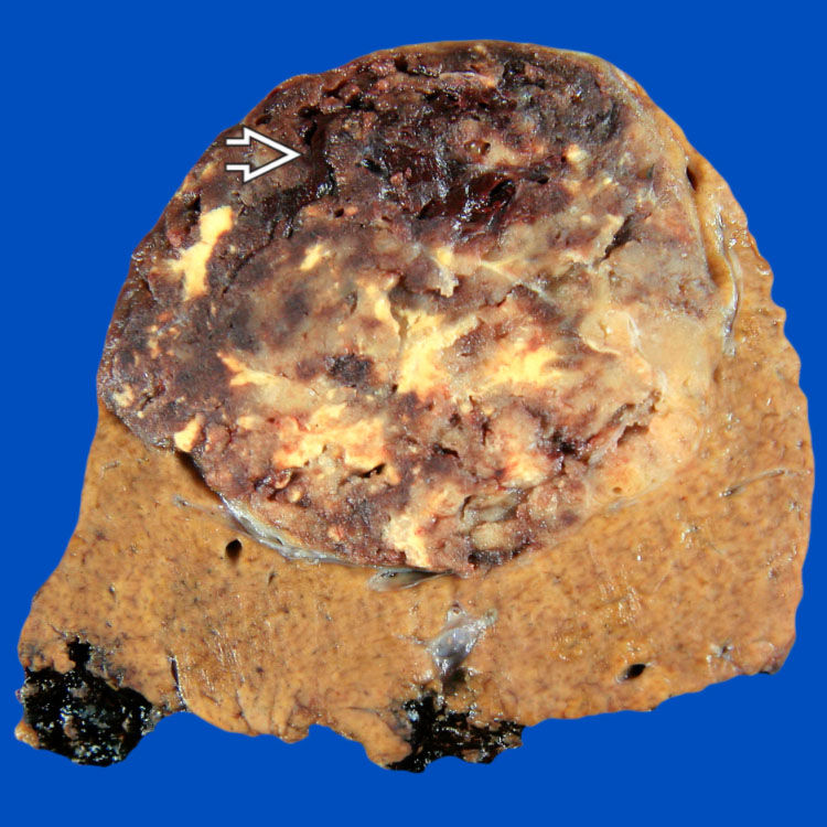
 . Note that the background liver is not cirrhotic.
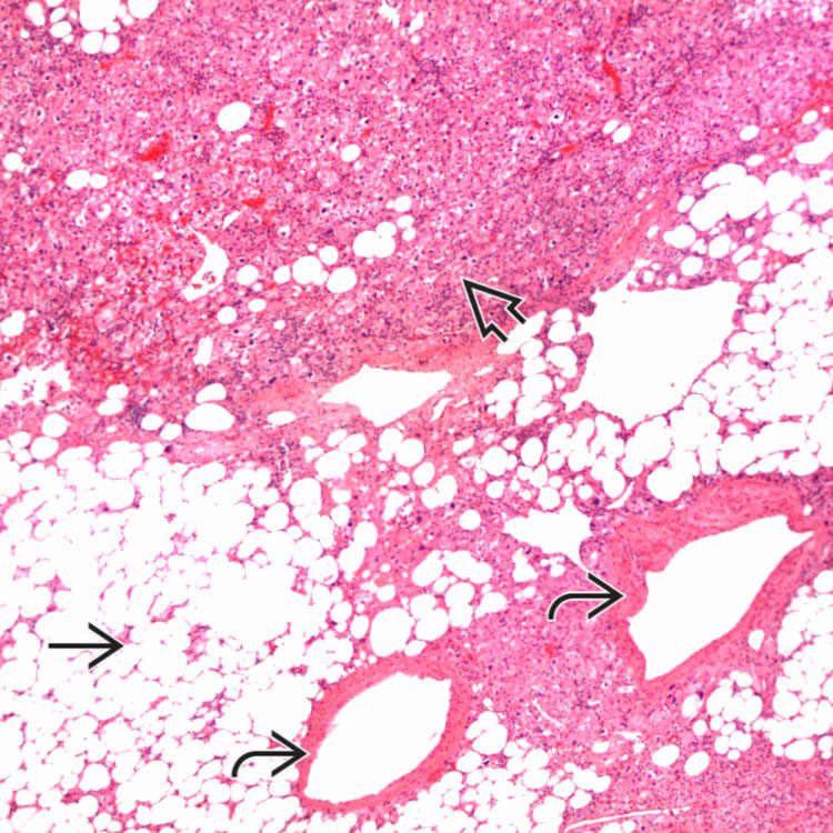
 , vessels
  , and myoid cells
  , which in this case are plump and spindled.
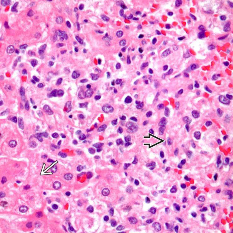
 of an angiomyolipoma contrasts with the more eosinophilic and granular hepatocyte cytoplasm
  .
.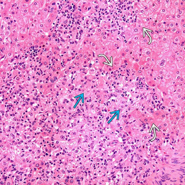
 at the interface with background liver
  is not an indication of malignancy.


