Anatomy of the Parotid Gland, Submandibular Triangle, and Floor of the Mouth
Aaron Ruhalter
The parotid gland is the largest of the paired salivary glands. The gland is wedged into the parotid space.
Parotid Space
The parotid space has a skeletal background created by the ramus of the mandible anteriorly, the styloid process medially, the mastoid process posteriorly, and the external acoustic meatus and the posterior part of the temporomandibular joint posterosuperiorly (Fig. 1). A soft tissue background is created by the muscles that are attached to these bony landmarks—the masseter, medial pterygoid, and temporalis muscles on the mandible; the stylohyoid, styloglossus, and stylopharyngeus muscles arising from the styloid process of the temporal bone; and the sternocleidomastoid and digastric muscles related to the mastoid process and the lateral portion of the posterior occipital line.
Parotid Gland
The superficial surface of the parotid gland is triangular in shape, with the apex pointing inferiorly (Fig. 2). The deep surface of the gland is wedged into this parotid space and presents anteromedial and posteromedial surfaces. The gland frequently extends beyond the limits of the parotid space. Glandular tissue may extend from the anterosuperior edge of the superficial surface, creating what is called the facial process, and is superior to the parotid duct. The extension of glandular tissue may be separate from the main portion of the gland. This isolated segment of gland (accessory parotid) has a duct that empties into the main duct. Frequently, extensions are found from the deep surface of the gland toward the pharynx or the medial pterygoid muscle.
The parotid gland is somewhat artificially divided into two lobes by the facial nerve as it passes through. Endofacial (deep) and exofacial (superficial) portions are created. Multiple communications exist between the lobes, however, created by bridges of glandular tissue. A large area of communication, referred to as the isthmus, is related to the proximal part of the intraparotid portion of the facial nerve.
The parotid duct is approximately 5 cm long and lies on the superficial surface of the masseter muscle, approximately 1 cm below the zygomatic arch. The transverse facial artery is interposed between the duct and the arch, whereas the buccal branches of the facial nerve can be found inferior and superior to the duct. When this conduit reaches the anterior margin of the masseter muscle, it turns sharply, penetrates the buccinator muscle, and ends in the vestibule of the oral cavity opposite the upper second molar tooth.
Fascial Relations
The gland is encased by a split in the investing layer of the deep cervical fascia. The deep layer passes superiorly and attaches to the base of the skull. A portion of this fascia between the tip of the styloid process and the angle of the mandible is thickened, creating the stylomandibular ligament. This ligament supports the temporomandibular joint and separates the parotid gland from the submandibular gland. The superficial layer of this fascial split is much thicker, invests the masseter muscle, and attaches to the zygomatic arch. Its thickness and unyielding nature are responsible for the severe pain that results from enlargement of the gland.
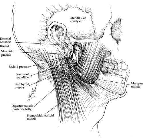 Fig. 1. Parotid bed. |
Neurovascular Relations
Neurovascular structures pass through the parenchyma of the gland and can conveniently be described in layers or planes. From deep to superficial are found the arterial, venous, and nerve layers.
Arterial Plane
The arterial layer includes the external carotid artery, which enters the parotid space after passing deep to the posterior belly of the digastric muscle (Fig. 3). At this point the external carotid artery gives rise to the posterior auricular artery, which gives off a stylohyoid branch that enters the stylomastoid foramen. This blood vessel is usually superficial to the facial nerve trunk as it exits from the skull by way of this same foramen. The posterior auricular artery then
continues posteriorly, running under cover of, and parallel to, the superior edge of the posterior belly of the digastric muscle. One should note that this muscle passes superficial to and protects almost all of the structures passing between the submandibular triangle superiorly and the carotid triangle inferiorly. This includes the internal jugular vein and the internal carotid artery in the carotid sheath, the last four cranial nerves, and the external carotid artery. The retromandibular vein or its branches, the cervical branch of the facial nerve, and the greater auricular nerve, however, pass superficial to the posterior belly of the digastric muscle.
continues posteriorly, running under cover of, and parallel to, the superior edge of the posterior belly of the digastric muscle. One should note that this muscle passes superficial to and protects almost all of the structures passing between the submandibular triangle superiorly and the carotid triangle inferiorly. This includes the internal jugular vein and the internal carotid artery in the carotid sheath, the last four cranial nerves, and the external carotid artery. The retromandibular vein or its branches, the cervical branch of the facial nerve, and the greater auricular nerve, however, pass superficial to the posterior belly of the digastric muscle.
The external carotid artery then pierces the medial surface of the parotid gland, and when it reaches the neck of the condylar process of the mandible it ends by giving rise to the maxillary artery and the superficial temporal artery. The maxillary artery passes medial to the condylar process of the mandible and enters the infratemporal fossa. The superficial temporal artery continues superiorly, accompanied by superficial temporal veins. The transverse facial artery, which arises from the proximal part of the superficial temporal artery, courses just superior to the parotid duct. The superficial temporal artery then enters the temporal region after passing between the external acoustic meatus and the temporomandibular joint. Accompanying these vascular structures at this level is the auriculotemporal nerve, which arises from the mandibular branch of the trigeminal nerve in the roof of the infratemporal fossa. It provides sensory innervation to the external acoustic meatus, external surface of the tympanic membrane, and temporomandibular joint, and cutaneous sensory innervation to the auricle and temporal scalp region.
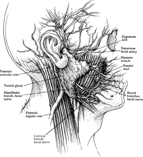 Fig. 2. Superficial view of the parotid region. |
Venous Plane
The venous plane is superficial to the arterial plane (Fig. 3). It includes the retromandibular vein and its branches. The retromandibular vein is created by the union of the superficial temporal and maxillary veins. The maxillary vein is formed by the union of veins that are part of a large plexus of veins surrounding the lateral pterygoid muscle. This venous plexus communicates with veins of the face as well as with the cavernous sinus within the skull. The parotid veins also communicate with the pterygoid venous plexus. This pterygoid venous plexus in turn represents a potential pathway for the spread of superficial cutaneous infections to the cavernous sinus. This is a potentially lethal condition.
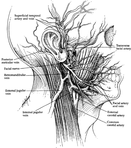 Fig. 3. Vascular background. |
The retromandibular vein passes inferiorly through the substance of the parotid gland and is found between the branches of the facial nerve and the arterial layer. The retromandibular vein terminates at the lower edge of the gland by giving off anterior and posterior branches. The posterior branch joins with the posterior auricular vein to form the external jugular vein, which continues inferiorly, superficial to the posterior belly of the digastric muscle, then passes obliquely across the sternocleidomastoid muscle. It is frequently found just anterior to the great auricular nerve, which is on its way to the skin overlying the parotid gland. The anterior branch of the retromandibular vein unites with the anterior facial vein, forming the common facial vein. This passes inferiorly, superficial to the digastric muscle, to empty into the internal jugular vein.
Nerve Plane
The nerve plane is created by the facial nerve and its branches (Fig. 4). The facial nerve (seventh cranial nerve) exits from the skull through the stylomastoid foramen. At this point it is found with the stylomastoid branch of the posterior auricular artery, which enters the foramen and supplies the mucosa of the tympanic cavity, the mastoid cells, and the semicircular canals. The artery is usually superficial to the facial nerve. After emerging from the foramen, two branches arise from the facial nerve before it enters the parotid gland. The first branch, the posterior auricular nerve, provides innervation to the posterior auricular muscles and the intrinsic muscles of the auricle. The second branch, arising from the extraparotid portion of the facial nerve, provides motor innervation to the posterior belly of the digastric and stylohyoid muscles. This portion of the nerve, approximately 1 cm in length, then penetrates the posteromedial surface of the parotid gland. This nerve trunk passes forward in the glandular parenchyma for a distance of approximately 1 cm or less, then divides into two branches—a larger temporofacial branch, which creates temporal and zygomatic nerves, and a smaller cervicofacial branch, which gives rise to buccal, marginal mandibular, and cervical nerves. An isthmus of glandular tissue separates the temporofacial branch from the cervicofacial branch.
The facial nerve provides motor innervation to the muscles of facial expression. The platysma is included in this category of muscle. The nerve branches, as they pass through the parotid gland, divide it into two portions. The part of the gland that is superficial to the nerves is referred to as the superficial (exofacial) lobe, and the portion of the gland that is internal to the nerve layer is referred to as the deep (endofacial) lobe. Multiple communications are found between the nerve branches as they pass through the gland. The zygomatic and temporal nerves are frequently multiple, whereas the mandibular and cervical branches are often single. The cervical and mandibular branches can extend below the mandible, whereas the cervical branch passes superficial to the posterior belly of the digastric muscle. The nerves become more superficial as they pass distally.
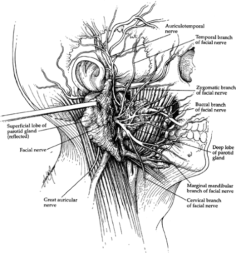 Fig. 4. Nerve plane. |
Innervation
The parotid gland receives postganglionic sympathetic fibers from a plexus of nerves that travel with the external carotid artery. Secretomotor postganglionic parasympathetic fibers reach the infratemporal fossa by way of the lesser petrosal nerve, synapse in the otic ganglion, and then travel to the parotid gland by way of the auriculotemporal nerve.
Lymphatic Drainage
The lymphatic drainage of the parotid gland is related to two systems. Superficial nodes in the superficial fascia (preauricular) drain into the superficial system of cervical nodes. These are related to the external jugular vein and pass to the supraclavicular nodes in the posterior triangle. The second set of nodes is found within the fascial covering of the parotid gland. These nodes drain into the deep cervical nodes and the jugular chain.
Anatomic Aids
The main trunk of the facial nerve can be found by tracing one of its branches proximally. The mandibular and cervical branches are more frequently used because they are often single and more convenient to find.
The parotid duct is approximately 1 cm inferior to the lower edge of the zygomatic arch. The pathway of the duct can be recreated by a line between the lower end of the tragus of the ear and the commissure of the mouth.
Two nerves may be found with the superficial temporal vessels. The auriculotemporal nerve is posterior, whereas the temporal branches of the facial nerve lie anterior to these vessels.
The great auricular nerve is frequently found posterior to the external jugular vein. This nerve and vein travel together until the inferior edge of the parotid gland is reached.
The nerve passes to the subcutaneous tissues superficial to the gland, providing sensory innervation to the skin overlying the parotid gland. The remainder of the face receives its sensory innervation only from the trigeminal nerve. The vein is seen to emerge from the substance of the gland.
The nerve passes to the subcutaneous tissues superficial to the gland, providing sensory innervation to the skin overlying the parotid gland. The remainder of the face receives its sensory innervation only from the trigeminal nerve. The vein is seen to emerge from the substance of the gland.
The external carotid artery is related to the medial boundary of the parotid space. The internal carotid artery is slightly deeper. The two vessels should not be confused. One must remember that the internal carotid artery does not have any branches in the neck. Separating the external carotid artery from the internal carotid artery are the styloid process or stylohyoid ligament, the stylopharyngeus muscle, and the glossopharyngeal nerve.
The stylohyoid muscle and the posterior belly of the digastric muscle diverge at their points of attachment to the skull. The main trunk of the facial nerve passes through this interval.
At the junction of the cartilaginous portion and the osseous portion of the auditory canal, a downward projection of cartilage frequently exists that points to the main trunk of the facial nerve.
When the external jugular vein is traced superiorly to its parent structure, the retromandibular vein, it leads to the interval between the superficial and deep lobes of the parotid gland. It is also a means of locating the cervical or mandibular branches of the facial nerve, because they pass superficial to the vein.
The stylomastoid branch of the posterior auricular artery enters the stylomastoid foramen and is superficial to the facial nerve trunk.
The internal jugular vein may be in contact with the deep surface of the gland.
Stay updated, free articles. Join our Telegram channel

Full access? Get Clinical Tree


