Intraampullary: Arise in ampulla (lined by pancreatobiliary epithelium)

Periampullary: Arise in duodenal surface of papilla (lined by intestinal epithelium)
Clinical Issues
•
5-year overall survival rate: 40%

Determined by histologic type, grade, stage, coexisting adenoma
Microscopic
•
2 main histologic types

Intestinal-type adenocarcinoma
–
Similar to adenocarcinoma of colon and small bowel

Pancreatobiliary-type adenocarcinoma
–
2nd most common (15-20%)
–
Similar to pancreatic ductal or bile duct adenocarcinoma
Ancillary Tests
•
Intestinal type

CK20, CDX-2, or MUC2 (+), MUC1(-)

CK20, CDX-2, and MUC2 (+), irrespective of MUC1
•
Pancreatobiliary type

MUC1(+), CDX-2 and MUC2 (-)
Top Differential Diagnoses
•
Adenocarcinomas of pancreas, distal common bile duct, and duodenum
TERMINOLOGY
Synonyms
•
Periampullary adenocarcinoma
Definitions
•
Malignant epithelial neoplasm originating in ampulla of Vater

Centered on, circumferentially surrounding, or completely replacing ampulla
ETIOLOGY/PATHOGENESIS
Histogenesis
•
Intraampullary: Arise in ampulla (lined by pancreatobiliary epithelium)
•
Periampullary: Arise in duodenal surface of papilla (lined by intestinal epithelium)
Predisposing Syndromes
•
Familial adenomatous polyposis, Lynch syndrome, type 1 neurofibromatosis
CLINICAL ISSUES
Epidemiology
•
Incidence

0.7 and 0.4 cases per 100,000 males and females, respectively

0.5% of all gastrointestinal malignancies
•
Age

Mean: 62 years (range: 29-85 years)
–
Younger in patients with predisposing syndromes
Presentation
•
Jaundice, abdominal pain, pancreatitis, weight loss
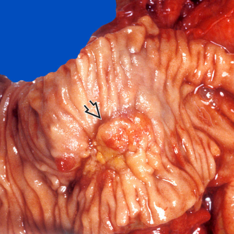
 involving the papilla and periampullary duodenal mucosa. Histologic examination confirms the diagnosis of ampullary adenocarcinoma.
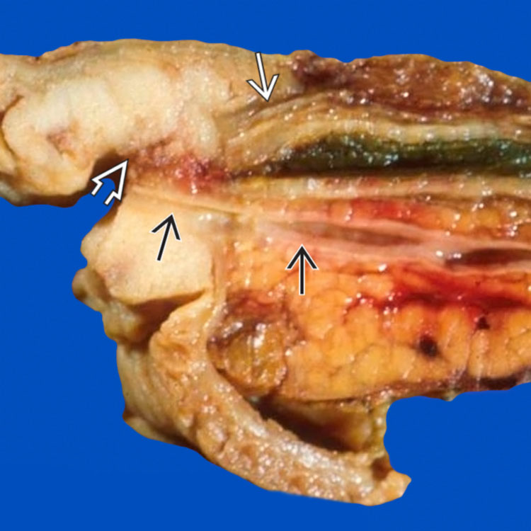
 of the common bile duct
  , which is dilated proximally. The pancreatic duct
  is not obstructed.
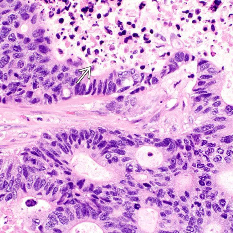
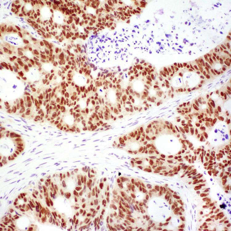
 . Tumor cells are columnar with stratified, elongated or oval nuclei, resembling adenocarcinoma of colon and small bowel.