Patient Story
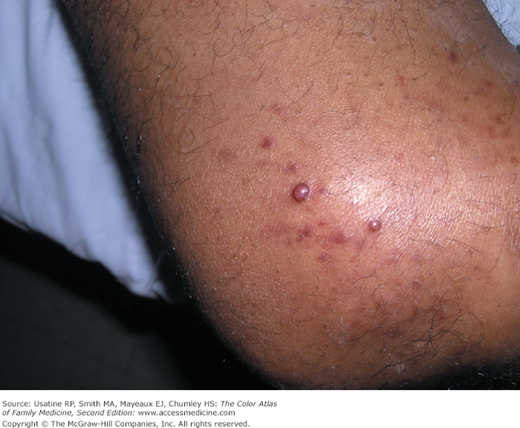
A 35-year-old gay man presented with papular lesions on his elbow (Figure 217-1). Shave biopsy demonstrated Kaposi’s sarcoma (KS). He subsequently tested positive for HIV and began treatment with antiretroviral combination therapy. The KS resolved with topical alitretinoin gel treatment.
Introduction
In the United States, KS is most often seen in patients with AIDS and patients on immunosuppressants after organ transplantation. KS can also be classic (older Mediterranean men) or endemic (young men in sub-Saharan Africa). KS is caused by Kaposi’s sarcoma-associates herpesvirus (KSHV), which promotes oncogenesis. KS cannot be cured, but treatment can result in improvement or disease stabilization. Current therapies improve the immune system or target KSHV. Therapies that modulate KSHV-mediated signaling are being studied.
Epidemiology
- KS can be classic (older Mediterranean men), endemic (young men in sub-Saharan Africa), epidemic (AIDS patients), or posttransplantation (organ recipients).1
- In the United States, 81.6% of KS is seen in patients with AIDS2 (Figure 217-1).
- In HIV-positive patients, the prevalence is 7.2/1000 person years; 451 times higher than general population.3
- In transplant patients, the prevalence is 1.4/1000 person years; 128 times higher than general population.3
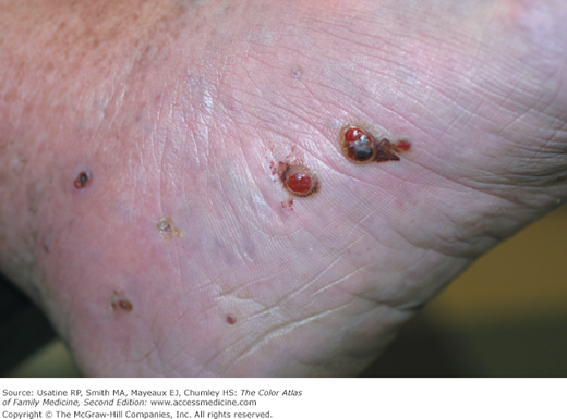
- The prevalence of classic KS in the general population of southern Italy is 2.5/100,0004 (Figure 217-2).
- The male-to-female ratio for epidemic KS in the United States is approximately 50:1 but is falling as the prevalence of AIDS increases among women.5 The male-to-female ratio has been approximately 10:1 for classic and endemic KS.
- KS is the most common malignancy seen in AIDS patients.
Etiology and Pathophysiology
- KS is caused by KSHV, also known as human herpes virus 8 (HHV-8). KSHV acts through host cell signal transduction to activate multiple oncogenic pathways.6
- KS is an angioproliferative neoplasm, with abnormal proliferation of endothelial cells, myofibroblasts, and monocyte cells.
- Lesions often begin as papules or patches and progress to plaques as proliferation continues.
- Some lesions ulcerate (nodular stage), and lymphedema can occur.
Risk Factors
Diagnosis
The diagnosis is often made clinically in a patient who has AIDS and a typical presentation of KS. In atypical presentations, diagnosis is made by biopsy.
- Cutaneous lesions are usually multifocal, papular, and reddish-purple in color (Figures 217-1 and 217-2).
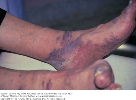
- Plaques or fungating lesions can be seen on the lower extremities, including the soles of the feet (Figure 217-3).
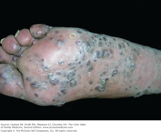
- Vascular-appearing papules on the feet and lower legs are typical of classic KS without AIDS (Figure 217-4).
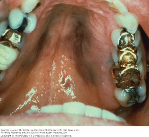
- Oral cavity lesions can be flat or nodular and are red to purple in color (Figure 217-5).
- GI lesions can be asymptomatic or can cause abdominal pain, nausea, vomiting, bleeding, or weight loss.
- Pulmonary lesions can cause shortness of breath or may appear as infiltrates, nodules, or pleural effusions on chest radiographs.
Figure 217-4
Classic Kaposi’s sarcoma on the foot of an 85-year-old Hispanic man from Mexico who is HIV-negative. He initially declined radiation and his lesions grew and multiplied over 3 years. Now he is receiving palliative radiation to improve his ability to walk. (Courtesy of Richard P. Usatine, MD.)
