Storage & Mobilization of Lipids
Histogenesis of White Adipose Tissue
Histogenesis of Brown Adipose Tissue
Connective tissue in which adipocytes or fat cells predominate is commonly called adipose tissue. These large cells are found isolated or in small groups within loose or dense irregular connective tissue but occur in large aggregates as adipose tissue or “fat” in many body regions and organs. Located throughout the body, adipose tissue normally represents 15%-20% of the body weight in men, somewhat more in women. Besides serving as storage depots for neutral fats (chiefly triglycerides, long-chain fatty acyl esters of glycerol), adipocytes function as key regulators of the body’s overall energy metabolism. With a growing worldwide epidemic of obesity and its associated health problems, including diabetes and heart disease, adipocytes and adipose tissue now constitute a major area of medical research.
Two properties of triglycerides explain their selection as the preferred form of nutrient storage against fluctuating availability and energy demands. Fats are insoluble in water and can be concentrated with no adverse osmotic effects on cells. Moreover, the caloric density of triglycerides (9.3 kcal/g) is twice that of proteins or carbohydrates, including glycogen, making these simple molecules the most efficient form of nutrient storage. Adipocytes specialize in concentrating triglycerides as lipid droplet(s), with other cells normally accumulating relatively little lipid.
Adipocytes are very active cells metabolically, responding to both nervous and hormonal stimuli. These cells release hormones and various other important substances, and adipose tissue is now recognized as an important endocrine tissue. With its unique physical properties, adipose tissue conducts heat poorly and helps thermally insulate the body. Adipose tissue also fills up spaces between other tissues and helps cushion and keep some organs in place. Subcutaneous layers of adipose tissue help shape the body surface, where pad-like deposits act as shock absorbers, chiefly in the soles and palms.
There are two types of adipose tissue with different locations: structures, colors, and pathologic characteristics. White adipose tissue, the more common type, is composed of cells that, when completely developed, contain one very large droplet of whitish-yellow fat in their cytoplasm. Brown adipose tissue contains cells with multiple lipid droplets interspersed among abundant mitochondria, which give these cells a darker appearance. Both types of adipose tissue have a rich blood supply.
WHITE ADIPOSE TISSUE
Specialized for relatively long-term energy storage, adipocytes of white adipose tissue are spherical when isolated but are polyhedral when closely packed in situ. Each cell is very large, between 50 and 150 μm in diameter, and contains a single huge droplet of lipid that fills almost the entire cell. White adipocytes are called unilocular because the triglycerides are stored in this single large droplet (Figure 6–1). Because lipid is removed from cells by xylene or other solvents used in routine histological techniques, unilocular adipocytes are often empty in standard light microscope preparations. The cells are sometimes said to have a signet-ring appearance, with the lipid droplet displacing and flattening the nucleus against the cell membrane (Figure 6–1d). This membrane and the thin rim of cytoplasm that remains after removal of the stored triglycerides may shrink, collapse, or rupture, distorting the tissue structure.
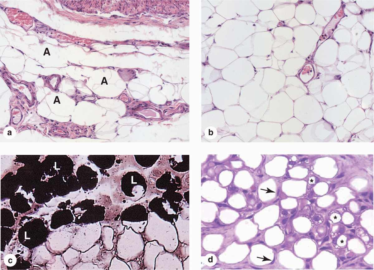
FIGURE 6–1 White adipose tissue.
 MEDICAL APPLICATION
Unilocular adipocytes can generate benign tumors called lipomas that are relatively common, although malignant adipose tumors (liposarcomas) occur infrequently. Fetal lipomas of brown fat are sometimes called hibernomas.
Most of the cytoplasm in a white adipocyte surrounds the nucleus and contains mitochondria, a small Golgi apparatus, a few cisternae of RER, and free polyribosomes. The thin, submembranous layer of cytoplasm surrounding the lipid droplet contains cisternae of smooth ER (SER) and pinocytotic vesicles. TEM studies reveal that most adipocytes, especially immature cells, contain minute lipid droplets in addition to the single large droplet seen with the light microscope. The lipid droplet-cytoplasm interface is reinforced by intermediate filaments of vimentin. Unlike other connective tissue cells, adipocytes are surrounded by a thin external lamina containing type IV collagen.
White adipose tissue is subdivided into incomplete lobules by partitions of connective tissue containing a vascular bed and nerve network. Fibroblasts, macrophages, and other cells make up about half the total number of cells. Reticular fibers form a fine interwoven network that supports individual fat cells and binds them together. The microvasculature between adipocytes may not always be apparent in tissue sections.
Almost all adipose tissue in adults is unilocular and it is found in and around many organs throughout the body. The distribution of this tissue changes significantly through childhood and adult life and is partly regulated by sex hormones, which control adipose deposition in the breasts and thighs. The color of freshly dissected white adipose tissue depends on the diet, varying from white to yellow with the amount of carotenoids dissolved in the lipid.
Storage & Mobilization of Lipids
Triglycerides stored by cells of white adipose tissue can be derived from dietary fats brought to adipocytes as circulating chylomicrons, from triglycerides synthesized in the liver and transported as very-low-density lipoproteins (VLDLs), and by the local synthesis of free fatty acids and glycerol from glucose.
Chylomicrons (Gr. chylos, juice + micros, small) are small particles of variable size, up to 1200 nm in diameter, formed in intestinal epithelial cells and transported in blood plasma and lymph. They consist of a central core, composed mainly of triglycerides and a small quantity of cholesterol esters, surrounded by a stabilizing monolayer of apolipoproteins, cholesterol, and phospholipids. Lipoproteins are also complexes of lipids and proteins, but are generally smaller than chylomicrons (providing a greater surface-to-volume ratio) and have much higher levels of lipoproteins, cholesterol, and phospholipids in the surface layer. Circulating lipoproteins are routinely measured in clinical tests for blood lipids; varying levels of surface apolipoprotein allow their categorization according to density, from VLDL to high-density lipoprotein (HDL).
In adipose tissue both chylomicrons and VLDL are hydrolyzed at the luminal surfaces of blood capillaries by lipoprotein lipase, an enzyme synthesized by the adipocyte and transferred to the capillary cell membrane (Figure 6–2). Free fatty acids enter the adipocyte by both active transport and diffusion. Within the adipocyte, the fatty acids combine with glycerol phosphate, supplied by glucose metabolism, to again form triglycerides, which are then deposited in the growing lipid droplet. Mitochondria and SER participate actively in the process of lipid uptake and storage.
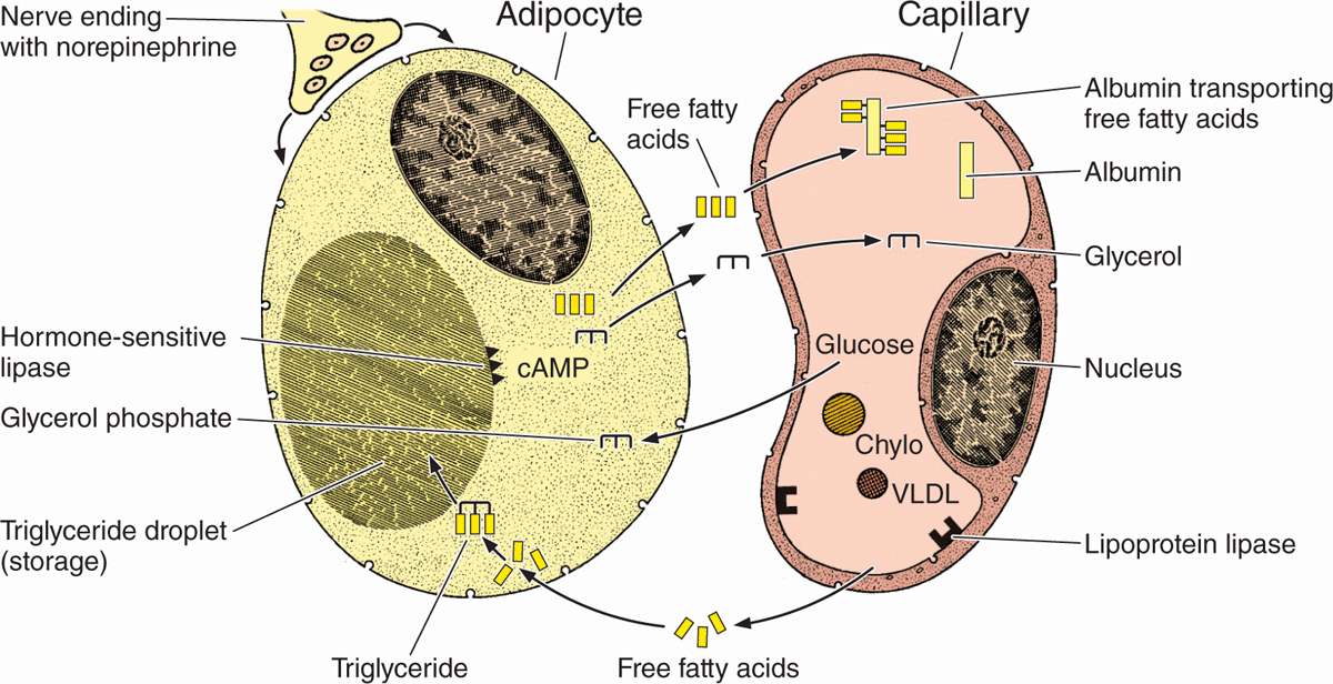
FIGURE 6–2 Lipid storage and mobilization from adipocytes.


