Clinical Photograph of Actinic Keratosis Clinical photograph of the scalp of an elderly patient shows multiple actinic keratoses (AKs) with prominent crusting. (Courtesy J. Wu, MD.)
Bowenoid Actinic Keratosis Bowenoid AK shows basilar to midepidermal involvement by atypical cells , with a mitotic figure . Overlying hyperkeratosis and parakeratosis are also present.
Classic Actinic Keratosis With Overlying Parakeratosis Low-magnification view of an AK shows basilar keratinocytic budding and atypia , with sparing of a central hair follicle. Areas of prominent overlying parakeratosis are also present.
Actinic Keratosis at High Magnification Another AK involving the superficial portion of a hair follicle shows basilar keratinocytic budding and atypia , with a mitotic figure . Parakeratosis is also present within and overlying the follicle.
TERMINOLOGY
Abbreviations
• Actinic keratosis (AK)
Synonyms
• Solar keratosis
• Often considered precancer or form of early squamous cell carcinoma (SCC) in situ
Definitions
• Atypical intraepidermal proliferation of keratinocytes typically confined to basilar portion of epidermis, with very low risk for progression to invasive SCC
ETIOLOGY/PATHOGENESIS
Solar Damage
• Ultraviolet light (primarily UVB) induces mutations in DNA, which leads to abnormal proliferation of intraepidermal keratinocytes
• TP53 mutations are most common genetic alteration identified
CLINICAL ISSUES
Epidemiology
• Incidence
Very common lesions, estimated to affect up to 10-40% of adult Caucasians; higher incidence in areas with heavy sun exposure (i.e., Australia)
• Age
Older adults typically affected
• Sex
Males more common than females
• Ethnicity
Mostly occurs in Caucasians; much less common in other races
Site
• Sun-exposed sites, especially face, head and neck, dorsal hands, and forearms
Presentation
• Scaly papules and plaques, often multiple
Natural History
• Minority of cases progress to invasive SCC
Treatment
• Options, risks, complications
Controversial whether treatment is necessary in all cases, but most clinicians opt for treatment to avoid potential development of SCC
• Surgical approaches
Conservative excision of lesions is not necessary in most cases (unless there is clinical suspicion for invasive SCC) but is curative
• Drugs
Topical therapy with drugs such as 5-fluorouracil, diclofenac, or imiquimod may be used
Liquid nitrogen (cryotherapy) frequently used
Photodynamic therapy is also emerging treatment that may be useful for extensive AK
Prognosis
• Excellent in vast majority of cases, as only ~ 2-3% progress to invasive SCC
• Most invasive SCCs arising in AK are low grade, but aggressive cases may also occur
MACROSCOPIC
Size
• Usually small (< 1 cm) papules, but larger lesions may occur
MICROSCOPIC
Histologic Features
• Intraepidermal proliferation of atypical keratinocytes, typically confined to basilar 1/3 of epithelium
Basilar budding of atypical cells
Cells show nuclear enlargement, hyperchromasia, and prominent nucleoli
Abundant eosinophilic-staining cytoplasm
Increased numbers of mitotic figures usually present
• Overlying parakeratosis present in vast majority of cases; hypogranulosis may also be present
• Lesional cells usually do not involve follicles (as opposed to Bowen disease) and adnexal ducts
Leads to alternating red and blue tiers of parakeratosis (overlying AK) and hyper/orthokeratosis (overlying follicles and eccrine ducts)
• Histologic subtypes
Hypertrophic AK
– Shows epidermal hyperplasia, often psoriasiform, with prominent overlying hyperkeratosis and parakeratosis
– Dermal fibrosis and vertical collagen bundles often present, suggesting lichen simplex chronicus changes (due to chronic excoriation) superimposed on AK
Only gold members can continue reading. Log In or Register to continue
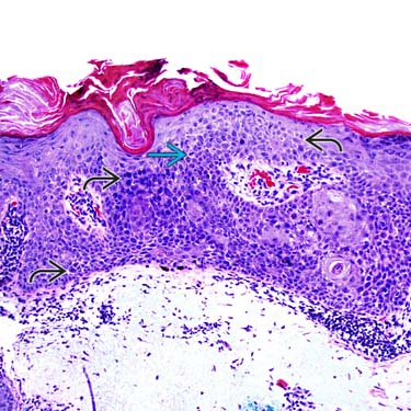
 , with a mitotic figure
  . Overlying hyperkeratosis and parakeratosis are also present.
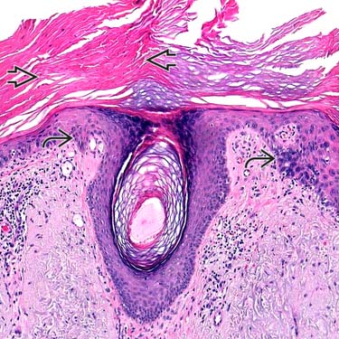
 , with sparing of a central hair follicle. Areas of prominent overlying parakeratosis
  are also present.
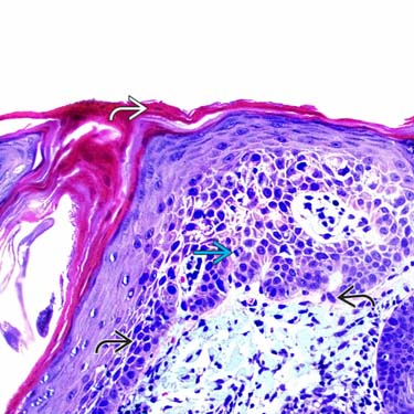
 , with a mitotic figure
  . Parakeratosis is also present
  within and overlying the follicle.


 Hypertrophic AK


