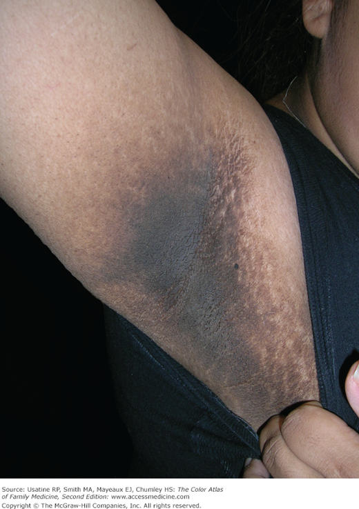
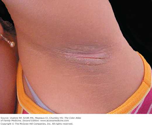
Patient Story
An adolescent girl with obesity and recently diagnosed type II diabetes mellitus (DM) presents to her family physician with concerns about a “dirty area” under her arms and on her neck that “couldn’t be cleaned” (Figure 220-1). She has irregular periods that often skip several months at a time. The physician makes the diagnosis of acanthosis nigricans.
Introduction
Epidemiology
- In a cross-sectional study conducted in a southwestern practice-based research network (N = 1133), AN was found in 17% of children and 21% of adults.1
- In 2 studies, AN was present in 36% of patients with newly diagnosed DM and 39% of children with obesity.2,3
- AN is sometimes associated with malignancy, primarily adenocarcinoma (60%) of the stomach, gallbladder, colon, ovary, pancreas, rectum, and uterus.4,5
- Although most cases are idiopathic, there are also genetic causes of AN.5
- A condition of hyperandrogenism (HA), insulin resistance (IR), and AN called HAIR-AN syndrome occurs in approximately 1% to 3% of women with HA.6 This syndrome may also be seen in patients with autoimmune disorders like Hashimoto thyroiditis.
- AN can be an adverse effect from hormonal therapies.7
Etiology and Pathophysiology
- AN results from of long-term exposure of keratinocytes to insulin.
- Keratinocytes have insulin and insulin-like growth receptors on their surface and the pathogenesis of this condition may be linked to insulin binding to insulin-like growth receptors in the epidermis.
- Fibroblast growth factor receptor 3 (FGFR3) gene mutations should be considered in patients with coexistent AN and skeletal dysplasia.8
Diagnosis
The diagnosis of AN is made clinically in a patient with or at risk for IR who has the characteristic lesions.
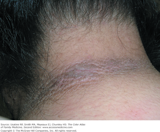
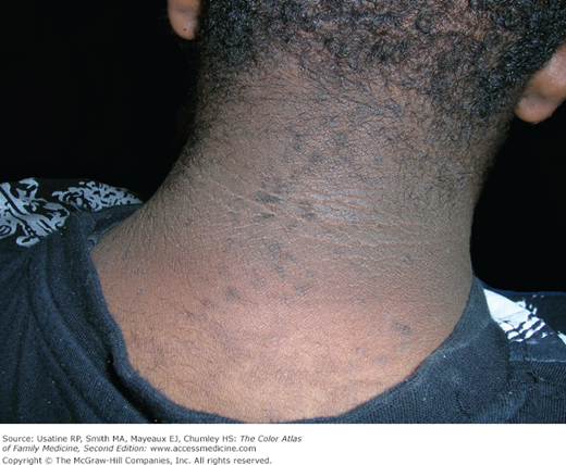
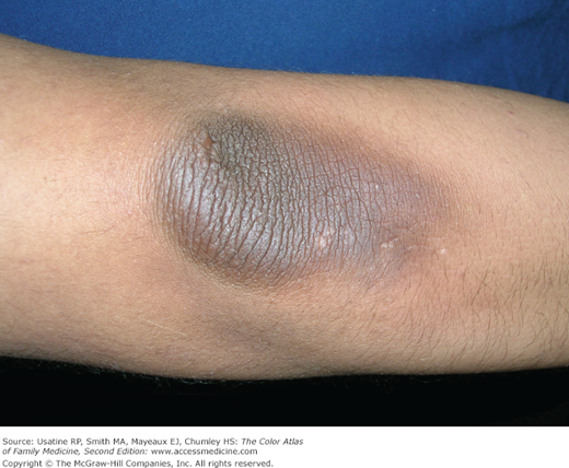
- AN ranges in appearance from diffuse streaky thickened brown velvety lesions to leathery verrucous papillomatous lesions (Figures 220-1, 220-2, 220-3, 220-4, 220-5, 220-6, 220-7, 220-8, and 220-9).
- Women with HAIR-AN syndrome have evidence of virilization (e.g., increased body hair in male distribution, enlarged clitoris) in addition to AN.6