Abnormalities of the red cell membrane

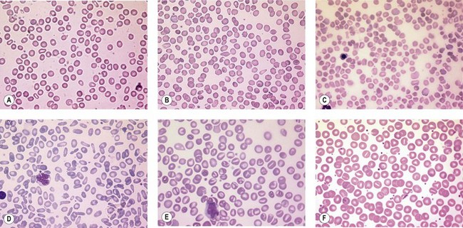
CHAPTER 7 Abnormalities of the red cell membrane


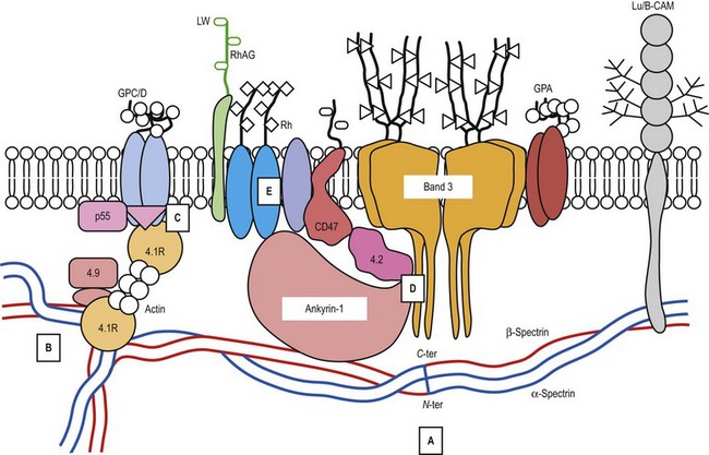
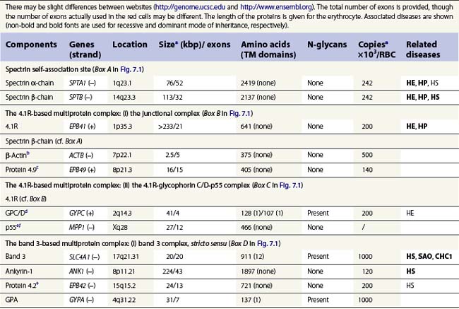
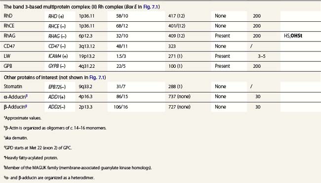
A number of hereditary hemolytic anemias result from mutations affecting the quality and/or amount of proteins that belong to the red cell membrane, its skeleton, or the attachment systems (nexuses) of the latter to the former. Most proteins participate in complexes (Fig. 7.1). They play a role in erythrocyte resilience and elastic deformability, either mechanically, through the skeleton and its attaching systems, or osmotically, through a variety of transporters and pumps. Major proteins and their genes are listed in Table 7.1. The ever increasing number of mutations, too numerous to detail here, have given insight into the function of such protein domains and the regulatory regions of some genes. A selection of abnormally-shaped red cells is shown in Fig. 7.2.


image

Fig. 7.1 Major proteins of the red cell membrane and their organization in complexes. The major proteins, usually belonging to complexes, are represented. Not all proteins mentioned are shown. Box A: the spectrin self-association site. Spectrin α2β2 tetramers form a network lining the inner surface of the lipid bilayer. The α- and β-chains are antiparallel and contain 22 and 17 repeats, respectively. Two dimers associate side-by-side, a process set off at the nucleation sites on both chains, near the C– and N-terminal regions of the α- and β-chains, respectively. Dimers associate head-to-head, α-chain N-terminal region vs. β-chain C-terminal region, at the self-association site, in order to generate tetramers, or higher order oligomers. Spectrin, through its α4 repeat (away from Box A), interacts with the Lu-BCAM protein. Box B: the 4.1R-based multiprotein complex: (i) the junctional complex. Several converging spectrin tetramers interact with oligomeric β-actin, whose length is limited by tropomodulin. 4.1R strengthens this interaction through its 10 kDa domain, which binds to a site located in the spectrin β-chain N-terminal region. Many additional proteins participate in the junctional complex: dematin (protein 4.9), tropomyosin, α- and β-adducin, and several others. Box C: the 4.1R-based multiprotein complex: (ii) the 4.1R-glycophorin C/D-p55 complex. 4.1R interacts through its 30 kDa domain with transmembrane glycophorin C/D and p55 in a triangular fashion. Box D: the band 3-based multiprotein complex: (i) the band 3 complex, stricto sensu. Band 3 appears as a tetramer. The bulky part of each band 3 monomer represents 12 transmembrane segments of band 3. The stalky part accounts for its cytoplasmic domain which serves as an anchor to ankyrin-1, protein 4.2 and many cytoplasmic proteins. Ankyrin-1, in turn, binds to spectrin β-chain (C-terminal region). Recently, band 3 has also been demonstrated to be present in the 4.1R multiprotein complex, making the interactions much more complicated (not shown). Box E: the band 3-based multiprotein complex: (ii) the Rh complex. It includes the Rh polypeptides (RhD/RhCE) and the RhAG protein (Rh-associated glycoprotein), being arranged as a trimer; the latter is associated with CD47, the Landsteiner–Wiener glycoprotein (LW, also called ICAM-4) and glycophorin B.


It should be pointed out that although the genes involved are expressed in a wide range of cell types as isoforms (spliceoforms in particular), genetic disorders are usually confined to the erythroid line. Naturally affected animals and animals with experimentally invalidated genes are helpful, although they do not necessarily mirror the human diseases.


Hereditary spherocytosis


Hereditary spherocytosis (HS) is the most common genetic disorder of the red cell membrane in Western countries. Its incidence has been estimated as 1 in 2000 live births and there is a wide spectrum of clinical severity. In typical cases the hemolytic anemia is moderate, with an increased reticulocyte count a reticulocytosis, intermittent jaundice, gallstones and splenomegaly. Severe cases are rare and may cause death in utero or shortly after birth. In contrast, patients with mild HS may be over 60 years of age when diagnosed. Parvovirus B19 infection commonly occurs. Blood films show a variable percentage of spherocytes. The diagnosis relies on an increased percentage of hyperdense cells and a reduction in osmotic resistance, and on a number of tests, including polyacrylamide gel electrophoresis of the red cell membrane proteins in the presence of sodium dodecylsulfate (SDS-PAGE). The main treatment is splenectomy, though the need for this should be carefully weighed owing to its complications, namely severe infections and a statistically significant increase in thromboembolic accidents.1 Transfusions may be necessary.


Most cases of HS result from reduced or absent proteins. Consequently, the lining of the inner surface of the lipid bilayer by the skeletal meshwork is less dense. Microvesicles bud out of naked bilayer patches. The surface area shrinks and the normal discoid cells gradually turn into spherocytes. The six genes most commonly involved in HS are discussed below.


ANK1 gene mutations


Ankyrin-1 is encoded by ANK1.2 It connects the skeleton to band 3, i.e. the anion exchanger-1. Approximately 60% of HS are due to ANK1 gene mutations and have reduced ankyrin-1. HS due to ANK1 mutations is relatively severe and has a dominant inheritance pattern, although de novo mutations may occur. Homozygosity is bound to be lethal. (One case has been recently described, however.) This may not be evident due to the elevated reticulocyte count, as young cells have a higher ankyrin-1 content. Spectrin α- and β-chains, and protein 4.2, interacting with ankyrin-1, are secondarily decreased.

Only gold members can continue reading. Log In or Register to continue

Stay updated, free articles. Join our Telegram channel

Feb 19, 2017 | Posted by in PATHOLOGY & LABORATORY MEDICINE | Comments Off on Abnormalities of the red cell membrane

Full access? Get Clinical Tree

Get Clinical Tree app for offline access