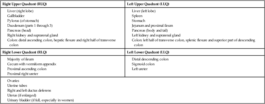
• Lies between the diaphragm and pelvic inlet • Largest cavity in the body and is continuous with pelvic cavity • Lined with parietal peritoneum, a serous membrane • Bounded superiorly by the diaphragm • Spleen, liver, part of stomach, and part of kidneys lie under dome and are protected by lower ribs and costal cartilages. • Lower extent lies in greater pelvis. • Anterior and lateral walls composed of muscle • Posterior wall made up of vertebral column, lower ribs, and associated muscles • Anterior ends of lower six ribs (ribs 7 to 12) (see Section 3.3, Thorax—Body Wall) • Costal margin: formed by medial borders of 7th through 10th costal cartilages • From xiphoid process and 5th through 7th costal cartilages → pubic symphysis and pubic crest • Contains rectus abdominis muscle (see Section 4.2, Abdomen—Body Wall) • Slight indentation that can sometimes be seen extending from xiphoid process to pubic symphysis • Fibrous raphe where aponeuroses of external and internal abdominal oblique and transversus abdominis muscles on either side unite • Semilunar line (linea semilunaris) • Vertical indentation seen as curved line from tip of 9th rib cartilage to pubic tubercle on each side in well-muscled individuals • Transverse attachments between anterior rectus sheath and rectus abdominis muscle • May be seen as transverse grooves in skin on either side of midline (“six-pack”) • Mainly in right upper quadrant, behind ribs 7 through 11 on right side • Crosses midline to reach toward left nipple (see Section 4.5, Abdomen—Viscera [Accessory Organs]) • Urinary system—kidneys and ureters • Organs that develop within abdominal cavity and then become retroperitoneal • All the rest of the organs are peritoneal • Clinicians usually divide the abdomen is into four quadrants for descriptive purposes, using the following planes: • Median plane: imaginary vertical line following line alba from xiphoid process to pubic symphysis • Transumbilical plane: imaginary horizontal line at level of umbilicus • These lines or planes create four quadrants • Clinicians may divide the abdomen into nine regions • Horizontal planes (in descending order): • Subcostal plane: passes through lower border of 10th costal cartilage on either side • Sometimes transpyloric plane is used instead of subcostal; passes through pylorus on right and tips of 9th costal cartilage on either side • Transumbilical plane: passes through umbilicus at level of L3/4 intervertebral disc • Transtubercular (intertubercular) plane: passes through tubercles of iliac crests and the body of L5 • These planes create nine abdominal regions: • Right and left hypochondriac regions, superiorly on either side • Right and left lumbar (flank) regions, centrally on either side • Right and left inguinal (groin) regions, inferiorly on either side • Epigastric region superiorly and centrally • Umbilical region, with umbilicus as its center • Hypogastric or suprapubic region, inferiorly and centrally • Descriptive quadrants and regions are essential in clinical practice. • Each area represents certain visceral structures • Allow correlation of pain and referred pain from these areas to specific organs. • Regions and quadrants are palpated, percussed, and auscultated during clinical examination. Contents of the Abdominal Quadrants • McBurney’s point is a surface landmark that roughly indicates the location of the appendix, located approximately one third of the way along a line from the anterior superior iliac spine to the umbilicus. • Appendicitis is inflammation of the appendix. Pain first presents in the epigastric region, moves to the umbilical region, and then localizes in the right lower quadrant. Rupture of the appendix leads to peritonitis (inflammation of the peritoneum). This presents with severe pain, fever, and abdominal rigidity. • Muscle-splitting incision (of McBurney) is used to access the appendix. Each muscle layer is split in the direction of the fiber orientation. If the incision extends too far laterally the ascending branch of the deep circumflex iliac artery may be severed. • Superficial fascia: two layers in abdomen • Fatty superficial layer (Camper’s fascia) • Deeper membranous layer (Scarpa’s fascia) • Deep fascia—very thin layer investing most superficial muscles • Transversalis fascia (endoabdominal fascia) • Endoabdominal fat separates the transversalis fascia from the parietal peritoneum. • Can compress abdominal contents, thus raising intra-abdominal pressure, such as in sneezing, coughing, defecating, micturating, lifting, and childbirth • Four paired muscles make up anterolateral abdominal wall: three flat muscles and single vertical muscle. • Largest and most superficial • Fibers run inferiorly and medially and end in aponeurosis that contributes to rectus sheath. • Inferior border of its aponeurosis forms inguinal ligament, where it thickens and folds back on itself. • Innervated segmentally by T6–T12 spinal nerves and subcostal nerve • Fibers run inferiorly and laterally and end in an aponeurosis that contributes to rectus sheath. • Inferior aponeurotic fibers join with those of rectus abdominis to form conjoint tendon, inserting onto pubic crest. • Innervated segmentally by ventral rami of T6–T12 spinal nerves • Innermost of three flat muscles • Fibers run transversely and medially and end in an aponeurosis that contributes to rectus sheath. • Innervated segmentally by ventral rami of T6–T12 spinal nerves • Vertical muscle = rectus abdominis • Separated by linea alba in midline • Wider superiorly than inferiorly • Typically composed of four segments connected by tendinous intersections that attach anteriorly to sheath of this muscle • Innervated segmentally by ventral rami of T6–T12 spinal nerves • Have the superior epigastric and inferior epigastric arteries running inferiorly and superiorly, respectively, on their deep surfaces. • Tough, fibrous sheath composed of aponeuroses of three flat muscles • Extends from xiphoid process and 5th through 7th costal cartilages to pubic symphysis and crests • Contains superior and inferior epigastric vessels, lymphatics, and branches of ventral primary rami of T7–T12 • Encloses rectus abdominis and pyramidalis muscles • Has crescent-shaped line—the arcuate line—on its posterior wall approximately three fourths of way down wall. • Anterior wall composed of aponeurosis of external abdominal oblique and anterior layer of aponeurosis of internal abdominal oblique. • Posterior wall composed of posterior layer of aponeurosis of internal abdominal oblique, aponeurosis of transversus abdominis, transversalis fascia of abdomen, and parietal peritoneum. • Aponeuroses of all three flat muscles pass anterior to rectus muscle, reinforcing anterior wall. • Posterior wall composed of just transversalis fascia and parietal peritoneum. • Vessels and nerves enter sheath at its lateral edge—the semilunar line—to supply rectus muscle. • Found between internal abdominal oblique and transversus abdominis • Contains vessels and nerves supplying skin and muscles of anterior and lateral abdominal wall. • Nerves and vessels are transversely oriented and segmental • Anterior cutaneous branches of ventral primary rami of T7–T11 • T7–T9 supply skin above umbilicus • T10 supplies skin around umbilicus • T11 (plus subcostal and ilio-inguinal and iliohypogastric nerves) supplies skin below umbilicus • Subcostal nerves (T12) supply skin below umbilicus • Iliohypogastric and ilio-inguinal nerves (terminal branches of L1) supplies skin below umbilicus • Superficial fascia: single layer • Deep fascia— very thin layer investing the most superficial muscles • Transversalis fascia (endoabdominal fascia) • Endoabdominal fat separates transversalis fascia from parietal peritoneum • Fascia covering psoas muscle • Attaches to lumbar vertebrae and pelvic brim • Thickened superiorly to form medial arcuate ligament— site of origin of muscle of diaphragm • Fascia of quadratus lumborum • Fuses medially with psoas fascia • Thickened superiorly to form lateral arcuate ligament—site of origin of muscle of diaphragm • Thoracolumbar fascia (see Section 2, Back and Spinal Cord) • Lies lateral and is attached to lumbar vertebrae • Tendon passes deep to inguinal ligament to lesser trochanter of femur • Together with iliacus forms iliopsoas muscle, which flexes the hip, helps maintain erect posture • Origin of most of arteries supplying posterior wall • Begins anterior to body of T12 and ends at bifurcation of common iliac arteries at L4 • Follows medial border of psoas • Divides into internal and external iliac arteries at pelvic brim • Gives off inferior epigastric and deep circumflex arteries • Exits under inguinal ligament as the femoral artery • Unpaired visceral branches of abdominal aorta • Unpaired parietal branch: Median sacral artery arising just above aortic bifurcation • Run inferiorly on surface of quadratus lumborum • Supply external abdominal oblique and skin of anterolateral abdominal wall • Ilio-inguinal and iliohypogastric nerves (L1) • Enter abdomen posterior to medial arcuate ligament • Pierce transverse abdominis near anterior superior iliac spine (ASIS) • Lateral femoral cutaneous nerve (L2/3) • Emerges from lateral aspect of psoas muscle • Enters thigh posterior to inguinal ligament and medial to ASIS • Emerges from lateral border of psoas • Passes beneath inguinal ligament on surface of iliopsoas muscle • Greater (T5–T9), lesser (T10–T11), and least (T12) thoracic splanchnic nerves • Convey presynaptic sympathetic fibers to celiac, superior mesenteric, and aorticorenal sympathetic ganglia • Rise of abdominal sympathetic trunks • Convey presynaptic sympathetic fibers to inferior mesenteric, intermesenteric, and superior hypogastric plexuses • Prevertebral sympathetic ganglia • Contain preganglionic sympathetic and parasympathetic fibers, postganglionic sympathetic fibers, sympathetic ganglia (prevertebral), and visceral afferent fibers • Some named for major blood vessels (periarterial): celiac, superior mesenteric, inferior mesenteric, intermesenteric, aorticorenal • Superior hypogastric plexus—continuous with inferior mesenteric and intermesenteric plexuses at aortic bifurcation • Lined by parietal peritoneum • Five peritoneal folds, inferior to umbilicus • Peritoneal fossae are formed between the umbilical folds. • Supravesical fossae: between median and medial folds • Medial inguinal fossae: between medial and lateral folds • Oblique canal, approximately 4 cm long at inferior margin of anterior abdominal wall • Parallel and superior to medial half of inguinal ligament • Deep inguinal ring: internal entrance to canal • Entrance to canal through transversalis fascia • Located 1.25 cm superior to midpoint of inguinal ligament • Superficial ring: external exit of canal • Anterior wall: external oblique aponeurosis (and internal oblique laterally) • Transversalis fascia laterally • Internal oblique and conjoint tendon (joint insertion of aponeuroses of internal oblique and transverses abdominis) medially • Roof: arching fibers of internal oblique • Floor: inguinal ligament, reinforced medially by lacunar ligament
Abdomen Study Guide
4.1 Topographic Anatomy
Guide
Abdomen—Overview
Bony Landmarks of the Abdomen
Abdominal Contents
Abdominal Quadrants and Regions
Right Upper Quadrant (RUQ)
Left Upper Quadrant (LUQ)
Right Lower Quadrant (RLQ)
Left Lower Quadrant (LLQ)

Clinical Points
Mcburney’s Point and Appendicitis
4.2 Body Wall
Guide
Anterolateral Abdominal Wall
Fascial Layers
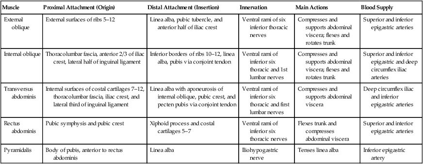
Muscles
Rectus Sheath
Muscle
Proximal Attachment (Origin)
Distal Attachment (Insertion)
Innervation
Main Actions
Blood Supply
External oblique
External surfaces of ribs 5–12
Linea alba, pubic tubercle, and anterior half of iliac crest
Ventral rami of six inferior thoracic nerves
Compresses and supports abdominal viscera; flexes and rotates trunk
Superior and inferior epigastric arteries
Internal oblique
Thoracolumbar fascia, anterior 2/3 of iliac crest, lateral half of inguinal ligament
Inferior borders of ribs 10–12, linea alba, pubis via conjoint tendon
Ventral rami of inferior six thoracic and 1st lumbar nerves
Compresses and supports abdominal viscera; flexes and rotates trunk
Superior and inferior epigastric and deep circumflex iliac arteries
Transversus abdominis
Internal surfaces of costal cartilages 7–12, thoracolumbar fascia, iliac crest, and lateral third of inguinal ligament
Linea alba with aponeurosis of internal oblique, pubic crest, and pecten pubis via conjoint tendon
Ventral rami of inferior six thoracic and first lumbar nerves
Compresses and supports abdominal viscera
Deep circumflex iliac and inferior epigastric arteries
Rectus abdominis
Pubic symphysis and pubic crest
Xiphoid process and costal cartilages 5–7
Ventral rami of inferior six thoracic nerves
Flexes trunk and compresses abdominal viscera
Superior and interior epigastric arteries
Pyramidalis
Body of pubis, anterior to rectus abdominis
Linea alba
Iliohypogastric nerve
Tenses linea alba
Inferior epigastric artery

Innervation
Posterior Abdominal Wall
Fascial Layers
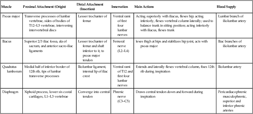
Muscles
Muscle
Proximal Attachment (Origin)
Distal Attachment (Insertion)
Innervation
Main Actions
Blood Supply
Psoas major
Transverse processes of lumbar vertebrae, sides of bodies of T12–L5 vertebrae, intervening intervertebral discs
Lesser trochanter of femur
Ventral rami of first four lumbar nerves
Acting superiorly with iliacus, flexes hip; acting inferiorly, flexes vertebral column laterally; used to balance trunk in sitting position; acting inferiorly with iliacus, flexes trunk
Lumbar branch of iliolumbar artery
Iliacus
Superior 2/3 iliac fossa, ala of sacrum, and anterior sacro-iliac ligaments
Lesser trochanter of femur and shaft inferior to it, to psoas major tendon
Femoral nerve (L2–L4)
lexes thigh at hips and stabilizes hip joint, acts with psoas major
Iliac branches of iliolumbar artery
Quadratus lumborum
Medial half of inferior border of 12th rib, tips of lumbar transverse processes
Iliolumbar ligament, internal lip of iliac crest
Ventral rami of T12 and first four lumbar nerves
Extends and laterally flexes vertebral column, fixes 12th rib during inspiration
Iliolumbar artery
Diaphragm
Xiphoid process, lower six costal cartilages, L1–L3 vertebrae
Converge into central tendon
Phrenic nerve (C3–C5)
Draws central tendon down and forward during inspiration
Pericardiacophrenic musculophrenic, superior and inferior phrenic arteries

Arteries of Posterior Abdominal Wall (see Section 4.6, Abdomen—Visceral Vasculature)
Nerves of Posterior Abdominal Wall (see Section 4.7, Abdomen—Innervation)
Internal Features of Anterior Abdominal Wall
Inguinal Canal: Feature of Anterior Abdominal Wall.
Abdomen Study Guide


