50 CASE 50
PATHOPHYSIOLOGY OF KEY SYMPTOMS
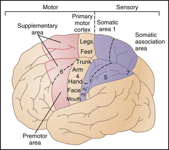
The cortical regions involved in muscle control are the primary motor cortex, the premotor area, and the supplemental motor areas. The premotor area coordinates patterns of movement and is interconnected with the basal ganglia, cerebellum, somatosensory areas, thalamus, and primary motor cortex (Fig. 50-1).
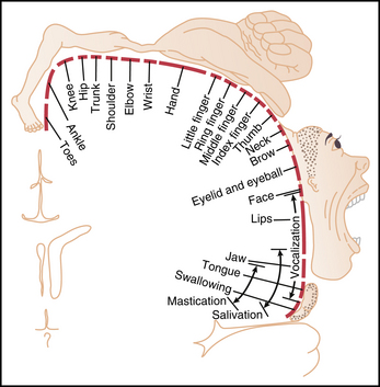
The primary motor cortex exerts executive control of voluntary movement. It is located in the frontal lobes anterior to the central sulcus (precentral gyrus). The neurons of the primary motor cortex have a somatotopic organization, with the feet and legs located midline and the head and face extending toward the lateral (Sylvian) fissure. Axons from the neurons of the primary motor cortex (upper motor neurons) descend and synapse within the spinal cord (corticospinal tract) or synapse within the motor nuclei of the brain stem (corticobulbar tract). Corticorubral fibers project to the red nucleus of the mesencephalon, which then sends a projection into the spinal cord as the rubrospinal tract. Motor signals are also transmitted to the basal ganglia and cerebellum. Lower motor neurons are the final common pathway to skeletal muscles. They are the motor neurons of the cranial nerve nuclei that innervate skeletal muscles as well as the motor neurons of the ventral horn of the spinal cord that innervate the rest of the body’s musculature (Fig. 50-2).

FIGURE 50–2 Degree of representation of the different muscles of the body in the motor cortex.
(Redrawn from Penfield W, Rasmussen T: The Cerebral Cortex of Man: A Clinical Study of Localization of Function. New York, Hafner, 1968.)
Stay updated, free articles. Join our Telegram channel

Full access? Get Clinical Tree


