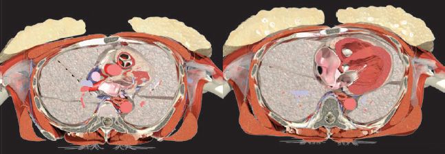
The upper part of the trunk is called the thorax, or chest. It lies between the neck and abdomen, and is mainly formed by the bony ribcage. This structure protects the contents of the thoracic cavity (the space inside the ribcage), as well as moving to allow us to breathe.
The thorax contains the heart, lungs, and large blood vessels, along with other structures moving between the neck above, and the abdomen below. The thoracic cavity is separated from the abdominal cavity by the muscular diaphragm. Numerous muscles that assist with breathing and movements of the arms and trunk are attached to the rib cage. The breasts are found at the front of the thorax.

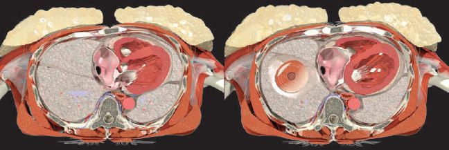
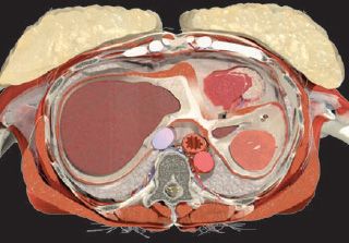
BONES OF THE THORAX
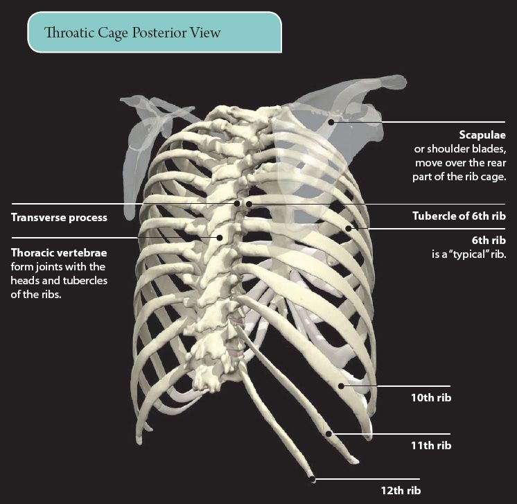
The ribs, sternum, and thoracic vertebrae form the bony framework of the thorax, the thoracic cage. There are twelve pairs of ribs. They form joints at the back with the thoracic vertebrae. At the front, most are connected to the sternum by flexible costal cartilages.
The rib cage is mobile, with the ribs able to move up and down. This movement expands the thoracic cavity and allows us to breathe. A typical rib has a head, neck, tubercle, and body.
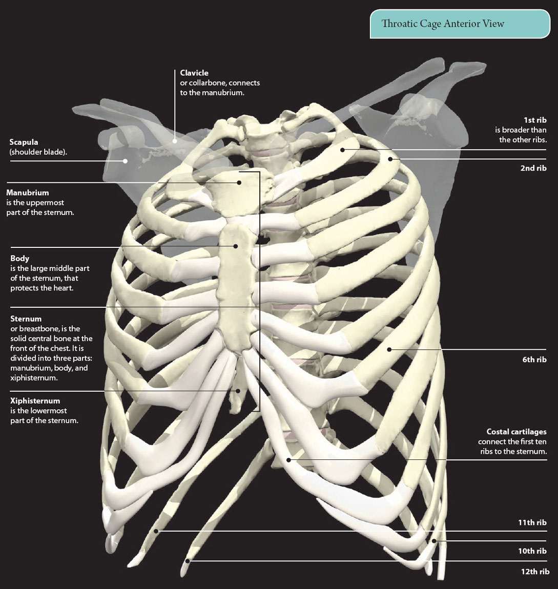
Ribs and Sternum
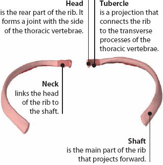
Rib 6
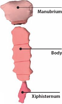
Sternum, manubrium, and xipisternum
Ribs
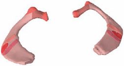
Rib 1
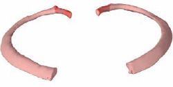
Rib 2
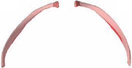
Rib 10
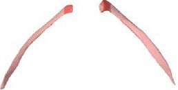
Rib 11
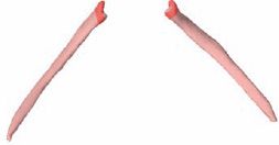
Rib 12
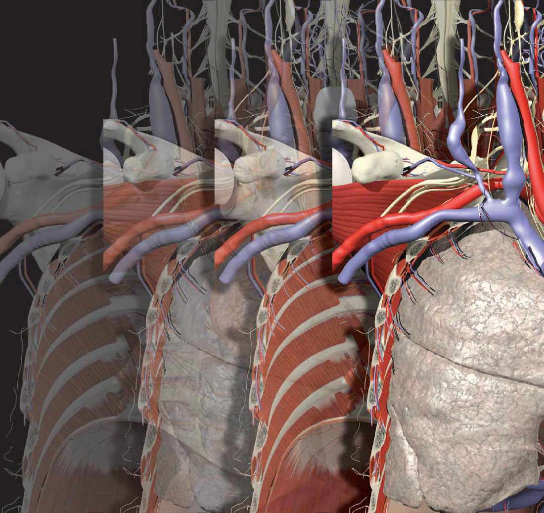
MUSCLES OF THE THORAX
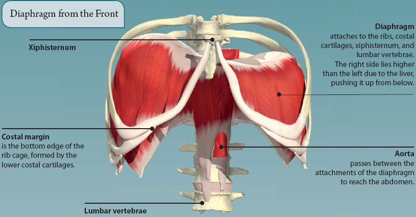
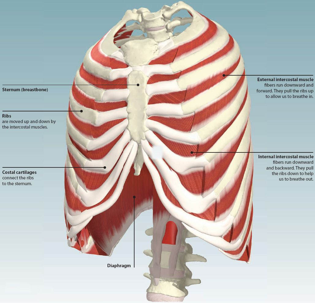
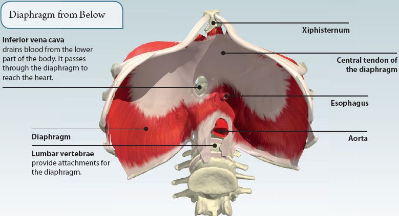
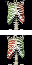
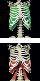
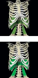
The main muscles of the thorax are the diaphragm and intercostal muscles. They are called muscles of respiration, as their main role is to help us breathe.
The intercostal muscles lie between the ribs, and move the rib cage up and down.
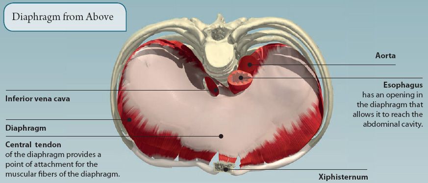
The diaphragm is a large dome-shaped sheet of muscle that separates the thoracic and abdominal cavities. When it contracts, it flattens and moves downward. This increases the size of the thoracic cavity and helps us to breathe in.
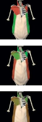
Other muscles that lie on the surface of the rib cage can help when we need to breathe hard and fast, for example, during exercise.
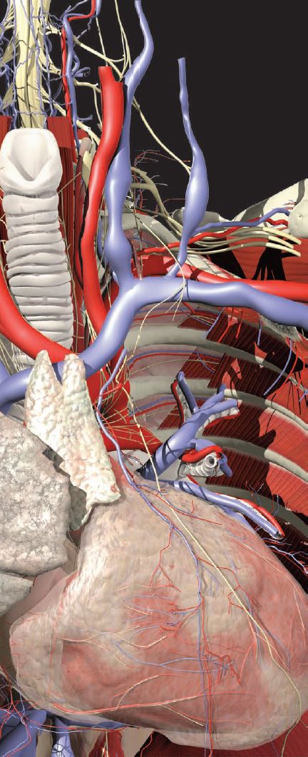
NEUROVASCULAR STRUCTURES OF THE THORAX
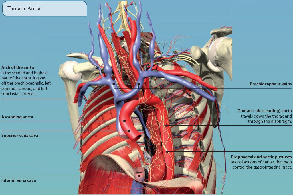
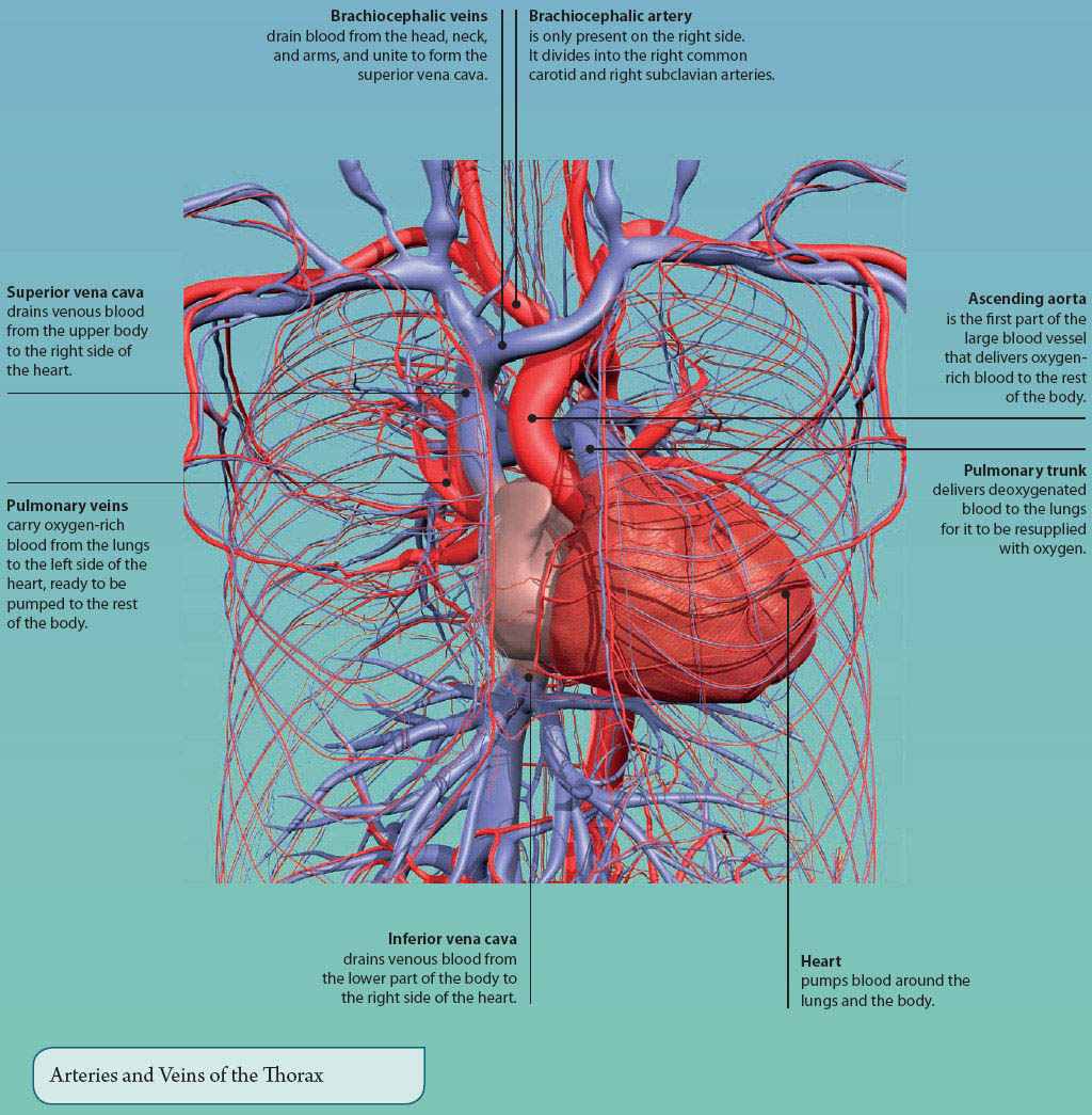
The thorax contains the large blood vessels that enter and leave the heart, along with numerous other branches.
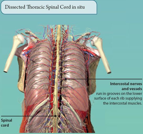
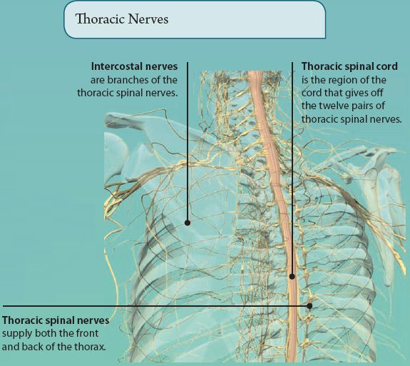
Twelve pairs of thoracic spinal nerves innervate the skin of the thorax and intercostal muscles.
THE HEART
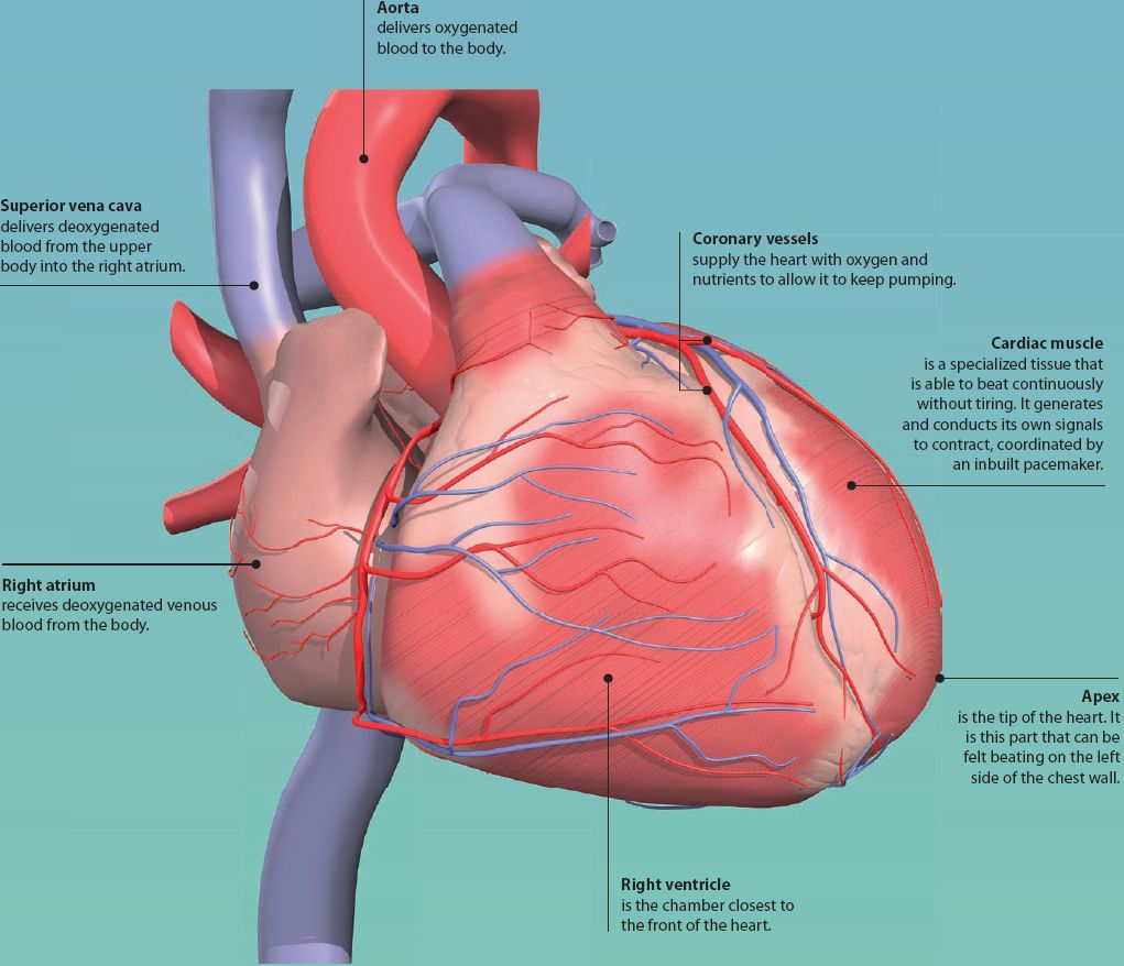
The heart is a fist-sized, muscular organ. It is located in the chest between the lungs, and is tilted to the left side. It continuously pumps blood around the lungs and body. This provides the cells and tissues of the body with the oxygen and nutrients they need to stay alive.
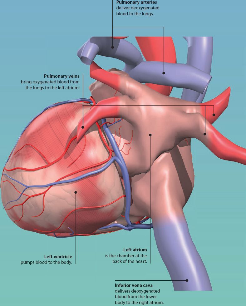
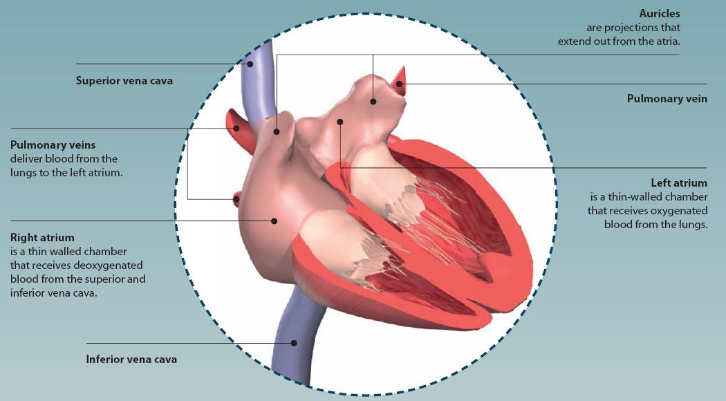
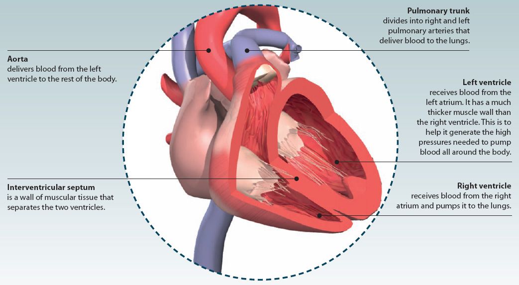
The heart is divided into right and left sides, which pump blood around different circuits. The right side of the heart receives deoxygenated blood from the body and pumps this to the lungs. The left side of the heart receives oxygenated blood from the lungs and pumps this to the rest of the body. Each side of the heart has two chambers: an atrium (priming chamber) and a ventricle (pumping chamber).
CHAMBERS OF THE HEART
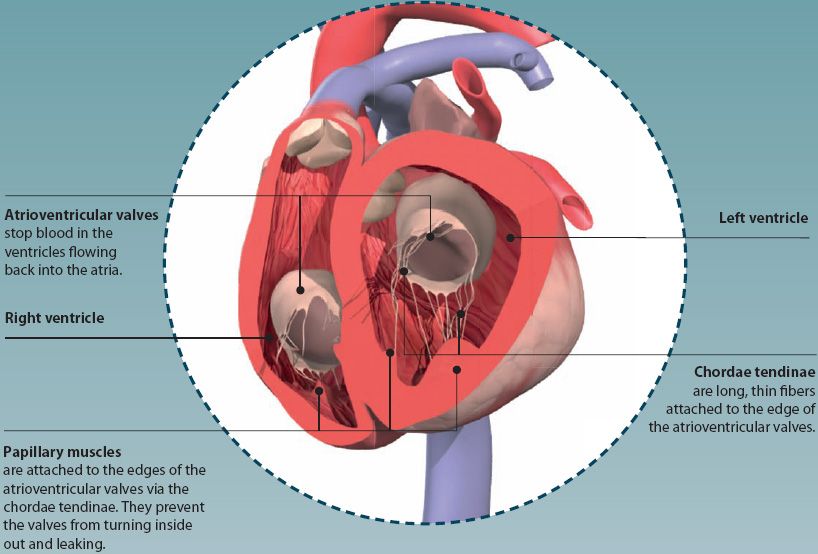
The heart has four chambers (two atria and two ventricles) in which blood is collected and pumped. The chambers of the right and left side of the heart are separated by a fibromuscular wall called the septum.
Atrioventricular valves lie between the atria and ventricles on each side. These ensure that blood only flows in one direction through the heart.
Did you know?
On average, the human heart beats about 100, 000 times a day; that’s approximately 35 million times every year without a rest!
Stay updated, free articles. Join our Telegram channel

Full access? Get Clinical Tree


