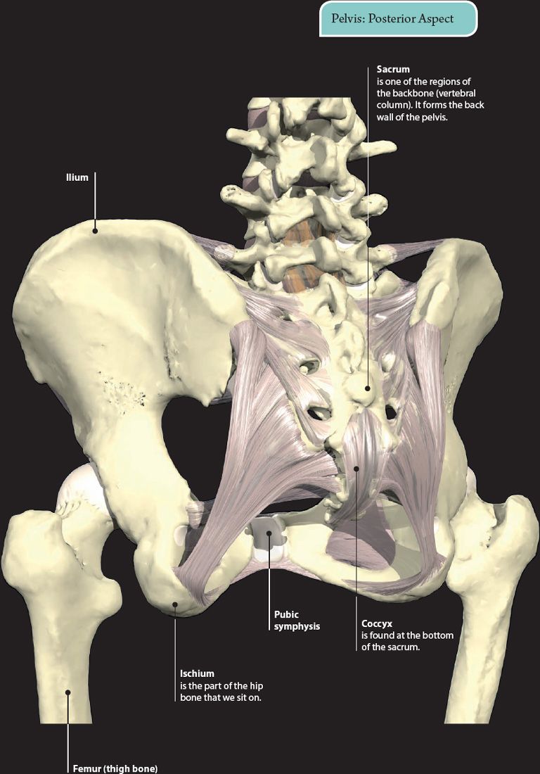
The pelvis is the strong, bowl-shaped area at the base of the trunk, where the legs attach to the rest of the body. It is formed from the hip and lower back bones. It contains and protects the reproductive organs and lower parts of the urinary and digestive systems. A muscular sheet provides support to these organs and forms the pelvic floor.
The pelvis provides attachment for various muscles that move both the trunk and the legs. The shape and position of the pelvis helps us in standing, walking, and keeping an upright position.
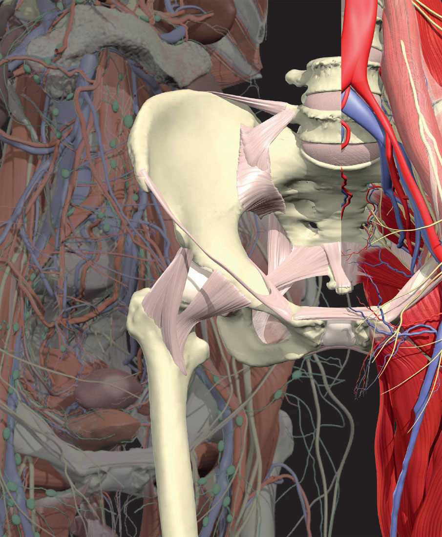
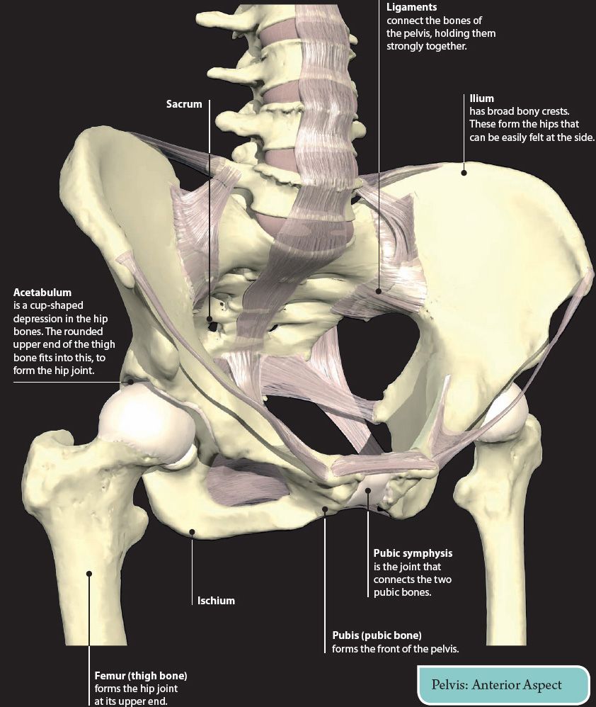
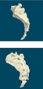
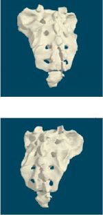
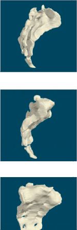
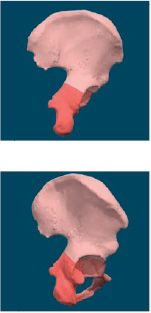
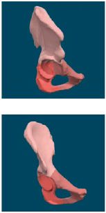
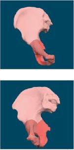
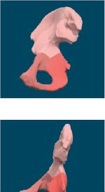
HIP BONE
The pelvis is formed by the two hip bones, and the sacrum and coccyx at the back. The hip bone is formed by three separate bones (ilium, ischium, and pubis), which fuse together. They all meet at the acetabulum. The central bowl-shaped region protected by the bones is known as the pelvic cavity, and contains the pelvic organs.
The hip bones form the pelvic girdle, which is the point of attachment of the lower limbs to the rest of the body.
In diseases that affect the blood cells, it is often helpful for doctors to analyze a sample of the bone marrow. A common site for obtaining this sample is either side of the lower back, where the hip bone is near the surface. A special needle is passed into the bone, and a sample of marrow is obtained.
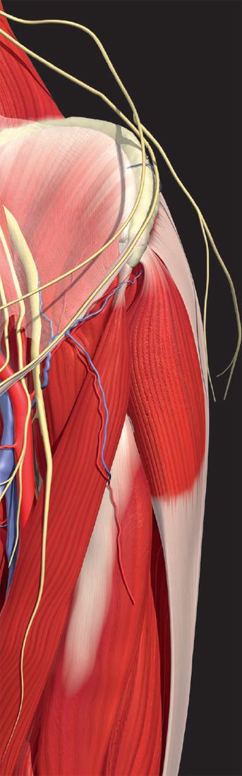
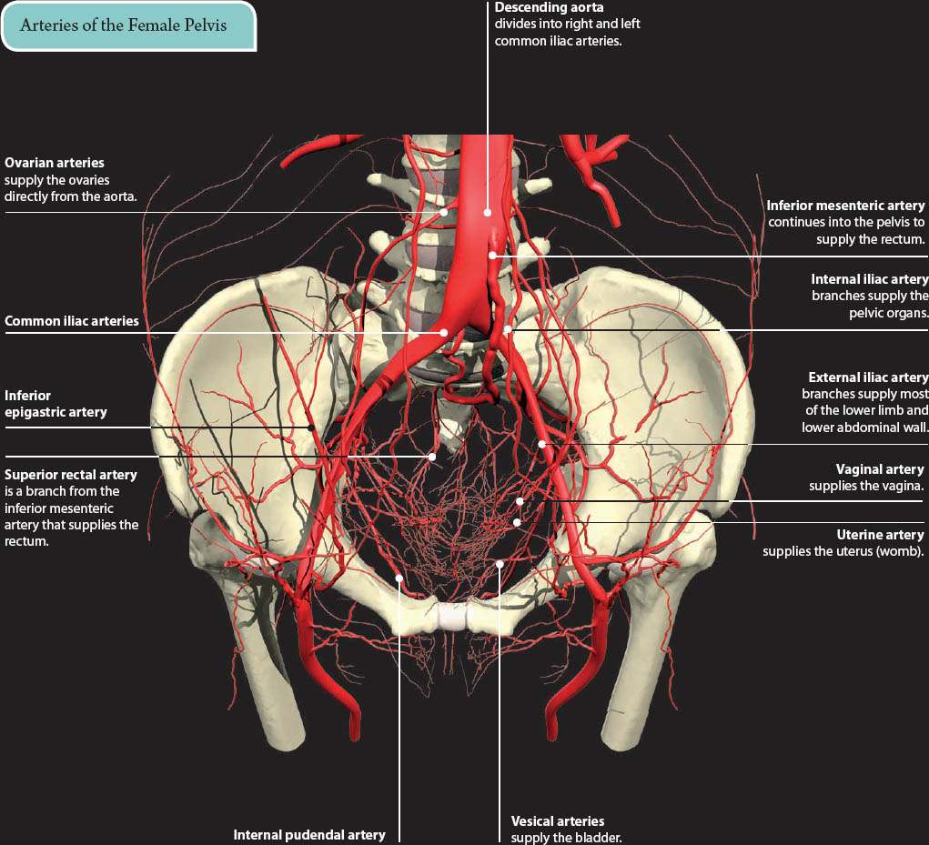
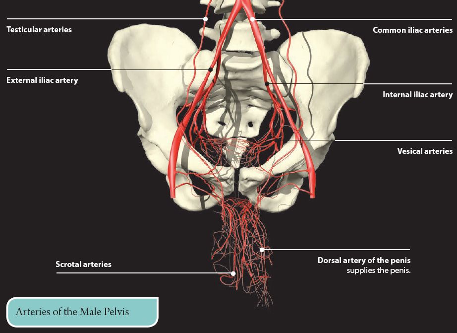
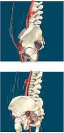
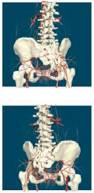
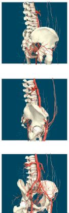
VESSELS OF THE PELVIS
The descending aorta splits into two common iliac arteries just above the pelvis. These vessels and their branches supply the pelvic organs and the lower limbs.
Although most blood vessels are the same in males and females, there are some variations that reflect the differences in the reproductive organs.
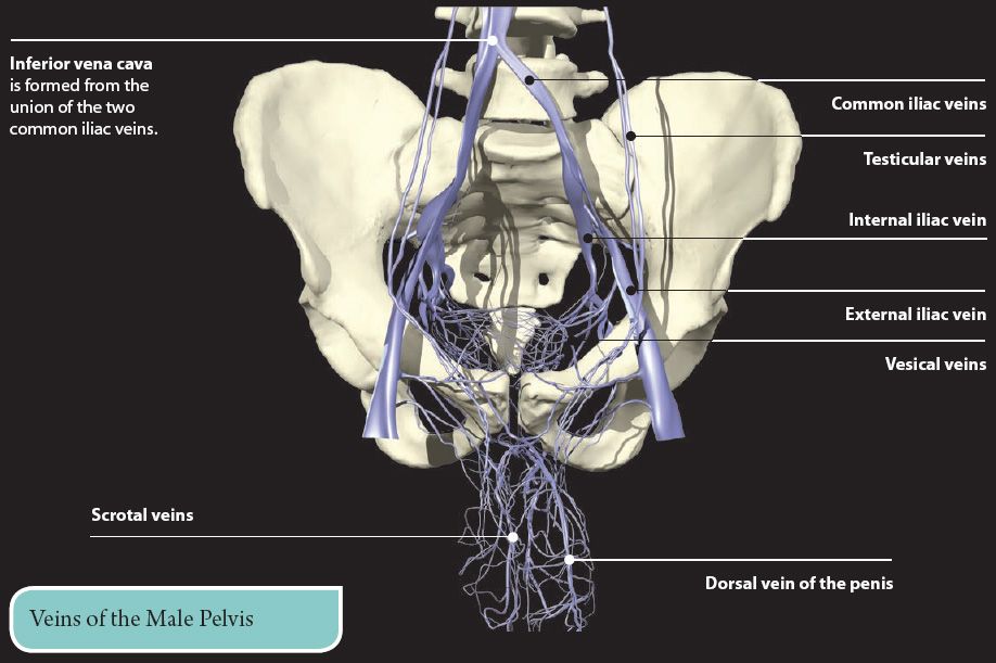
In general, the veins of the pelvis follow the same pattern as the arteries.
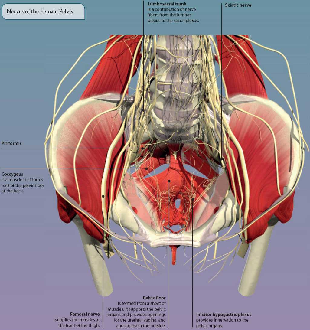
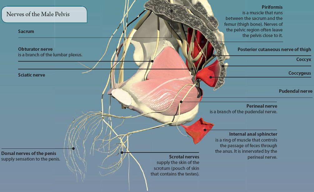
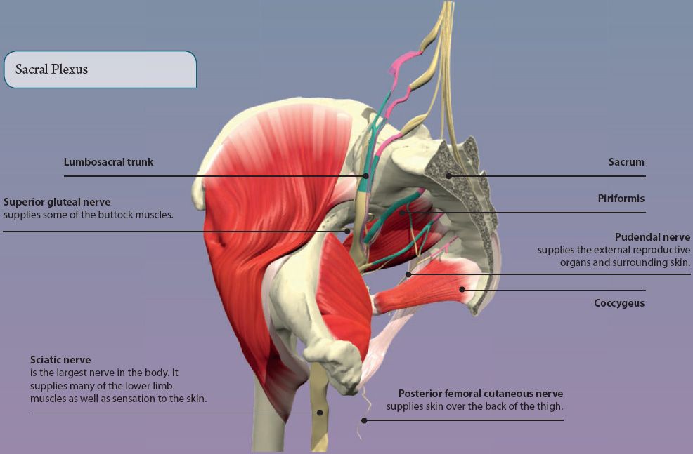
NERVES OF THE PELVIS
The pelvis contains the nerves that form the sacral plexus, along with branches from the lumbar plexus. Between them, these collections of nerves supply the organs, muscles, and skin of the pelvis and lower limb.
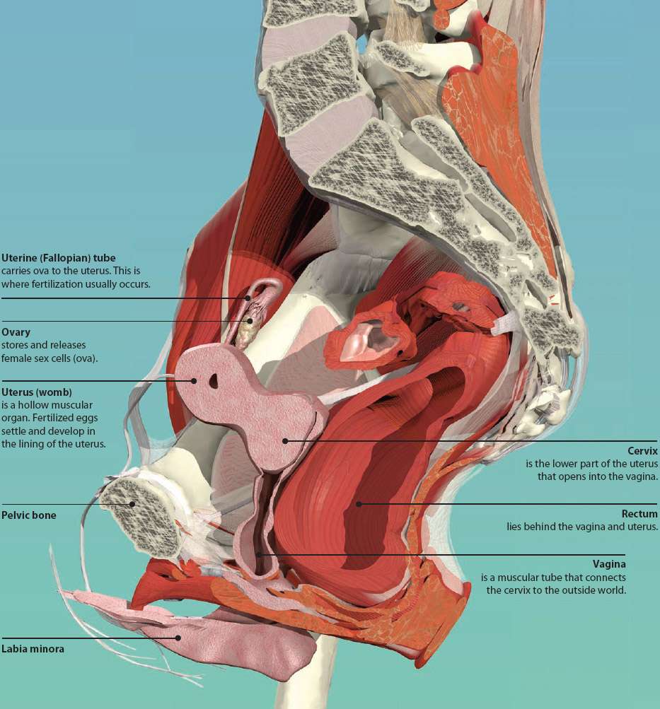
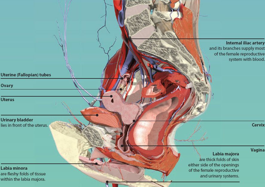
FEMALE REPRODUCTIVE SYSTEM
The female reproductive system includes the ovaries, uterine (Fallopian) tubes, uterus, and vagina. Every month from puberty (usually early teens) to menopause (usually early fifties), it undergoes a series of events that prepare it for the possibility of pregnancy. These changes are brought about by hormones, such as estrogen and progesterone.
Every month, a female sex cell (ovum) is released from the ovaries into the uterine tubes. If the ovum is fertilized by a sperm (male sex cell) it settles into the lining of the uterus (womb), where it grows and develops into a baby. After nine months, strong contractions of the uterus push the baby out in the process of childbirth.
Infection of the uterine tubes is called salpingitis. Following infection, scar tissue may form, which blocks the uterine tubes and prevents the egg and sperm from meeting, resulting in infertility.
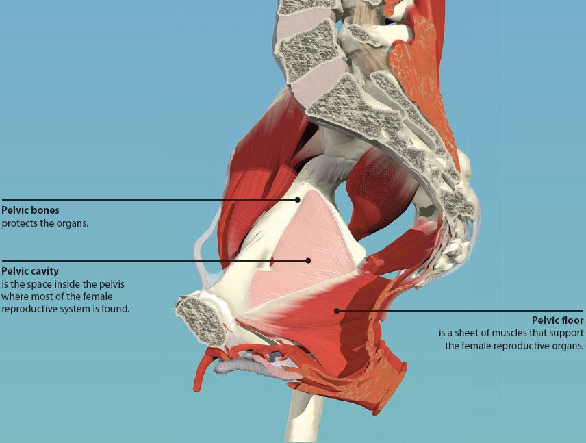
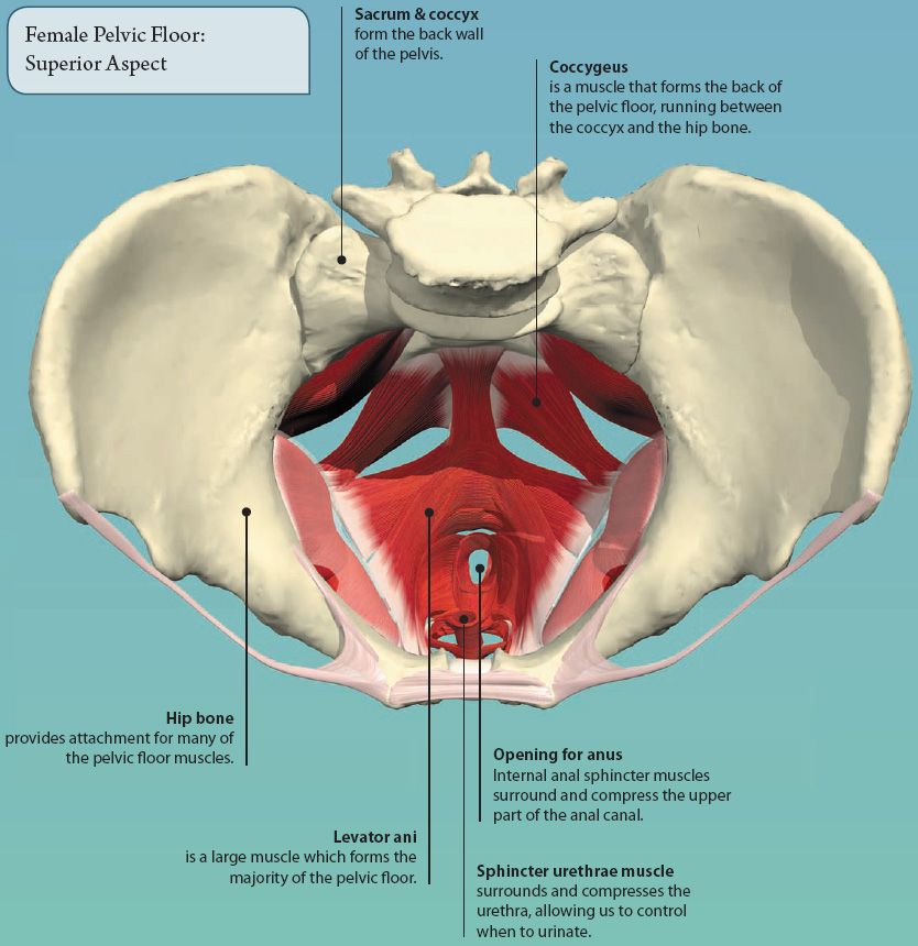
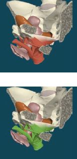
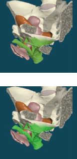
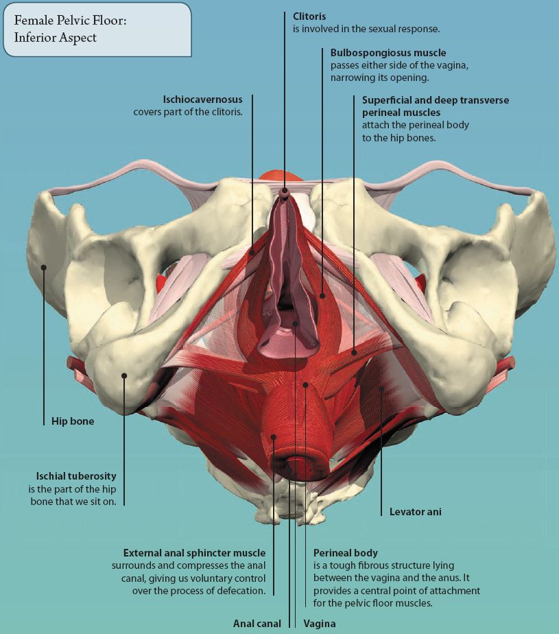
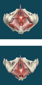
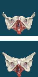
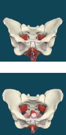
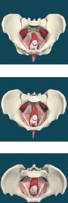
FEMALE PELVIC FLOOR
The pelvic floor is a muscular sheet that forms the base of the pelvic cavity. It is made up of numerous muscles, suspended from the pelvic bones. These form a hammocklike support for the pelvic organs. In the female it has openings for the urethra, vagina, and anus.
OVARIES
The ovaries are paired almond-shaped structures located within the peritoneal cavity, close to the entrance to the uterine tubes. At birth, the ovaries contain all the developing female gametes, called ova (eggs). These are surrounded by specialized cells to form follicles. Every month during the reproductive years, a few follicles mature and grow until one is large enough to rupture, releasing its ovum in a process called ovulation.
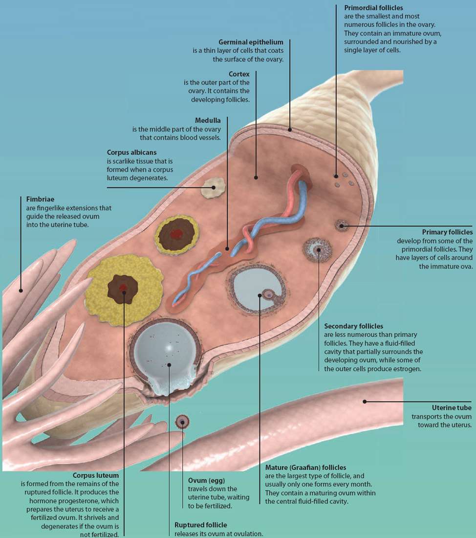
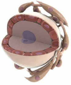
Primordial Follicle
Stay updated, free articles. Join our Telegram channel

Full access? Get Clinical Tree


