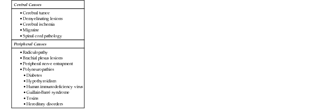
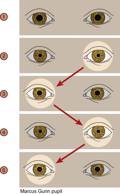
38 A 32-Year-Old Female With Bilateral Hand Numbness


Case 38

A 32-Year-Old Female With Bilateral Hand Numbness


Albert Huang, John Khoury


A 32-year-old female presents to an outpatient clinic with numbness in her hands. It started several days prior and was not associated with any specific incident. She describes additional tingling as pins and needle sensations. The symptoms have been constant and she is unable to identify anything that makes it better or worse.


How can occupational history contribute to the evaluation in this case?


A potential cause of hand numbness is peripheral nerve entrapments involving the radial, ulnar, or more commonly the median nerve within the carpal tunnel, also referred to as carpal tunnel syndrome (CTS). It is important to inquire about work or recreational activities that result in repetitive actions. In today’s technological society, heavy use of mobile devices or poor ergonomic wrist placement while typing can result in hand numbness due to CTS. Other activities that result in median nerve damage include frequent wrist flexion while operating machinery or regularly using tools such as wrenches, screwdrivers, and even surgical instruments. Examples of occupations that can lead to repetitive wrist or hand motions include secretaries, mechanics, and surgeons.


Step 1/2/3


Basic Science/Clinical Pearl


The median nerve innervates both sensory and motor components of the hand. Its sensory dermatome involves the anterolateral aspect of the hand. Muscles involved cause flexion of the thumb, index, and middle fingers, as well as flexion of the wrist and pronation of the forearm. Because the sensory fibers are smaller and more sensitive to damage, sensory deficits typically occur first. The presence of weakness and motor deficits is an indication of severe median nerve damage.


What additional questions are important to ask related to the numbness and tingling in her hands?


Other common causes for upper extremity paresthesias include cervical spine pathology, such as a disc herniation or arthritic facet joint spaces leading to narrowed neuroforamen and ultimately damaged nerves exiting the cervical spine. Chronic conditions can result in damage of smaller nerves fibers distally and raise the question of hypothyroidism or diabetes mellitus. However, in this particular case, cervical spine pathology would more commonly present unilaterally and a metabolic cause is less likely considering the patient’s younger age (see Table 38.1).


Upon further questioning, the patient states the numbness and tingling is not associated with actual weakness and does not seem to worsen or improve with any particular movements. Inquiring about her past medical history, she mentions a visit to the emergency department for increased blurry vision in her left eye. She was diagnosed with optic neuritis at that time, and her vision returned to normal 2 weeks later. She is currently employed as a fitness instructor and unable to recall activities that require repetitive actions at her wrist. The remaining review of systems is unremarkable.


How is optic neuritis commonly tested on physical exam?


The swinging light test is commonly utilized to assess both afferent and efferent function of the optic nerves. This test is conducted by swinging a pen light from one eye to the other and assessing for symmetrical bilateral pupil dilation. Despite light being shown in only one eye, the normal response is bilateral pupil constriction. When there is unequal pupil constriction, the examiner must determine whether the cause is due to an afferent or efferent defect. If there is an afferent defect, both pupils demonstrate a symmetrical decreased constriction of the pupils when the light is shone in the affected eye. If the defect is efferent, only the affected eye demonstrates an unequal and diminished pupillary constriction when compared to the unaffected eye no matter which side the light is shone. An afferent pupillary defect is also known as a Marcus Gunn pupil and is commonly seen in cases of optic neuritis (see Fig. 38.1).


Step 2/3


Clinical Pearl


Presence of a Marcus Gunn pupil indicates the optic nerve is still innervated despite decreased function. This dysfunction can be due to inflammation, a demyelinating process, or compression by a tumor. In this case, the affected eye will still demonstrate a bilateral pupillary constriction although decreased when compared to the unaffected side. In contrast, a complete optic nerve lesion or detachment would result in no response when light is shone into the affected eye.


On exam, her temperature is 36.8 °C (98.3 °F), pulse rate is 94/min, blood pressure is 122/84 mm Hg, and respiration rate is 20/min. The cardiac and pulmonary exams are unrevealing. Sensation is present but decreased throughout her right arm and unchanged on the left. The strength in her right arm is slightly deceased to 4+/5 as compared to the left. There is also notable weakness in her legs, approximately 4−/5. Hoffman’s reflex is positive on the right and negative on the left. Tinel’s and Phalen’s signs are negative bilaterally. Flexion of her neck results in an electric-like pain that extends down her back.


Step 1/2/3


Basic Science/Clinical Pearl


Hoffman’s reflex is an upper motor sign akin to the Babinski reflex. It is elicited by holding the middle or ring finger and flicking the distal phalanx. The presence of flexion of the remaining digits is a positive sign and indicative of a possible upper motor neuron lesion. Because it can be seen in normal individuals, a positive sign is only relevant when accompanied by additional history and exam findings consistent with an upper motor neuron lesion.


Jun 15, 2016 | Posted by in GENERAL & FAMILY MEDICINE | Comments Off on 38 A 32-Year-Old Female With Bilateral Hand Numbness

Full access? Get Clinical Tree

Get Clinical Tree app for offline access