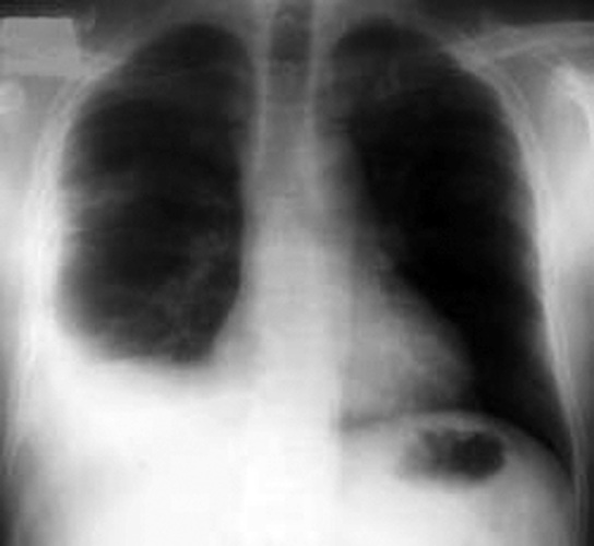
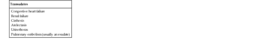
Walter Chou, Raj Dasgupta, Ahmet Baydur A 63-year-old male presents to the emergency department with 3 months of progressive dyspnea on exertion, nonproductive cough, night sweats, and 15 pounds of unintentional weight loss. He denies lower extremity edema, chest pain, hemoptysis, fevers, chills, or animal contact. He has no known past medical history. He has a 30-pack-year history of smoking. He has recently emigrated from Vietnam. On physical exam, his blood pressure is 128/72 mm Hg, pulse rate is 90/min, respiration rate is 20/min, and oxygen saturation is 97% on room air. He has dullness to percussion and decreased breath sounds of the right lower chest. The rest of his physical exam is normal. A chest radiograph (CXR) reveals a moderate right-sided opacity of the right lower lung consistent with a pleural effusion (see Fig. 49.1). A pleural effusion is an excess of fluid in the pleural space between the parietal and visceral pleura of the thorax. A variety of clinical conditions, particularly infection and malignancy, can cause a pleural effusion. Classifying a pleural effusion as exudative or transudative often aids in the diagnosis of the cause of a pleural effusion. Causes of both exudative and transudative effusions are summarized in Table 49.1. Light’s criteria, which use pleural fluid lactate dehydrogenase (LDH), pleural fluid protein, serum LDH, and serum protein levels, are used to evaluate for exudative effusions. Given the various causes of pleural effusions, a thorough history and physical exam often play a key role in diagnosing the etiology of pleural effusions. Congestive heart failure (CHF) usually causes bilateral pleural effusions and can be accompanied by other signs of volume overload such as lower extremity edema, orthopnea, and elevated jugular venous distension. Hepatic cirrhosis, often with concomitant abdominal ascites, can cause a unilateral, typically right-sided, pleural effusion. Various malignancies can cause pleural effusions, either as a primary presentation or in conjunction with other symptoms. Pneumonia can lead to a parapneumonic effusion, which can be simple or complicated. If left untreated, these can lead to frank pus in the pleural space, also called an empyema. A thoracentesis can be both therapeutic and diagnostic but is not always indicated. Relative contraindications include small volume (<10 mm on ultrasound or decubitus radiograph), positive pressure ventilation, systemic anticoagulation or coagulopathies, altered mental status, or an overlying cutaneous infection at the proposed entry site. A therapeutic thoracentesis is indicated to relieve symptoms of dyspnea but should not be performed in patients where previous thoracentesis has not improved their symptoms. A diagnostic thoracentesis should be performed whenever the cause of an effusion is unclear. For example, if a patient is known to have an acute CHF exacerbation and has bilateral pleural effusions, observation with appropriate therapy is acceptable. However, if that same patient’s effusion does not resolve despite 3 days of appropriate therapy, is unilateral, or the patient has atypical features like fever or chest pain, a diagnostic thoracentesis is indicated. There are many options for laboratory testing of pleural fluid (Table 49.2
A 63-Year-Old Male With a Unilateral Pleural Effusion
What can cause pleural effusions?
When should a thoracentesis be performed?
What tests should be performed on pleural fluid?
![]()
Stay updated, free articles. Join our Telegram channel

Full access? Get Clinical Tree


49 A 63-Year-Old Male With a Unilateral Pleural Effusion
Case 49
