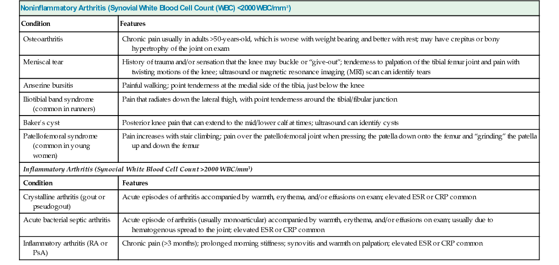
36 A 68-Year-Old Male With Right Knee Pain


Case 36

A 68-Year-Old Male With Right Knee Pain


R. Michelle Koolaee


A 68-year-old male is evaluated for a 2-year history of progressive right knee pain accompanied by morning stiffness lasting 20 minutes. He describes his pain as worse with ambulation (particularly going up and down stairs) and better with rest. He takes naproxen, which minimally relieves the pain. He works as a mail carrier and states that he has had to take several days off of work recently due to the increased severity of pain.


How does the history help to narrow your differential diagnosis?


Table 36.1 summarizes some common causes of knee pain. There are several key questions to always ask anyone who presents to you with knee pain:


On physical exam, vital signs are normal. Body mass index (BMI) is 29 kg/m2. The right knee has a small effusion, with tenderness at the medial joint space; the knee is not erythematous or warm. There is no worsening of pain when compressing and grinding the patella upon the femur. Upon ambulation, a slight valgus deformity is noted. Range of motion of the knee elicits crepitus without any flexion contractures. Laboratory tests reveal a normal complete blood count and an erythrocyte sedimentation rate (ESR) of 6 mm/h. Synovial fluid is aspirated from the knee and reveals 750 white blood cell (WBC)/mm3.


TABLE 36.1


Common Causes of Knee Pain


Noninflammatory Arthritis (Synovial White Blood Cell Count (WBC) <2000 WBC/mm3)
Condition Features
Osteoarthritis Chronic pain usually in adults >50-years-old, which is worse with weight bearing and better with rest; may have crepitus or bony hypertrophy of the joint on exam
Meniscal tear History of trauma and/or sensation that the knee may buckle or “give-out”; tenderness to palpation of the tibial femur joint and pain with twisting motions of the knee; ultrasound or magnetic resonance imaging (MRI) scan can identify tears
Anserine bursitis Painful walking; point tenderness at the medial side of the tibia, just below the knee
Iliotibial band syndrome (common in runners) Pain that radiates down the lateral thigh, with point tenderness around the tibial/fibular junction
Baker’s cyst Posterior knee pain that can extend to the mid/lower calf at times; ultrasound can identify cysts
Patellofemoral syndrome (common in young women) Pain increases with stair climbing; pain over the patellofemoral joint when pressing the patella down onto the femur and “grinding” the patella up and down the femur
Inflammatory Arthritis (Synovial White Blood Cell Count >2000 WBC/mm3)
Condition Features
Crystalline arthritis (gout or pseudogout) Acute episodes of arthritis accompanied by warmth, erythema, and/or effusions on exam; elevated ESR or CRP common
Acute bacterial septic arthritis Acute episode of arthritis (usually monoarticular) accompanied by warmth, erythema, and/or effusions on exam; usually due to hematogenous spread to the joint; elevated ESR or CRP common
Inflammatory arthritis (RA or PsA) Chronic pain (>3 months); prolonged morning stiffness; synovitis and warmth on palpation; elevated ESR or CRP common


image


CRP, c-reactive protein; ESR, erythrocyte sedimentation rate; PsA, psoriatic arthritis; RA, rheumatoid arthritis.


What is your differential diagnosis?


This is a 68-year-old overweight male with no history of trauma who presents with chronic right knee pain that is worse with weight-bearing activity, medial joint space tenderness with a small effusion, and noninflammatory synovial fluid.


Synovial fluid aspiration is a critical part of narrowing down the differential diagnosis for anyone with joint pain. Noninflammatory fluid, as in this case, is characterized by a synovial fluid WBC count of less than 2000 WBC/mm3. A WBC count of 750 WBC/mm3 essentially rules out any type of inflammatory arthritis; this includes crystalline arthritis, RA, PsA, or septic arthritis, to name a few. Even if synovial fluid analysis were not available, the chronicity of the patient’s knee pain would make crystalline or acute bacterial septic arthritis unlikely. As mentioned earlier, the short duration of morning stiffness would make RA or PsA unlikely.


Table 36.1 lists common causes of knee pain with noninflammatory fluid. The clinical presentation here is most consistent with OA, although it is important to also be aware of other conditions that can often be present concurrently in someone with a history of OA. The lack of a history of trauma or sensation that the knee is going to “give out” or buckle makes the possibility of an acute meniscal or ligament tear very unlikely. The medial location of the knee pain makes iliotibial band syndrome or a baker’s cyst unlikely as well. A negative patellofemoral grind test makes patellofemoral syndrome unlikely. Anserine bursitis is a cause of focal tenderness at the medial tibial plateau, just below the knee; however, this patient’s pain is at the medial joint space.


Step 2/3


Clinical Pearl


Musculoskeletal ultrasound can be a useful screening tool for evaluation of meniscal/ligament tears (when suspected). Although not as specific as magnetic resonance imaging (MRI), large tears can often be identified.


Step 2/3


Clinical Pearl


As a cause of unexplained joint pain, always think about referred pain from pathology in either the joint one above or one below the affected joint (i.e., in someone with knee pain, think about referred pain from pathology in either the hip or the ankle joints).


Radiographs of the right knee reveal severe medial compartment joint space narrowing with subchondral sclerosis and osteophytes.


Step 2/3


Clinical Pearl


When ordering knee radiographs, be sure to specify that they be weight-bearing films (otherwise the evaluation for joint space narrowing will be inaccurate). This is a common oversight, particularly in the inpatient setting.


Diagnosis: Osteoarthritis of the right knee


Step 1/2/3


Basic Science/Clinical Pearl


Plain films are not necessary to diagnose OA. In the majority of cases, history and physical exam are sufficient to establish a diagnosis.


Jun 15, 2016 | Posted by in GENERAL & FAMILY MEDICINE | Comments Off on 36 A 68-Year-Old Male With Right Knee Pain

Full access? Get Clinical Tree

Get Clinical Tree app for offline access