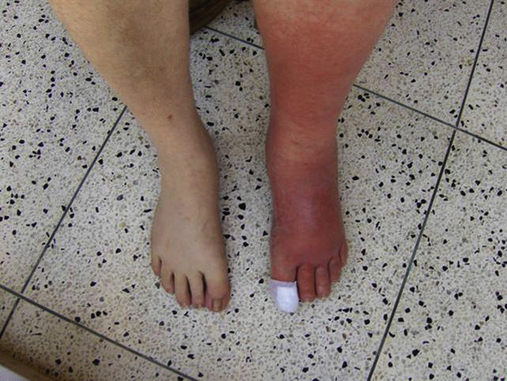
32 A 43-Year-Old Male With Left Leg Erythema and Pain


Case 32

A 43-Year-Old Male With Left Leg Erythema and Pain


Arthur Jeng, Arzhang Cyrus Javan


What is your differential diagnosis?


In a patient who has fever and an extremity that is erythematous, swollen, warm, and tender, cellulitis should be the primary concern. Cellulitis is an infection of the dermal layers of the skin and the immediate subcutaneous tissue. The leukocytosis with left shift (immature neutrophils, such as bands) is consistent with this infectious process and the left inguinal mass represents swelling of the draining lymph node(s) (lymphadenitis), a common sequelae of cellulitis.


Other conditions for which an extremity can appear erythematous, swollen, and/or painful include:


Deep vein thrombosis (DVT): Thromboses of the deep veins can cause swelling, warmth, pain, erythema, and even fevers. Clinically, DVT can be indistinguishable from cellulitis. Some of the features in this case that are less typical of DVT include the lymphadenopathy noted on the left inguinal exam and the leukocytosis with left shift. In the absence of the latter two features, a patient could have either cellulitis or DVT. Therefore, a Doppler ultrasound of the leg veins is commonly performed in the emergency department to distinguish between the two conditions.


Contact dermatitis: If the patient came in contact with a substance to which the skin has an inflammatory reaction, it will appear erythematous and swollen and develop vesicles, which can ooze serous fluid that may be mistaken for pus. The latter will crust over with time. The affected areas may be pruritic but can be painful, stinging, or burning. The key features of contact dermatitis are the well-demarcated edges of the erythema (demonstrating precisely where the substance came in contact with the skin) and the lack of systemic symptoms (fever, leukocytosis, or left shift). Further questioning of the patient should reveal the offending substance the skin came in contact with.


Stasis dermatitis: In patients with chronic venous insufficiency or lymphedema, the affected area can appear swollen and darker-hued and can be painful. Inflammatory papules can appear, as can erosions and ulcerations, which can be colonized with bacteria, making superficial swabs for bacterial culture confusing to the health care provider, who will commonly attribute the findings to an infection. To distinguish from cellulitis, careful history taking should reveal a history of chronic edema of the extremity from an underlying condition (congestive heart failure, liver disease, kidney disease, proteinuria, venous insufficiency, or prior soft tissue infection of the ipsilateral extremity). Furthermore, stasis dermatitis does not cause fever, leukocytosis, nor left shift.


Arterial insufficiency: With the lack of adequate blood flow to the extremities, the skin can appear shiny, atrophic, hairless, and have reactive hyperemia, where the redness blanches and abates with leg elevation. The affected area is very painful, and ulcers can develop, which are often mistaken for the nidus of infection that leads to the surrounding erythema. Detailed history may reveal claudication with use of that extremity. Demonstration that the erythema resolves with elevation of the leg can help distinguish this condition from cellulitis. As with the other conditions, fever and leukocytosis/left shift would not be seen with arterial insufficiency.


Step 2/3


Clinical Pearl


In all of these noninfectious mimics of cellulitis, erythema, swelling, and/or pain may be present. In DVTs, even fever can be present, which makes these diagnoses clinically difficult to distinguish. The presence of leukocytosis and, especially, left shift would be a feature specific for cellulitis. Although not always seen, presence of lymphangitic spread/streaking and/or lymphadenopathy in the ipsilateral inguinal region would also clinch the diagnosis of cellulitis.


Which pathogens should you be worried about causing cellulitis?


Gram-positive bacteria cause cellulitis. Historically, beta-hemolytic streptococci (Streptococcus pyogenes or group A streptococcus/GAS, Streptococcus agalactiae or group B streptococcus/GBS, and Streptococcus dysgalactiae or groups C and G streptococcus/GCS/GGS) have been shown to be the primary culprits. Staphylococcus aureus (Staph.aureus) can also cause soft tissue infections, although in the absence of a purulent focus (such as an abscess or furuncle), it is a less common cause than streptococci. In the absence of neutropenia or specific exposure to animal bites or water, gram-negative and anaerobic bacteria are extremely uncommon causes of cellulitis.


In the presence of a purulent focus, such as an abscess or furuncle, the reverse is true, with Staph.aureus being the primary culprit, including methicillin-resistant Staphylococcus aureus (MRSA). Streptococci do not tend to form pus and therefore are less commonly involved in purulent soft tissue infections.


Step 1


Basic Science Pearl


Both streptococci and Staph.aureus are gram-positive cocci. The distinguishing feature on gram-stain is that when streptococci replicate, they form chains, whereas Staph.aureus forms clusters. Hence, a quick gram-stain from the wound (if there is any) or of colony growth from a culture plate can distinguish between these two gram-positive cocci.


Which life-threatening emergency conditions should you consider when evaluating someone with a presumptive diagnosis of cellulitis?


Necrotizing fasciitis: This is a life-threatening infection of the fascial layers in the soft tissue that surrounds the muscles. In this condition, the initial symptom is severe pain, but there may be minimal visible signs of infection on the skin surface. It is thus extremely difficult to diagnose necrotizing fasciitis at this early stage. Over time, erythema will appear and may be indistinguishable from cellulitis. However, the pain is excruciating and the patient will appear very ill, often with septic physiology (tachycardia, high fevers, and, commonly, shock). Over a period of hours to days, the skin will continue to change color, becoming darker, and bullae may form, which signifies deep tissue destruction. Rapid spread, despite antibiotic administration, is a hallmark of this condition. The main cause of necrotizing fasciitis is GAS. A mixed infection comprised of non-beta-hemolytic streptococci (usually Streptococcus viridans group), Enterobacteriaceae family gram-negative bacilli (e.g., Escherichia coli, Klebsiella spp., Proteus spp.), and anaerobes can also occur; this entity is termed mixed synergistic necrotizing fasciitis. Immediate surgical debridement of all infected fascial tissue (fasciotomy with wide tissue excision) is critical for survival of the patient. Antibiotics play an adjunctive role in necrotizing fasciitis.


Myonecrosis: This is a life-threatening infection of the muscle, usually caused by Clostridium perfringens, an anaerobic spore-forming gram-positive rod. The initial symptom is extreme pain in the muscle, which may feel wooden. However, the superficial skin exam may be unrevealing, making diagnosis at this early stage difficult. Over time, the skin will change color, and eventually bullae may develop, signifying extensive muscle destruction. Crepitus may be appreciated on exam. The key feature that differentiates clostridial myonecrosis from GAS necrotizing fasciitis is the presence of gas in tissue, which the former anaerobic bacteria produce in abundance. Management, as in necrotizing fasciitis, is immediate muscle debridement, and antibiotics play an adjunctive role.


Step 2/3


Clinical Pearl


“Pain out of proportion with exam” is a red flag and should prompt one to consider necrotizing fasciitis and myonecrosis. Other red flags for these life-threatening conditions include rapid spread of “cellulitis,” especially when the patient is on appropriate antibiotics, and/or the presence of severe sepsis/septic shock, particularly if the area of skin infection is not correspondingly severe.


Step 1/2/3


Basic Science/Clinical Pearl


In GAS necrotizing fasciitis, patients are usually in toxic shock syndrome (TSS) and not septic shock. TSS is caused by the many superantigens that GAS possesses (Streptococcal pyrogenic exotoxins or Spe), which nonspecifically recruit T-cell activation with the antigen presenting cells, causing uncontrolled inflammatory response and shock. In TSS, there is some evidence for using intravenous immunoglobulin (IVIG), the pooled antibodies of which may bind and inactivate the bacterial superantigens. Additionally, ribosomal-active antibiotics that inhibit protein production (e.g., clindamycin) can also decrease the superantigen production and are used adjunctively in GAS necrotizing fasciitis and/or TSS.


Jun 15, 2016 | Posted by in GENERAL & FAMILY MEDICINE | Comments Off on 32 A 43-Year-Old Male With Left Leg Erythema and Pain

Full access? Get Clinical Tree

Get Clinical Tree app for offline access