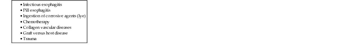
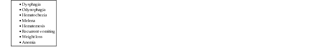
Monisha Bhanote, Wen Chen, Daniel Martinez Chest pain is one of those red-flag symptoms that require a clinician to at least consider several life-threatening pathologies, including acute coronary syndrome, aortic dissection, pneumothorax, and pulmonary embolism. Because the tests to rule out these conditions are costly, time consuming, and include radiation exposure, a detailed history and physical exam is the most important first step in evaluation. It is important to know all the risk factors for these conditions, ask questions relating to them, and evaluate them specifically on physical exam with each patient presenting with chest pain. Given that the patient is comfortable appearing, not tachycardic, not tachypneic, and this has been a chronic problem for many years, these acute life-threatening etiologies are very unlikely. The patient describes a substernal chest pain associated with acidic regurgitation, precipitated by large meals and lying flat, and initially relieved by over-the-counter antacids. Given this history, the most likely diagnosis is gastroesophageal reflux disease (GERD). When a relatively young patient without risk factors for other conditions has this classic set of symptoms, it is appropriate to start empiric treatment for GERD with close follow up to ensure resolution of symptoms. However, you should first evaluate whether the patient has any risk factors for other conditions on the differential diagnosis or any alarm symptoms (see Table 27.1 and Table 27.2). Other gastrointestinal causes of his chest pain include other types of esophagitis such as infectious, eosinophilic, and pill esophagitis. Infectious esophagitis typically presents in immunocompromised individuals and can be caused by viruses, fungi, and bacteria. The most common pathogens include Herpes simplex, Cytomegalovirus, and Candida species. These patients typically present with chest pain, odynophagia, and endoscopic findings of ulcerations. Eosinophilic esophagitis also typically presents with odynophagia, and patients may report that they have a sensation that food is getting stuck in their throat. Patients with eosinophilic esophagitis do not respond to antireflux therapy and tend to have normal pH monitoring. Pill esophagitis can be caused by long-standing use of medications. This has a similar presentation as other forms of esophagitis with odynophagia, dysphagia, and retrosternal pain and is typically seen in patients who are taking medications such as nonsteroidal antiinflammatory drug (NSAIDs), bisphosphonates, and antibiotics (Table 27.3). The initial management consists of an empiric trial of acid-reducing agents as well as lifestyle and dietary modifications. These can include eating smaller meals, avoiding late-night meals or snacks 2 to 3 hours before bed, avoiding the common precipitating foods, stopping smoking, losing weight, and elevating the head of the bed for sleeping. Physiologically, the gastroesophageal junction (GEJ) is a barrier formed by the lower esophageal sphincter (LES) and the diaphragm that prevents the acidic contents of the stomach from flowing back into the esophagus (Fig. 27.1). In the act of swallowing, the GEJ relaxes momentarily to allow food to pass from the esophagus into the stomach before closing to reestablish the barrier between the esophagus and the stomach. GERD is caused by the abnormal reflux of acidic gastric contents back into the esophagus and can be precipitated by physical abnormalities within the GEJ such as a hiatal hernia or abnormal GEJ tone as can be seen in scleroderma. Other contributing factors include a defective LES, delayed gastric emptying, and increased gastric acid production. Risk factors include abnormalities in the GEJ such as a hiatal hernia, obesity, pregnancy, and abnormal GEJ tone. Obesity is thought to increase intragastric pressure as well as increase the frequency of transient lower esophageal sphincter relaxations. Pregnancy is thought to increase GERD through the effects of high serum levels of estradiol and progesterone, which can relax the smooth muscles of the GEJ.
A 35-Year-Old Male With Substernal Chest Pain
How should you approach chest pain?
What is most likely in this patient?
What other conditions should be considered?
How do you manage a patient who presents with GERD?
What is the mechanism of reflux in GERD?
What are some risk factors and/or precipitating factors for GERD?
![]()
Stay updated, free articles. Join our Telegram channel

Full access? Get Clinical Tree


27 A 35-Year-Old Male With Substernal Chest Pain
Case 27