The Kidney and Lower Urinary Tract


The Kidney and Lower Urinary Tract


Laura S. Finn, M.D.

Aliya N. Husain, M.D.


Rapid advances in the field of genetics and molecular biology are leading to a better understanding of normal embryology, congenital malformations, glomerular and tubulointerstitial diseases, and neoplasia of the kidney and lower urinary tract. Approximately one-third of all congenital malformations are found in the urogenital system, many of which are part of complex multisystem anomalies with cumulative effects that are lethal in the neonatal period. Almost 80% of congenital uropathies seen in second trimester fetuses are associated with other anomalies—both chromosomal and nonchromosomal, either syndromic or in casual combination (1). Malformations of the bladder are often accompanied by major anomalies of the male and female genital tract because of the interrelated embryologic development of these organ systems. Glomerular diseases, reflux nephropathy, and infections are important causes of morbidity in childhood. Although cancer of the kidney is relatively uncommon in the pediatric age group, accounting for about 7% of childhood malignancies, 5-year survival from Wilms tumor has increased from 73% in patients diagnosed in 1975 to 1977 to 92% in the period 1996 to 2002, thus establishing a successful model for national multicenter study groups. However, 24% of survivors have severe chronic health conditions 25 years postdiagnosis, thus emphasizing the need to continuously improve treatment protocols (2).


EMBRYOLOGY

Functionally, the urinary and the genital systems can be divided into two entirely separate systems; however, embryologically and anatomically, they are intimately interwoven. Both develop from a common mesodermal ridge (intermediate mesoderm) along the posterior wall of the abdominal cavity, and initially, the excretory ducts of both systems enter a common cavity, the cloaca. In humans, three separate but temporally overlapping renal systems form. The pronephros, which is the most caudal and nonfunctional, regresses completely by the end of the 4th week of gestation, during which time the first excretory tubules of the mesonephros appear that may function for a short period. While the caudal tubules are still differentiating, the cranial tubules and glomeruli show degenerative changes, and by the end of the second month, most have disappeared. In the male, a few of the caudal tubules and the mesonephric duct persist and participate in the formation of the genital system, but they disappear in the female, leaving only a few vestigial structures.

The metanephros, or permanent kidney, appears in the 5th week at the level of the upper sacral segment, with its blood supply coming from the lateral sacral branches of the aorta. By the eighth week, it “ascends” to the lumbar region, mainly secondary to differential growth of the embryo, and derives its blood supply from progressively higher levels of the aorta. In the pelvic ectopic kidney, the renal arteries arise from a lower level of the aorta or from the iliac arteries. The nephrons develop from the caudal end of the metanephric blastema (metanephric mesenchyme), while the renal excretory system (collecting duct, calyces, pelvis, and ureter) develops from the ureteric bud, which is an outgrowth of the mesonephric duct close to its entrance into the cloaca. The proximal tip or the ampulla of the ureteric bud grows dorsally and cranially, pushes the metanephric mesenchyme, and undergoes a series of dichotomous branching; the ampulla of each branch ultimately induces nephron formation. Each division proceeds more rapidly at the poles, so that the kidney acquires its characteristic shape. Ureteric bud branching is complete by 22 weeks of gestation, and the first few generations of branches coalesce to form the renal pelvis and calyces, whereas subsequent generations give rise to collecting ducts (3).

Nephrons form from condensation of the metanephric blastema, which develops a cyst-like cavity, elongates, and folds back to become S-shaped. One end fuses with the ampulla that induced it, while at the other end, a mesh of capillaries develops and invaginates the nephrogenic vesicle to form a glomerulus. The upper and middle limbs of the nephrogenic vesicle elongate and differentiate into the proximal and distal convoluted tubules and the loop of Henle (4).

The process of nephrogenesis can be divided into four periods. From 7 to 14 weeks of gestation, the ureteric bud branches dichotomously for six to eight generations, with each branch inducing the formation of a new nephron.
From 14 to 22 weeks, nephron arcades are formed, with the innermost nephron formed first (juxtamedullary nephron) (eFigure 17-1). From 22 to 36 weeks, no branching of the ureteric bud occurs. The ampulla extends to the subcapsular cortex to induce four to seven nephrons (eFigure 17-2). Thus, the nephrons formed last are subcapsular (the nephrogenic zone seen in sections of fetal kidneys) (Figure 17-1). From 36 weeks of gestation through birth and up to maturity, the nephrons grow, but no new nephrons are formed. In extremely premature infants, nephrogenesis continues after birth until the kidney reaches maturity. However, in this setting, renal maturation appears to be accelerated and associated with an increased number of morphologically abnormal and large glomeruli, suggestive of hyperfiltration and predicting fewer functioning nephrons in postnatal and later life (5).


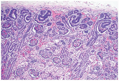
FIGURE 17-1 • Early third trimester kidney with subcapsular nephrogenic zone. (Hematoxylin-eosin stain, original magnification ×100.)

Studies indicate that nephrons in the developing metanephros may begin functioning as early as the eleventh or 12th week after conception. In fact, it has been suggested that the formation of a tubule fluid is essential to ensure the normal development of the renal pelvis and calyces.


Molecular Regulation of Kidney Development

As with most organs, differentiation of the kidney involves epithelial-mesenchymal interactions, which are under the control of multiple gene regulatory networks that act as inducers or inhibitors. Briefly, epithelium of the ureteric bud from the mesonephros interacts with mesenchyme of the metanephric blastema. The mesenchyme expresses WT-1, a transcription factor that makes this tissue competent to respond to induction by the ureteric bud. WT-1 also regulates production of glial-derived neurotrophic factor (GDNF) and hepatocyte growth factor (HGF or scatter factor) by the mesenchyme, and these proteins stimulate branching and growth of the ureteric buds. The tyrosine kinase receptors RET, for GDNF, and MET, for HGF, are synthesized by the epithelium of the ureteric buds, establishing signaling pathways between the two tissues. In turn, the buds induce the mesenchyme via fibroblast growth factor 2 and bone morphogenetic protein 7. Both these growth factors block apoptosis and stimulate proliferation in the metanephric mesenchyme while maintaining production of WT1. Conversion of the mesenchyme to an epithelium for the nephron formation is also mediated by the ureteric buds, in part through modification of the extracellular matrix. Thus, fibronectin, collagen I, and collagen III are replaced with laminin and type IV collagen, characteristic of an epithelial basal lamina. In addition, the cell adhesion molecules, syndecan and E-cadherin, which are essential for condensation of the mesenchyme into an epithelium, are synthesized. Regulatory genes for conversion of the mesenchyme into an epithelium appear to involve PAX2 and WNT4 (4).


CONGENITAL MALFORMATIONS OF THE KIDNEY

If all malformations are considered, ranging from incidental findings with no clinical significance to major lethal anomalies, it is estimated that congenital abnormalities of the kidney and urinary tract are present in 10% of all newborns and interestingly, when unilateral, are more often on the left (6). Worldwide, a substantial percentage of children develop chronic kidney disease early in life, with congenital anomalies of the kidney and urinary tract (CAKUT) such as obstructive uropathy and aplasia/hypoplasia/dysplasia being responsible for almost one-half of all cases (7). Table 17-1 lists the relative frequency of malformations seen in two series of pediatric autopsies—one from a children’s hospital and the other from a tertiary care university medical center. Forty-one (38%) of the 107 renal malformations in series No. 2 were associated with major malformations of at least one other organ system.

A wide variety of renal malformations result in the oligohydramnios (Potter) sequence (i.e., characteristic facies, including low-set ears, beaked nose, prominent epicanthic folds, receding chin, limb deformities, growth retardation, and pulmonary hypoplasia) (Figure 17-2). These abnormalities are the result of decreased amniotic fluid rather than renal malformations per se and can result from even a relatively short duration of oligohydramnios, including persistent leakage of amniotic fluid. When these findings are associated with renal agenesis, the term Potter syndrome (as initially described by Potter in 1946) is used. Renal findings in children with oligohydramnios sequence are listed in Table 17-2. Most urogenital abnormalities are now diagnosed antenatally on high-resolution ultrasound scans, which have enabled recognition of those that are not compatible with survival and those that can benefit from intervention (8). CAKUT are responsible for approximately 40% to 50% of childhood chronic kidney disease worldwide (9). The classification of congenital and developmental anomalies of
the kidney has historically been based on morphologic criteria established decades ago; more recent systems attempt to incorporate new information about nephrogenesis and the pathogenesis of urinary tract malformations (10,11). It is well established that many cases of CAKUT have a genetic basis and many are associated with syndromes; nonsyndromic forms may also be causally linked to several developmental genes. Thus, neither approach is perfect as a given gene can result in a spectrum of anomalies, and the same anomaly can be caused by multiple genes. Nonetheless, it is worthwhile to consider the molecular mechanism of kidney and urinary tract development to better understand the malformation. The diversity of signaling pathways in nephrogenesis likely explains the remarkable locus heterogeneity found in CAKUT. Currently, mutations can be identified in approximately 10% to 20% of children, with HNF1B and PAX2 mutations being the most common (12). Table 17-3 lists the common malformations of the kidney and includes cystic diseases because many of these are inheritable disorders or are secondary to malformations of the kidney parenchyma and lower urinary tract (13).


TABLE 17-1 RENAL MALFORMATIONS SEN IN PEDIATRIC AUTOPSIES


No. of Cases


Anomaly


Series 1*


Series 2+


Total (%)


Renal agenesis, bilateral


16


13


29 (12)


Renal agenesis, unilateral


10


6


16 (6.6)


Renal dysplasia, bilateral


45


33


78 (32)


Renal dysplasia, unilateral


4


5


9 (2.1)


Renal dysplasia, unilateral, with contralateral renal agenesis


9


3


12 (5)


Autosomal recessive polycystic kidney disease


5


10


15 (6.2)


Autosomal dominant polycystic kidney disease


1


1


2 (0.8)


Renal fusion


20


12


32 (13.2)


Renal ectopia


4


1


5 (2.1)


Congenital hydronephrosis, bilateral


6


5


11 (4.5)


Congenital hydronephrosis, unilateral


4


8


12 (5)


Ureteral duplication


10


5


15 (6.2)


Renal hypoplasia


1


3


4 (1.7)


Others


0


6


6 (2.5)


Total


135


111


246 (˜100)


Series 1* compiled from 1442 consecutive autopsies performed at Minneapolis Children’s Medical Center from 1977 to 1987, including stillborn infants and children younger than 1 year of age.


Series 2+ compiled from 1242 pediatric autopsies performed at Loyola University Medical Center from 1978 to 1998, including stillbirths and children up to 18 years (Unpublished data from Aliya N. Husain, M.D.).


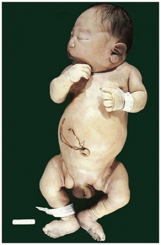
FIGURE 17-2 • Oligohydramnios (Potter sequence) is associated with low-set and deformed ears, beaked nose, receding chin, and lower limb positional deformity.


Renal Ectopia

Permanent malposition of one or both kidneys is seen in 2% of pediatric autopsies (Table 17-1). The incidence is even higher in perinatal autopsies because renal ectopia is commonly associated with multiple other malformations; however, the incidence in screening studies is approximately 1 in 683 infants (14). The ectopic kidney(s) may be located in the pelvis (most common), on the other side (crossed renal ectopia) with or without fusion, or even in the thorax (rare) (15). Prenatal ultrasonographic diagnosis of the pelvic kidney is possible, usually after 24 weeks of gestation, although postnatal ultrasound or CT is more effective at diagnosing
anomalies of the urinary tract (14). While renal function is normal in the neonatal period in patients with renal ectopia without other associated malformations, vesicoureteral reflux and hydronephrosis have been reported in approximately 20% to 50%, particularly in crossed renal ectopia (16). Pseudocrossed renal ectopia occurs when an enlarging retroperitoneal mass displaces the kidney to the contralateral side of the abdomen.


TABLE 17-2 Renal Findings in Children with Oligohydramnios Sequence


No. of Cases


Renal Abnormality


Series 1


Series 2


Total (%)


Bilateral renal agenesis


16


32


48 (30)


Bilateral cystic dysplasia


17


30


47 (29)


Unilateral agenesis with contralateral dysplasia


9


8


17 (10)


Obstructive uropathy


13


13 (8)


Autosomal recessive polycystic kidney disease


4


2


6 (4)


Renal ectopia and fusion


1


1


2 (1)


Autosomal dominant polycystic kidney disease


1


1


2 (1)


Others


13


13


26 (16)


Total


74


87


161 (˜100)


TABLE 17-3 CLASSIFICATION OF MALFORMATIONS OF THE KIDNEY, INCLUDING CYSTIC DISEASES


  1. Renal position and form


    1. Ectopia


    2. Fusion


    3. Supernumerary


  2. Renal quantity


    1. Agenesis (bilateral, unilateral)


    2. Hypoplasia, simple, or oligomeganephronic


    3. Renal tubular dysgenesis


    4. Renomegaly


    5. Duplication


  3. Hydronephrosis


  4. Renal dysplasia/cystic diseases (gross and/or microscopic)


    1. Renal dysplasia


      1. Sporadic (bilateral, unilateral)


      2. Hereditary


      3. With malformation syndromes


    2. Polycystic kidney disease


      1. Autosomal recessive (infantile type)


      2. Autosomal dominant (adult type)


    3. Medullary cysts


      1. Medullary sponge kidney


      2. Nephronophthisis (Types 1-3)


      3. Medullary cystic disease (Types 1 and 2)


    4. Cortical cysts


      1. Glomerulocystic disease(s)


      2. Simple cysts (acquired)


      3. Microcysts associated with syndromes


    5. Renal cysts with hereditary syndromes


Renal Fusion

Renal fusion, often with ectopia, was seen in 32 (1.2%) of 2684 pediatric autopsies (Table 17-1). The most common fusion anomaly is horseshoe kidney, in which both kidneys are normally lateralized but have fused lower poles (Figure 17-3) and are located in a lower than normal position. The incidence of horseshoe kidney is reported to be 1 in 600 to 700 in the general population (17). One-third of persons with this condition have associated congenital malformations of other organs, including Turner syndrome (18); two-third have a major urologic complication, most of which require surgery, although newer techniques such as laparoscopic roboticassisted management allow for minimally invasive procedures. Symptomatic hydronephrosis eventually develops in
more than half of patients with horseshoe kidney, secondary to ureteropelvic junction obstruction, reflux, or malrotation. Individuals with horseshoe kidney are at higher risk for the development of stones (19) and tumors (20). Rare cases of renal-adrenal fusion have been described, which may present as a renal mass in the upper pole (21).


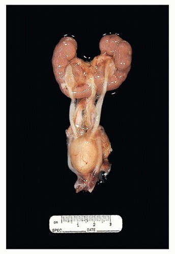
FIGURE 17-3 • Horseshoe kidney with fused lower poles.


Renal Agenesis/Hypoplasia

Inadequate renal tissue can be considered as a continuum, ranging from renal agenesis to subtle congenital nephron deficits. Renal agenesis (i.e., the complete absence of one or both kidneys) is commonly accompanied by other malformations of the genitourinary tract and various lower body defects and is often the result of one or more genetic mutations that leads to molecular dysregulation of nephrogenesis. Homozygous null mice for c-Ret , Gdnf , and Gfra-1 all exhibit bilateral renal agenesis due to the inhibition of ureteric bud growth and branching morphogenesis; they are less frequently implicated in human renal agenesis (22). Pax2 plays an integral role in the initiation and maintenance of the Ret/Gdnf pathway by not only activating the ligand of the pathway but also enhancing the expression of the pathway receptor Ret (4). Since an exhaustive review is beyond the scope of this chapter, one can focus on Pax2, one of the earliest genes expressed widely during fetal kidney development in the nephric duct, the metanephric mesenchyme, the ureteric bud, and in the S-shaped body. Early failure in the first two developmental stages (e.g., homozygous inactivation of Pax2) precludes formation of metanephric kidneys and causes bilateral renal agenesis, incompatible with life. Interference with the later stages affects the extent of branching morphogenesis (e.g., heterozygous Pax2 mutations). Although the resulting nephron deficits are compatible with life, they may be moderately severe and account for up to 40% of the children in dialysis and transplant units around the world. Finally, the effect of Pax2 on apoptosis in the branching ureteric bud seems to imply a quantitative process that is finely tuned. Modest changes in this program could account for subtle nephron deficits in “normal” humans and increased risk of hypertension or susceptibility to acquired renal disease later in life (23).


Bilateral Renal Agenesis

Uniformly fatal, bilateral renal agenesis, although less common than unilateral renal agenesis (URA), is seen more frequently in pediatric autopsies (1.1% of total autopsies in Table 17-1). The incidence of bilateral renal agenesis varies from 0.1/1000 to 0.3/1000 births (8). It accounts for one-third of births with the oligohydramnios sequence (Table 17-2). The male to female ratio is 2.5:1. It is usually associated with severe oligohydramnios (Potter syndrome), intrauterine growth restriction, extrarenal anomalies, and malpresentation. The ureters and renal arteries are also absent, and the urinary bladder is hypoplastic or absent. The adrenals are disc shaped secondary to lack of molding from the kidneys (eFigure 17-3). Forty percent of affected infants are stillborn, and the remainder die in the immediate postnatal period, generally of pulmonary hypoplasia.

Bilateral renal agenesis is usually sporadic, although familial cases have been described (22). Hereditary renal adysplasia (agenesis/dysplasia syndrome) is defined as URA in association with dysplasia of the contralateral kidney. The term adysplasia is often used more broadly to include dysplasia, absent kidneys, and almost any associated structural or positional defect of the kidney or lower urinary tract. However, hereditary adysplasia should be considered as any combination of unilateral or bilateral agenesis, unilateral or bilateral renal dysplasia, or dysplasia of one kidney and agenesis of the other, in different members of the same family for which autosomal dominant inheritance with varying expression has been suggested (9,24). An increased prevalence of congenital renal anomalies was identified in the relatives of index patients with bilateral renal agenesis/adysplasia (14.7%) compared to controls (2.2%), with a recurrence risk of 6.2 for first-degree relatives (25). But the occasional cases of agenesis or dysplasia affecting siblings with normal parents suggest that there are other types of inheritance. Some patients with adysplasia have PAX2 mutations. The genetic link between renal agenesis and some types of renal dysplasia points to a common pathogenetic mechanism and may result from varying degrees of failure of induction of the metanephric blastema by the ureteric bud.

Other reported malformations associated with bilateral renal agenesis include congenital pulmonary airway malformation type 2 (cystic adenomatoid malformation), left heart hypoplasia (26), sirenomelia (27), and urorectal septum malformation sequence (28). Limb reduction defects and renal agenesis have been reported in fetuses born to mothers with cocaine addiction and insulin-dependent diabetes (29).

The genetic mechanisms leading to bilateral renal agenesis remain largely unresolved. The absence of kidney development in a large number of mouse knockout models suggests a role for recessive mutations in various genes. Indeed, homozygous mutations in FGF20 have been shown in fetuses from consanguineous families with bilateral renal agenesis (30). More recently, the same researchers have demonstrated recessive mutations in integrin alpha-8 (ITGA8) as being responsible for renal agenesis (31). Integrins are transmembrane receptors expressed in metanephric mesenchyme that mediate biologic processes during organogenesis. Interruption of signaling pathway can thus lead to severe kidney developmental defects.


Unilateral Renal Agenesis

URA is a common developmental defect in humans, occurring at a frequency of approximately 1 in 2000 (32). Although compatible with normal life, URA is still seen twice as often in pediatric autopsies (0.6%) than in the general population (0.3%) owing to its frequent association with other complex malformations (33). The male to female ratio is about 1:1.5.
Long-term follow-up of patients with URA has shown that these patients are at higher risk for the development of proteinuria, hypertension, and renal insufficiency.

Malformations of the genitalia are commonly associated with URA. These include ipsilateral absence of fallopian tube, unicornuate and bicornuate uterus, cysts of the epididymis and seminal vesicle, agenesis of the vas deferens, cystic dysplasia of the testis and rete testis, ectopia of the vas deferens, and urorectal septum malformation sequence (ambiguous genitalia with absence of perineal and anal openings) (28,32,34,35,36).

Cystic dysplasia of the testis appears to be associated consistently with renal malformations, most frequently ipsilateral renal agenesis; both conditions could be explained by failure of development of the ureteric bud (36).

Genetic factors seem to play a significant role in URA, especially when it is part of a syndrome. Mutations in genes such as HNF1-β, PAX2, SALL1, WT1, SIX1, and EYA1 have been shown to cause some of these rare syndromes; however, a genetic basis for the VATER association, in which URA is a common feature, has not yet been identified (32).


Renal Hypoplasia

Renal hypoplasia, defined as histologically normal kidneys with a weight that is less than two standard deviations below the norm, is extremely rare. In the older literature, any small kidney was labeled hypoplastic, irrespective of the cause. Currently, small kidneys that are also dysplastic are considered with the dysplastic group, and those with scarring, inflammation, and hypertensive changes are end-stage kidneys with hypoplasia, or more correctly, atrophy, considered secondary to the underlying disease and thusly categorized. Segmental renal hypoplasia (so-called Ask-Upmark kidney), which may be unilateral or bilateral and is characterized by localized atrophic scarring, was originally regarded as a primary malformation is now known to be secondary to vesicoureteral reflux and considered a form of reflux nephropathy.

In true renal hypoplasia, the absolute number of nephrons is reduced, possibly as a consequence of inadequate branching of the ureteric ducts that may result in a decreased number (<5) of reniculi. Although the volume is reduced, the renal shape and differentiation are normal. Unilateral hypoplasia is a sporadic condition, only rarely associated with lower urinary tract anomalies; patients may present with hypertension and occasionally, when associated with malrotation, may be predisposed to reflux nephropathy. Bilateral hypoplasia results in renal insufficiency and early death in severe cases; less severe cases manifest growth retardation and chronic renal insufficiency likely a consequence of developing secondary focal segmental glomerulosclerosis (FSGS) and tubulointerstitial damage as a consequence of hyperfiltration (37).

Bilateral oligomeganephronic renal hypoplasia is a nonfamilial form of congenitally small kidneys characterized by slowly progressive renal insufficiency. The absolute number of nephrons is reduced. The characteristic feature is glomerulomegaly, unlike the normal glomerular size encountered in simple hypoplasia. Infection, dysplasia, and obstructive uropathy are absent, and patients present with polyuria, polydipsia, and salt wasting; proteinuria may develop. Although several causes, including toxic factors, renal infection, vascular insufficiency, and disseminated intravascular coagulation, have been mentioned, it is not known what factors arrest the development of the metanephric renal blastema, presumably between weeks 14 and 20 of fetal life. Mutations in hepatocyte nuclear factor-1beta (HNF-1β) and PAX2 have been reported in patients with oligomeganephronia (23,38). A nephron deficit has also been reported in premature and low-birth-weight infants and has been linked to the development of hypertension in adults (39).


Renal Tubular Dysgenesis

Familial renal tubular dysgenesis (RTD) is a rare autosomal recessive congenital disorder of the renin-angiotensin system (RAS) that causes the absence or marked reduction in the number of differentiated proximal tubules. Mutations in AGT, encoding angiotensinogen; REN, encoding renin; ACE, encoding angiotensin-converting enzyme; or AGTR1, encoding angiotensin 2 receptor type 1 (AT1 receptor) affect the production or efficacy of angiotensin 2 and result in the absence of a functional RAS and abnormal renin expression (40). Oligohydramnios is detected as early as 20 weeks’ gestation and persists, resulting in the Potter sequence. Fetuses may die in utero, but more often, infants die shortly after birth as a consequence of anuria and respiratory failure secondary to lung hypoplasia. Kidneys are normal or slightly enlarged and lack significant ultrasound abnormalities. Intrauterine growth is normal in the genetic form (41). Associated skull ossification defects, attributed to bone hypoxia, spontaneously improve after birth. In most patients, no proximal tubules can be identified by anti-CD10 or anti-CD15 antibodies and glomeruli are closely packed. The medulla contains few loops of Henle, and collecting ducts are atrophic and collapsed and surrounded by abundant mesenchyme. Interlobular arteries and afferent arterioles show thick and disorganized muscular walls, but larger interlobular arteries are normal.

The lack of proximal tubules is not specific for autosomal recessive RTD and has been seen in animals and humans with reduced renal blood flow of various etiologies, confirming the importance of a functional RAS in the maintenance of fetal blood pressure and renal blood flow (41). Secondary RTD has been described in humans with major cardiac malformations, renal artery stenosis, and extensive ischemic necrosis of the placenta and in conjunction with twin-twin transfusion syndrome, neonatal hemochromatosis, and in utero exposure to RAS blockers and nonsteroidal antiinflammatory drugs (NSAIDs) (41,42). Despite the lack of normal proximal tubules, the major site of water resorption
in the kidney, the principal clinical manifestations are caused by fetal and neonatal oliguria.


Renomegaly

The most common form of renal enlargement is compensatory hypertrophy, in which a single functioning kidney may reach twice the normal size and can be detected in utero. Bilateral renomegaly secondary to an increased number or size of normally developed nephrons is seen in growthrelated disorders such as Beckwith-Wiedemann syndrome, hemihypertrophy, Perlman syndrome, and congenital nephrosis of the Finnish type (43,44).


Renal Duplication (Duplex Kidney)

Duplex kidneys are the most common anomalies of the upper urinary tract in childhood with an estimated incidence of 0.8% (45). The term renal duplication denotes the presence of two separate pelves in the same kidney accompanied by complete or partial duplication of the ureter (45) (Figure 17-4). These kidneys usually have greater than normal number of reniculi. The anatomical and functional divisions between upper and lower moieties of duplex kidney are extremely variable. The underlying pathologic condition associated with a lower moiety is usually massive vesicoureteral reflux to the lower collecting system and only rarely, obstruction. The nonfunctioning upper moiety is usually associated with obstructive ectopic ureter (with or without ureterocele) and may show chronic tubulointerstitial injury and hypoplastic or dysplastic changes (46). In mice, a model of congenital kidney and urinary tract anomalies, severe early gestational hypoxia resulted in reduced β-catenin signaling and formation of duplex kidneys. This mirrors the defects caused by suppression of canonical Wnt/β-catenin signaling at that stage of development (47).


FIGURE 17-4 • Renal duplication (duplex kidney) with two separate pelves in the same kidney and more than the normal number of reniculi.


Supernumerary Kidney

Supernumerary kidney is one of the rarest of renal malformations with fewer than 100 cases reported so far. In addition to the normal two kidneys, an additional, usually small, kidney is present within the renal fascia caudal to and completely separate from the ipsilateral kidney. Few cases of multiple supernumerary kidneys are described (48,49). Most cases are originally diagnosed as hydronephrosis, or pyelonephritis; thus, the true incidence may be higher than reported. The malformation is thought to result from greater than one ureteric bud divergently arising from different positions in the wolffian duct with induction of metanephric blastema and aberrant divisions resulting in at least two kidneys on one side.


Hydronephrosis

Hydronephrosis may be congenital or acquired, unilateral or bilateral, and mild to severe. Renal dysplasia may or may not be present. Hydronephrosis is readily seen on antenatal ultrasonography but does not necessarily imply obstruction. Although most cases will resolve spontaneously, the probability of a significant pathology is related to the degree of pyelectasis, as seen on the third trimester ultrasound study. Criteria of obstruction are difficult to define with precision, but two that are well accepted are size of the renal pelvis (>15 mm) and relative renal function (50).

Hydronephrosis is the most common cause of an abdominal mass of genitourinary tract origin in neonates (51). It is most frequently caused by obstruction of the ureteropelvic junction, which leads to dilatation of the renal pelvis and calyceal system. Depending on the severity and timing of the obstruction, the appearance of the renal parenchyma varies from relatively normal to markedly atrophic, with fibrosis and a scant chronic inflammatory infiltrate (52). One hypothesis for ureteropelvic obstruction is impaired smooth muscle differentiation, aberrant smooth muscle arrangement, and abnormal pyeloureteral innervation, which results in impaired peristalsis and subsequent hydronephrosis (52,53). FOXF1 is the only gene so far associated with UPJ obstruction in humans (53).

The specimen most commonly seen in surgical pathology is a portion of the ureteropelvic junction that shows remarkably little pathology on microscopic examination. When end-stage nonfunctioning hydronephrotic kidneys are removed, marked dilatation of the pelvis and calyces with only microscopic foci of residual renal parenchyma can be
seen (Figures 17-5 and 17-6). Neonatal hydronephrotic kidneys seen at autopsies usually have a histologically normal, although grossly compressed, cortex and medulla.


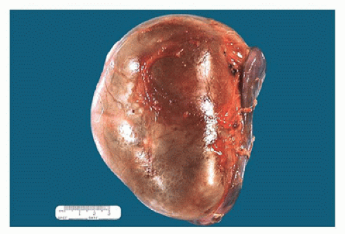
FIGURE 17-5 • Nephrectomy specimen from a 5-year-old who presented with a unilateral renal mass. The kidney appears to be one large cystic structure.

Hydronephrosis is often associated with renal dysplasia, so that the definition of these two entities often overlaps. Also, because urinary tract obstruction is a common underlying condition, it is best to consider them as the opposite ends of a spectrum. When severe obstruction occurs early in fetal development, it results in renal dysplasia; when it occurs late, one sees hydronephrosis; when it develops in between, both hydronephrosis and dysplasia are apparent to varying degrees. Hydronephrosis associated with reflux disease is discussed later in this chapter in the section on tubulointerstitial diseases (52).

Although the vast majority of cases of hydronephrosis are sporadic, some syndromic associations have been reported (53,54,55). Hydronephrosis should also be distinguished from the rare disorder of megacalycosis (Puigvert disease), which is characterized by calyceal dilatation, an increased number of calyces, hypoplasia of the pyramids of Malpighi, a normal renal pelvis, and, most importantly, normal renal function (56).


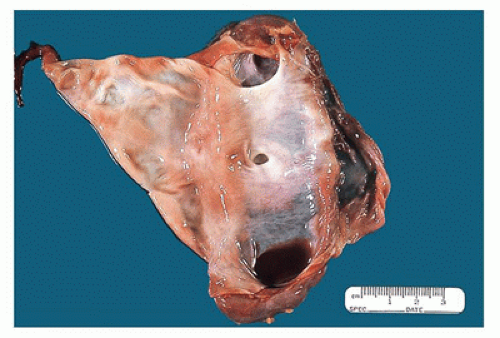
FIGURE 17-6 • Cut section of the kidney in Figure 17-5 shows a markedly dilated pelvis and calyces and very little residual renal parenchyma.


Renal Dysplasia/Cystic Diseases

Cystic diseases of the kidney are a heterogeneous group of congenital (sporadic and inherited) and acquired disorders characterized by multiple cysts. In view of our better understanding of the genetic basis for some of the cystic renal diseases, the original “Potter classification” with division into types I to IV is no longer widely used (57). There is no universally accepted classification for cystic diseases with categorization dependent upon cyst location, onset (congenital versus acquired), or known etiology (genetic or syndromic association); even renal dysplasia has been variably classified under abnormal renal differentiation or developmental defects or as a unique cystic disease (10).

By convention, the term multicystic is reserved for a category of renal dysplasia characterized by multiple unilateral or bilateral cysts, while polycystic is conventionally used for hereditary autosomal recessive and autosomal dominant kidney diseases.


Renal Dysplasia

Multicystic dysplastic kidneys are the most common type of malformed kidneys seen in pediatric autopsies (Table 17-1), with bilateral dysplasia accounting for 32% and unilateral dysplasia (with or without contralateral agenesis) for 7.1% of patients with renal malformations. It may involve one or both kidneys or a portion of one kidney, with or without enlargement of the affected kidney and with or without grossly visible cysts. Thus, the dysplastic kidney may be smaller or larger than normal and grossly misshapen or reniform. Renal dysplasia is one of the most common causes of an abdominal mass in children younger than 1 year; although often diagnosed antenatally, it may present in older children and adulthood (58).

The most common form of dysplasia is sporadic, although genetic causes are being increasingly identified (9,11,33). It is typically associated with obstruction of the ureteropelvic junction and enlarged distorted kidneys that are no longer reniform (Figure 17-7). Cysts of varying sizes can be
appreciated through the capsule and on sectioning are seen to be irregularly distributed throughout the parenchyma, with no residual identifiable cortex or medulla (Figure 17-8).


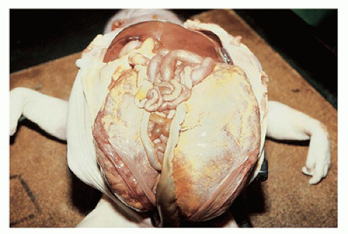
FIGURE 17-7 • Massively enlarged cystic dysplastic kidneys.


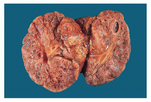
Figure 17-8 • Cut surface of cystic dysplastic kidney with multiple small, variably sized cysts involving both cortex and medulla.

The microscopic hallmark is the presence of immature dysplastic-appearing tubules surrounded by collarettes of condensed mesenchyme (Figure 17-9). The tubules are lined by a single layer of primitive undifferentiated cuboidal to columnar epithelium that often appears to be excessive, so that it is folded and may fill the lumen (Figure 17-10). The cells tend to have a relatively high nuclear to cytoplasmic ratio, and the tubular basement membrane may be thick and eosinophilic. A myxoid, moderately cellular condensation of spindle cells encircles the tubules. Ducts and tubules are fewer in number than normal, and the remaining connective tissue is loose and contains many blood vessels, lymphatics, and peripheral nerves. Medullary pyramids often lack vasa recta and loops of Henle. The cortex is usually thin and may contain islands of immature-appearing hyaline cartilage, but their presence is not required for the diagnosis of dysplasia (Figure 17-11). Cysts of varying sizes are formed by the dilated, dysplastic tubules and lined by a markedly flattened, often inapparent, epithelium. Cysts can occur in any part of the nephron. Varying numbers of normal glomeruli and tubules can be identified between the dysplastic areas.


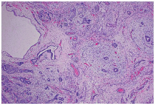
FIGURE 17-9 • Cystic dysplastic kidney with disorganized renal parenchyma in which immature tubules are surrounded by collarettes of condensed mesenchyme. (Hematoxylineosin stain, original magnification ×40.)


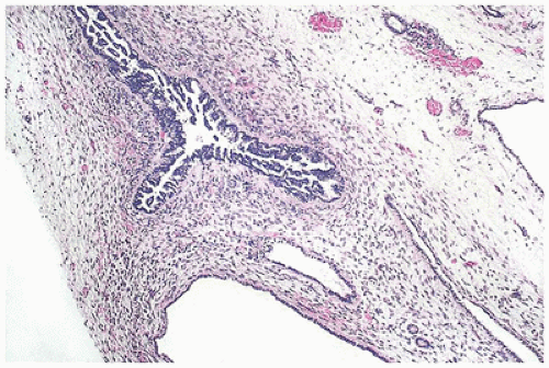
FIGURE 17-10 • Dysplastic tubules with an excessive amount of lining epithelium, which is thrown into papillary folds. (Hematoxylin-eosin stain, original magnification ×40.)

The terms renal adysplasia (aplastic dysplasia) and hypoplastic dysplasia are used to describe small kidneys with limited nephron development and extensive dysplasia (Figure 17-12) that are totally nonfunctioning or minimally functioning, respectively. The essential histologic features are the same regardless of the size of the kidney or the extent of involvement.

In patients who survive the immediate postnatal period, clinical complications include hypertension, febrile urinary tract infection (UTI), vesicoureteral reflux, progressive scarring, and renal failure. Approximately 50% to 60% of cases undergo involution by 10 years, the rate partly influenced by size on postnatal ultrasound and side (59,60). In 3% to 5% of cases of dysplastic kidney, nodular renal blastema is also present, and although Wilms tumor developing in dysplastic kidney has been reported, a systematic review showed no increased risk of development of WT (61).While the
majority of multicystic dysplastic kidneys (MCDK) can be an isolated finding and is uncommonly bilateral, it is often associated with other (contralateral) anomalies of the kidney and urinary tract. The vast majority of MCDK are associated with congenital urinary tract obstruction, which is often at the ureteropelvic junction but may occur at any level. Several animal models of renal dysplasia after gestational ureteral obstruction have been described (62).


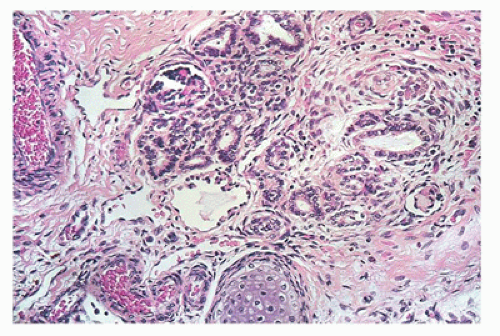
FIGURE 17-11 • Disorganized renal parenchyma and island of immature cartilage in cystic dysplastic kidney disease. (Hematoxylin-eosin stain, original magnification ×100.)


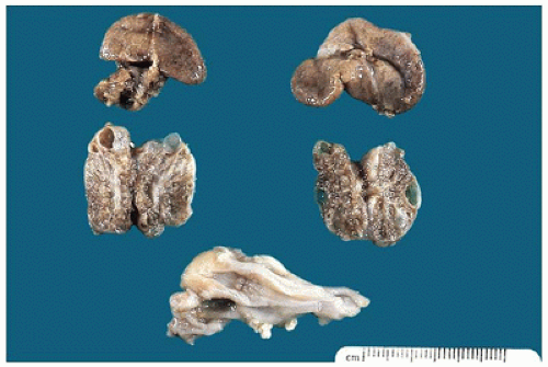
FIGURE 17-12 • Cystic dysplastic kidneys, shown bisected in the middle of the picture, are smaller than the adrenals above.

Renal dysplasia is also seen as part of a syndrome in association with extrarenal manifestations. Also, it may be diagnosed sporadically or described with familial aggregation in up to 15% of the cases. When familial, the mode of inheritance is most often autosomal dominant with variable expressivity and reduced penetrance. Epigenetic modifications may explain some of the variability; factors as improbable as maternal diet have been shown to regulate embryonic renal gene expression (63).

Due to the central role of GDNF-RET signaling pathway in ureteric budding, it is not surprising that mutations in genes that regulate the pathway, including transcription factors PAX2, EYA1, and SALL1, have been implicated in dysplasia (9,12,24). Mutations in other genes including DSTYK CHD1L; SIX 1, SIX 2, or SIX 5; and HNF1-β have also been demonstrated in patients with dysplasia, and a recent large genetic screening study of 749 individuals with various nonsyndromic renal abnormalities identified SALL1, PAX2, and HNF1-β as the most prevalent disease-causing genes (24). An excellent and exhaustive tabulation of these can be found at the end of Chapter 27 in Potter Pathology of the Fetus, Infant, and Child (64). A brief summary is provided in Table 17-4.

Earlier work had shown that apoptosis is prominent in undifferentiated cells around dysplastic tubules, which perhaps explains the tendency of these organs to regress. In contrast, apoptosis was rare in dysplastic epithelia, thought to be ureteric bud malformations. A high rate of proliferation has been demonstrated postnatally in dysplastic tubules, and PAX2, a potentially oncogenic transcription factor, is expressed in these epithelia. In contrast, both cell proliferation and PAX2 are down-regulated during normal maturation of human collecting ducts. Ectopic expression of BCL2, which encodes a protein that prevents apoptosis during renal mesenchymal to epithelial conversion, has been observed in dysplastic kidney epithelia. Thus, dysplastic cyst formation may be understood in terms of aberrant temporal and spatial expression of master genes that are tightly regulated in normal human nephrogenesis.


Polycystic Kidney Disease

According to current concepts, the term polycystic kidneys should be used to describe only two forms of inherited disease, autosomal recessive (ARPKD) and autosomal dominant (ADPKD) polycystic kidney disease, and not for any other disease in which multiple renal cysts are present. Despite different patterns of inheritance, clinical presentation, and typical appearance of the kidneys, these two diseases have some similarities. Both diseases are caused by mutations in proteins located in primary cilia, resulting in renal concentrating defect, and both are characterized by increased rates of tubular epithelial proliferation, apoptosis, and secretion as well as elevated levels of tissue cAMP (65). Elucidation of the pathogenic mechanisms of PKDs has been aided by the availability of several animal models. Rodent models have arisen by spontaneous mutation, random mutagenesis, transgenic technologies, or gene-specific targeting. Many of the proteins encoded by these mutated genes are expressed in the primary cilium or the centrosome—indicating the importance of the ciliary-centrosomal axis to normal tubular epithelial cell differentiation—or at sites of cell-cell or cell-matrix adhesion. Cytogenesis is partly the result of loss-of-function mutations in these genes or from loss-of function or gainof-function mutations in genes that encode downstream signaling molecules and transcription factors in the cystogenic pathway (65,66).


Autosomal Recessive Polycystic Kidney Disease

ARPKD is rare, with an incidence of 1 in 20,000 live births, and shows extreme variability in its severity. It is caused by mutations in the polycystic kidney and hepatic disease (PKHD1) gene, named for the consistent hepatic involvement, located on chromosome 6p21.1-p12 (67). The gene encodes fibrocystin/polyductin that is expressed predominantly in the kidney (mostly collecting ducts and thick ascending loops of Henle), liver (bile duct epithelium), and pancreas. Fibrocystin/polyductin localizes to apical membranes, the primary cilia/basal body, and the mitotic spindle (68).

In ARPKD, nephrogenesis proceeds normally, and the earliest abnormality involves the medullary ducts. Oligohydramnios occurs subsequently (usually before 20 to 21 weeks of gestation). These observations suggest that in severe fetal ARPKD, medullary collecting duct dilatation occurs first and is successively followed by cortical collecting duct dilatation, increased renal echogenicity, and diminution of urine production (67,69,70,71,72).


TABLE 17-4 SYNDROMES AND DISEASES ASSOCIATED WITH RENAL DYSPLASIA


Chromosomal


Congenital


Name


Heredity


Defect


Major Features


Hepatic Fibrosis


Meckel-Gruber syndrome


Joubert


AR


MKS1-MKS10, JBTS1-JBTS20


Posterior encephalocele, polydactyly, microcephaly


Yes


Zellweger syndrome


AR


PEX 1-3; 5-6; 10-11; 13;14;16;19; 26


Peroxisomal deficiency, cerebrohepatorenal syndrome


Yes


Ivemark (II) syndromea


AR


NPHP3; NEK8


Renal-hepatic-pancreatic dysplasia


Yes


Jeune syndrome


AR


IFT80 (ATD2), DYNC2H1 (ADT3), ADT1, ADT4, ADT5


Asphyxiating thoracic dystrophy


Yes


Carnitine palmitoyltransferase deficiency


AR


CPT2


Myopathy


No


Short rib-polydactyly syndrome


AR


NEK1, DYNC2H1


Lethal skeletal dysplasias, multiple anomalies


Yes


Hereditary renal adysplasia


AD, XL


Various; see text


URA with contralateral dysplasia


No


Nail-patella syndrome


AD


LMX1b mutation


Hypoplastic nails, absent patellae, glomerular changes


No


Tuberous sclerosis complex


AD


TSC1:9q34


TSC2:16p13.3


Tumors of the skin, brain, heart, and kidney


No


von Hippel-lindau syndrome carcinoma, pheochromocytoma, pancreatic islet cell tumors


AD mutations


VHL


Retinal and CNS hemangioblastomas, renal cysts-clear cell carcinoma


No


Beckwith-Wiedemann syndrome


Usually sporadic


11p15.5 alterations


Organomegaly, nephroblastomatosis, WT


No


DiGeorge syndrome


Sporadic


DGS1 del(22q11)


DGS2 del(10p)


Hypoplasia of thymus and aortic arch defects


No


Prune belly sequence


Sporadic


UK


Deficient abdominal wall musculature, urinary tract dilatation, cryptorchidism


No


Trisomies 13, 18, 21


Risk factor: advanced maternal age


13, 18, 21


No


Fetal alcohol syndrome


In utero exposure


UK


CNS dysfunction, growth deficits


No


Diabetic embryopathy


In utero exposure


UK


Macrosomia, congenital malformations, stillbirth


No


a Not to be confused with asplenia and cardiac malformations, also known as Ivemark (I) syndrome.


AR, autosomal recessive; AD, autosomal dominant; XL, X-linked; UK, unknown; CNS, central nervous system.


Hartung EA, Guay-Woodford LM. Autosomal recessive polycystic kidney disease: a hepatorenal fibrocystic disorder with pleiotropic effects. Pediatrics 2014;134:e833-e845; Guay-Woodford LM, Bissler JJ, Braun MC, et al. Consensus expert recommendations for the diagnosis and management of autosomal recessive polycystic kidney disease: report of an international conference. J Pediatr 2014;165:611-617.


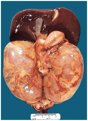
FIGURE 17-13 • Autosomal recessive polycystic kidney disease with massively enlarged symmetric reniform kidneys.

Thirty to fifty percent of patients present with oligohydramnios (Potter sequence): massively enlarged, symmetric, reniform kidneys (Figure 17-13) and pulmonary hypoplasia. Death occurs in the perinatal period. The gross and microscopic hallmark is the presence of tubular cysts with a diameter of 1 to 2 mm arranged radially. The cysts are uniformly distributed and can be appreciated through the capsule of the markedly enlarged kidneys, which retain their shape (Figure 17-14). On cut section, the cortex and the medulla are often unrecognizable. The cysts represent tubular dilatation of presumably normally formed collecting ducts; normal glomeruli and tubules are seen between the cysts (Figure 17-15). In the medulla, the cysts are more rounded. Significant fibrosis, inflammation, and obstruction are absent (72).


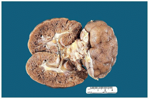
FIGURE 17-14 • Cysts of autosomal recessive polycystic kidney disease can be appreciated on the cortical surface. The cut section shows radially oriented cysts in the cortex and more rounded cysts in the medulla.


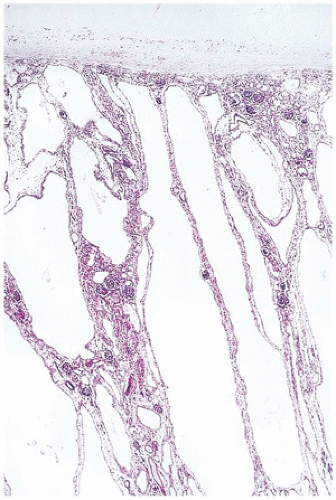
FIGURE 17-15 • Radially arranged cysts of autosomal recessive polycystic kidney disease. Normal glomeruli and tubules are seen between the cysts. (Hematoxylin-eosin stain, original magnification ×40.)

In cases with a later presentation, the degree of renal enlargement is less and the cystic change is less diffuse. However, all forms of ARPKD are associated with congenital hepatic fibrosis (ductal plate malformation), although the clinical expression of liver disease varies widely (see Chapter 15). Dilatation of the interlobular bile ducts is associated with a variable degree of portal fibrosis (73). The lobular architecture of the liver is preserved, but all portal areas are expanded and contain tortuous, slightly dilated ducts at the periphery with blood vessels in the middle (Figure 17-16). Stereologic studies have indicated that what appear as ducts on histologic section are in fact cisterns communicating with each other. Similar hepatic changes are seen in Meckel, Zellweger, and Jeune syndromes, medullary cystic disease complex, and tuberous sclerosis (67,71).

Mutational analysis of ARPKD presenting as infants and congenital hepatic fibrosis presenting in later childhood or adulthood with minimal or no renal disease has defined a broader spectrum of ARPKD. Genotype-phenotype correlations for the type of PKHD1 mutation have been shown. Patients with two truncating mutations usually display a severe phenotype with peri- or neonatal demise, whereas patients surviving the neonatal period bear one hypomorphic missense mutation that is expected to allow for the expression of a minimal amount of full-length fibrocystin
protein. From these data, researchers have concluded that the milder mutation defines the phenotype. No significant clinical differences have been observed between patients with two missense mutations and those with a truncating mutation in trans to a missense mutation (67).


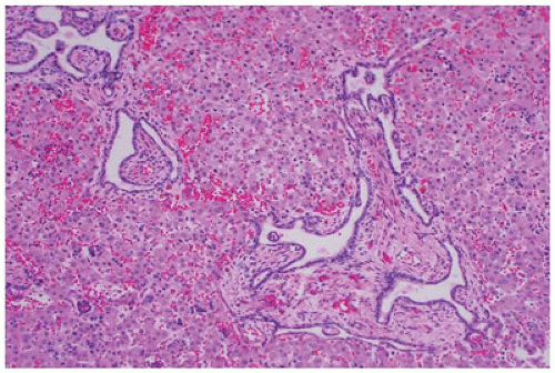
FIGURE 17-16 • Ductal plate malformation of the liver with expanded portal area, peripheral tortuous dilated bile ducts, and blood vessels in the middle. (Hematoxylin-eosin stain, original magnification ×40.)

Up to 30% of patients die in the perinatal period (72). The clinical course of children with ARPKD who survive the neonatal period is variable with ESRD developing in infancy, early childhood, or adolescence. Improved outcomes with kidney and/or liver transplantation are reported (74). A small percentage of long-term survivors are at increased risk of developing benign and malignant liver tumors, particularly cholangiocarcinoma (75). Early detection and appropriate management of renal failure and systemic portal hypertension are important.


Autosomal Dominant Polycystic Kidney Disease

Commonly referred to as adult PKD because the vast majority of cases become symptomatic in the fourth and into the fifth decade of life, ADPKD is more common than ARPKD, occurs worldwide, and affects all races with a prevalence estimated to be between 1:400 and 1:1000 (76). ADPKD accounts for about 5% of cases of ESRD.

ADPKD has two disease loci, PKD1 and PKD2, that encode the membrane glycoproteins, polycystin-1 and polycystin-2 (PC1 and PC2, respectively), which have been localized to the primary cilia of the renal epithelial cells (77). The primary cilia are fingerlike projections on the surface of all kidney cells, except acid-base transporting intercalated cells in the collecting duct. Renal cilia bend in response to fluid flow, which activates PC1, that results in activation of PC2, which leads to calcium influx and changes in gene transcription (78). Abnormal cilia structure or function or both may lead to abnormalities in cell proliferation and tubular differentiation, ultimately leading to cyst formation. Recent studies however have highlighted the complexity of cyst formation. Although abnormal or absent cilia are demonstrated in many “ciliopathies,” cilia are not required for cyst formation and the presence of intact cilia may actually cause worsening of cystic kidney disease in the presence of abnormal PKD proteins. Noncilia-associated proteins also influence cilia structure and function (77,79,80).

Approximately 85% of affected families have mutations in PKD1 gene, which has been mapped to chromosome 16p13.3, and the remaining 15% have mutations in PKD2, which has been localized to chromosome 4q22. Affected persons in these families appear to have a phenotypic similar to that in PKD1 families, but the onset of cystic disease, hypertension, and renal insufficiency is delayed (66). A third gene, PKD3, has been suspected since approximately 10% of families have disease apparently unlinked to PKD1 or PKD2; recent vigorous re-evaluation of previous “PKD3” families has identified PKD1 or PKD2 mutations in four of the five families (81). Mistaken classifications or failure to detect linkage may result from the use of nonflanking markers, the presence of de novo mutations, mosaicism, or complex inheritance of hypomorphic alleles (81).

Autosomal dominant polycystic kidney disease diagnosed in utero or in the first year of life is associated with more severe renal cystic disease and an increased risk of hypertension and early loss of kidney function. Gross hematuria, truncating PKD1 mutations, male sex, childhood onset of hyperfiltration, microalbuminuria or frank proteinuria, elevated copeptin levels (surrogate of circulating arginine vasopressin), and elevated total kidney volume have all been associated with rapid ADPKD progression (82).

Although the majority of ADPKD infants survive, they tend to have more significant hypertension and a more rapid decline in renal function than do their affected adult relatives (83). Children can present with acute or chronic abdominal, back or flank pain, hypertension, proteinuria, or hematuria. Urolithiasis is more common in children with ADPKD than the general population. Very extensive structural disease is required for any measurable decrease in eGFR, and the majority of affected children maintain normal eGFR. The kidneys vary in size from normal to enlarged, and rounded cysts range in size from microscopic (in asymptomatic children with disease detected on screening performed because of a positive family history) to about 3 cm in diameter. The kidneys may lose their reniform shape and become distorted by multiple cysts. Some infants and children present with unilateral renal cysts (83). Glomerulocystic disease may be the most common appearance of early-onset ADPKD (84). Residual parenchyma is compressed by the enlarging cysts and eventually becomes atrophic and fibrotic. In contrast to the cysts seen in ARPKD, these cysts occur in any part of the nephron and are present in both the cortex and the medulla (Figure 17-17, eFigure 17-4). Hepatic, pancreatic, and splenic cysts can occur in childhood ADPKD, most commonly teenagers, but usually only as a few small cysts. The prevalence of mitral valve prolapse is about 12% in ADPKD children (83).


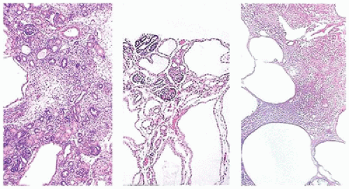
FIGURE 17-17 • Low-power photomicrographs illustrate the differences between cystic dysplastic kidney (left), autosomal recessive polycystic kidney disease (middle), and autosomal dominant polycystic kidney disease (right). (Hematoxylin-eosin stain, original magnification ×40.)


Medullary Cysts

Cysts in the medulla can occur as part of several cystic kidney diseases (e.g., multicystic dysplasia, ARPKD, and ADPKD). The term medullary cystic disease generally conjures three clinically and pathologically distinct entities.


Medullary Sponge Kidney

Also referred to as precalyceal canalicular ectasia, medullary sponge kidney is generally considered a sporadic disease; however, a few pedigrees have been described with an apparent autosomal dominant inheritance. A recent study demonstrated that 50% of stone-forming patients with MSK have affected relatives with milder forms of the disease (85). Reduced penetrance and variable expressivity imply that the disorder is not an uncommon cause of calcium kidney stones, just uncommonly recognized. It most commonly presents in adults, although some cases have been described in children and even infants. MSK has been associated with other renal and extrarenal malformations supporting the assumption that it is a congenital disorder (86). Medullary sponge kidney is a developmental disorder characterized by ectatic and cystic malformation of the papillary collecting ducts in the renal medulla; the condition is most often bilateral, but it may involve only one kidney or only one or several reniculi. Medullary sponge kidney affects both genders and remains symptomless unless complicated by UTI, renal stones, or hematuria—hence its presentation in later life (86). It is best diagnosed by intravenous pyelography (IVP), which shows dilated medullary tubules and the so-called papillary blush or bouquet of flowers; however, IVP has become an obsolescent test, having been replaced as a screening tool for renal colic by noncontrast CT, which has a low sensitivity for the diagnosis. MSK can be recognized by CT urography with contrast and even ultrasound interpreted by knowledgeable radiologists (87). Heterozygous GDNF mutations have been identified in some patients with MSK in regions of the gene that bind to PAX2, which has a crucial role in nephrogenesis (86,88).


Nephronophthisis

Originally described as two separate diseases, nephronophthisis (NPH) and medullary cystic kidney disease (MCKD) were subsequently considered to be part of the same complex, with similar clinicopathologic features; they are both inherited as progressive tubulointerstitial diseases characterized by medullary cyst formation and secondary glomerular sclerosis. Newer molecular techniques have proven that NPH and MCKD are clearly genetically distinct entities (89).

NPH is an autosomal recessive disease and is one of the most common genetic causes of ESRD in children and adolescents (90). Nearly 20 genes have been implicated and almost all of the gene products, including those of the nephrocystin protein family, are expressed in primary cilia, leading to the classification of NPH as a ciliopathy. NPH occurs as an isolated kidney disease, but approximately 15% of patients have extrarenal manifestations such as retinal degeneration in Senior-Loken syndrome, cerebellar vermis aplasia in Joubert syndrome, cone-shaped epiphyses of the phalanges in Mainzer-Saldino syndrome, or liver fibrosis and situs inversus (91). Several syndromal ciliopathies including Bardet-Biedl syndrome, Meckel-Gruber syndrome (MKS), and Jeune syndrome can present with renal NPH. Gene locus heterogeneity, allelism, and modifier genes influence the phenotype with renal manifestations ranging from isolated “degenerative” (tubulointerstitial damage with progressive medullary cysts) disease to “dysplastic” (perinatal multicystic dysplasia) disease with multiorgan involvement, the latter attributed to ubiquitous expression of NPHP proteins (92).

NPH is characterized by polyuria, polydipsia, salt wasting, secondary enuresis, and anemia and proceeds to end-stage renal disease. Three clinical forms are distinguished as the most common variant, juvenile NPH (type 1) in which ESRD occurs at a mean age of 13 years, but symptoms may present as early as 6 years of age; the rare infantile NPH (type 2) with onset of ESRD prior to 4 years of age; and adolescent NPH (type 3) that has a mean age of ESRD of 19 years. Symptoms in early stages may be subtle, but hypertension, growth retardation, and anemia emerge as renal echogenicity and renal failure progress. With advanced disease, corticomedullary cysts are seen. The cysts, 1 to 15 mm in diameter, are located primarily at the corticomedullary junction and present in only 70% of the patients; the remaining patients have no cysts. All cases have in common nonspecific tubulointerstitial disease with tubular atrophy and basement membrane thinning, thickening, and lamellation plus interstitial fibrosis and inflammation, which is usually more severe than the cystic component. In contrast to types 1 and 3, the kidneys in infantile NPH (type 2) are often enlarged and demonstrate not only tubulointerstitial alterations but widespread cyst development that can mimic polycystic kidney disease (90,91,93).


Medullary Cystic Kidney Disease

In contrast to NPH, MCKD has a dominant inheritance and ESRD usually occurs in adulthood but is extremely variable, ranging from 20 to 70 years. They share the same clinical renal presentation, except for the growth retardation and extrarenal malformations, which are absent in MCKD. Two forms are recognized: MCKD1 and MCKD2, and the genetic defects in both have recently been elucidated. MCKD1 is caused by a mutation in MUC1, which encodes mucoprotein 1 that is expressed in skin, breast, lung, and the gastrointestinal tract. In MCKD1, patients develop hyperuricemia (gout) secondary to renal failure, rather than as a primary manifestation of the disease, as seen in MCKD2. The kidney shows marked tubulointerstitial damage but no cysts (94). MCKD2 is caused by mutations in the uromodulin gene, UMOD, which encodes uromodulin (Tamm-Horsfall protein), the most abundant protein in human urine. UMOD mutations are responsible for a group of hereditary autosomal dominant tubulointerstitial diseases (often referred to as uromodulinassociated kidney disease), encompassing MCKD2, familial juvenile hyperuricemic nephropathy, and glomerulocystic kidney disease. Patients with MCKD2 develop tubulointerstitial nephritis in adulthood, but the earliest symptoms of hyperuricemia and gout develop in adolescence. Renal imaging demonstrates corticomedullary cysts, and histology shows interstitial fibrosis with tubular atrophy (95,96,97). New mutations and incomplete penetrance explain the absence of family history in approximately 15% of patients (96).


Cortical Cysts

While cystic dilatation of various parts of the nephron can involve the renal cortex (i.e., cystic change of the collecting ducts in ARPKD), cortical cysts primarily refer to glomerular cysts, characterized as cystic dilatation of Bowman space. The glomerulocystic kidney was recognized in the 1800s (84). The term disease is generally reserved for the familial autosomal dominant or sporadic GCK in which a cause is not identified. Bernstein delineated the diagnosis by defining the glomerular cyst as dilatation of Bowman space in the plane of section of two- to threefold that of normal (Figure 17-18) and considered that the presence of glomerular tufts within at least 5% of cysts as evidence to support the diagnosis of glomerulocystic kidney (98). The only evidence of a glomerular tuft may be a cluster of cells adherent to the cyst wall, but the presence of tubular cysts does not preclude the diagnosis. The cysts are usually round, ovoid, or polygonal, range from less than 0.1 cm to greater than 1.0 cm, and can be empty or filled with proteinaceous material or debris.

It is now recognized that GCK is not a single entity but can be categorized as primary or secondary. The former includes (a) autosomal dominant GCKD such as those due to UMOD, REN, or TCF2 mutations; (b) GCK associated with familial nonsyndromic cystic disease (ADPKD and ARPKD); (c) GCK associated with heritable syndromes such as tuberous sclerosis (TSC), NPH (NPHP3 mutations), Jeune syndrome, orofacialdigital syndrome type 1, glutaric aciduria type 2, Zellweger syndrome, among others; and (d) various genetic abnormalities including trisomies 21, 18, 13, and 9 and monosomy X. In the latter two categories, glomerular cysts are often a minor component, for example, Jeune syndrome and NPH, better known for chronic progressive tubulointerstitial disease, and Zellweger syndrome where glomerular cysts are scattered within the cortex but usually inconsequential and only occasionally serious enough to affect renal function. In all the syndrome and genetic abnormalities, the cysts are inconsistently expressed. Secondary and acquired causes relate to (a) ischemia (postthrombotic microangiopathy— hemolytic-uremic syndrome/thrombotic thrombocytopenic purpura (HUS/TTP), vasculitis, or renal artery stenosis); (b) drugs (lithium, corticosteroids); (c) renal dysplasia; and (d) obstruction (10,84,99).


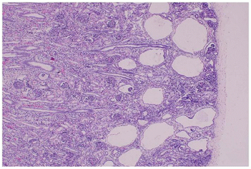
FIGURE 17-18 • GCKD with cystic dilatation of Bowman spaces; the medulla is uninvolved. (Hematoxylin-eosin stain, original magnification ×40.)

Most GCKD cases are transmitted according to an autosomal dominant mode of inheritance (84). This disease is often discovered in infants within the context of a familial history of ADPKD. The most common cause in neonates and young children in one series of 20 patients (aged 30 weeks’ gestation age to 78 years) was dysplasia (84). Adult presentation has been described along with unilateral disease, both typically associated with secondary etiologies (84). Ultrasonographically, minute cysts, smaller than those occurring in the usual autosomal dominant polycystic kidney disease, are seen in the echogenic renal cortex. No cysts are observed in the renal medulla, in ADGCKD, but may been seen with other etiologies. Kidneys in ADGCKD and ADPKD presenting with a GCK phenotype are bilaterally enlarged and diffusely cystic, in which the main microscopic finding is represented by glomerular cysts, but an asymmetric onset of this disease has also been seen. Kidneys in sporadic GCKD of non-ADPKD phenotype can have either clustered or diffuse cysts and are either slightly larger or normal in size.

Finally, in familial hypoplastic GCKD, the kidneys are smaller than normal and often associated with medullocalyceal abnormalities (100). Familial hypoplastic GCKD
is associated with mutations in TCF2, which encodes HNF1-β, and is considered the main cause of fetal hyperechoic kidneys (100) and, in some families, is associated with maturity-onset diabetes of the young, type V (MODY5) (101,102).Glomerular cysts in all types of GCKD are less than 1 cm in size and located in the cortex from the subcapsular zone to the inner cortex (eFigure 17-5), and are histologically similar to glomerular cysts seen in other disease conditions.


Simple Cysts

Simple cortical cysts, or retention cysts, which are very common in adults, are rarely seen in children. They are typically asymptomatic and incidentally discovered. They are important because they may present as an abdominal mass, or their appearance ultrasonographic or radiologic images may raise the diagnostic consideration of cystic WT. The diagnosis is established on the basis of typical radiographic findings with surrounding normal renal parenchyma, normal renal function, and the absence of systemic illness or disorders. Simple cysts arise from the cortex, are unilocular, contain yellow clear fluid, and are lined by a single layer of cuboidal epithelium. The cysts may slowly increase in size, at a rate averaging 0.3 mm and 1.0% per year, but can also disappear (103). In addition to excluding malignancy, follow-up should be undertaken to exclude an early presentation of ADPKD, especially when there are bilateral or multiple unilateral cysts (104,105).


Cysts Associated with Syndromes

Cysts of the cortex (sometimes referred to as pluricystic kidney), are usually asymptomatic, and have been described as a minor component of multiple malformation syndromes, both inheritable and noninheritable, including tuberous sclerosis; von Hippel-Lindau (VHL) disease; MKS; orofaciodigital syndrome type I; trisomies 9, 13, 18, and 21; short rib-polydactyly syndrome; Jeune asphyxiating thoracic dystrophy syndrome; Zellweger cerebrohepatorenal syndrome; VATER association; lissencephaly; renal-hepatic-pancreatic dysplasia; glutaric aciduria type II; Ellis-van Creveld syndrome; Elejalde syndrome; Peutz-Jeghers syndrome; Robert syndrome; phocomelia syndrome or pseudothalidomide syndrome; and Bardet-Biedl syndrome (13). In the following diseases, the renal cysts are often a dominant feature of the disease and histologically distinct from dysplasia.


Tuberous Sclerosis

Tuberous sclerosis complex is an autosomal dominant systemic malformation syndrome caused by mutations in TSC1 on chromosome 9q34 (encoding hamartin) and TSC2 on chromosome 16p13.3 (encoding tuberin). Hamartin and tuberin form a complex that acts as a tumor suppressor by inhibiting mammalian target of rapamycin (mTOR) signaling, which is implicated in proliferation, cell cycle control, and regulation of cell size (106). It is characterized by hamartomatous proliferations of the skin, brain, kidney, eye, bone, liver, and lung. In addition to the well-recognized association with renal angiomyolipomas, which occur in 40% to 80% of patients with tuberous sclerosis, characteristic cortical cysts are present in about 50% of patients. Unilateral renal cystic disease has been rarely reported, and glomerulocystic kidney disease is well known (106). The severe, very earlyonset polycystic phenotype is associated with mutations that involve the adjacent TSC2 and PKD1 genes on chromosome 16p13 and accounts for about 2% of patients (107). The extent of involvement varies; small cysts may be diagnosed on imaging, or “polycystic kidneys” may lead to renal failure. The cysts vary in size and are lined by hyperplastic epithelium, which is often multilayered and papillary, with abundant eosinophilic granular cytoplasm (Figure 17-19). The histologic findings are so characteristic as to be virtually diagnostic of tuberous sclerosis when seen in an early biopsy performed before the onset of other stigmata of the disease. Solid nodules of these cells may also form, and nuclear atypia and pleomorphism may be present. Mitotic activity evident in these cells may be related to the increased risk for neoplasia; cystic renal carcinomas with a granular eosinophilic macrocystic morphology have been considered a distinct pattern of TSC (108).


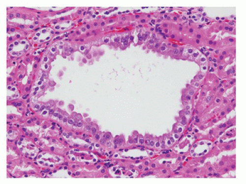
FIGURE 17-19 • Renal cysts of tuberous sclerosis lined by characteristic hyperplastic epithelium with abundant eosinophilic granular cytoplasm. (Hematoxylin-eosin stain, original magnification ×200.) (Courtesy of Dr. John Hicks, Houston, TX).


Von Hippel-Lindau Disease

VHL disease is an autosomal dominant multisystem (pre) neoplastic disorder genetically linked to a germline mutation of a tumor-suppressor gene (VHL gene) located on chromosome 3p25. The incidence of VHL disease is about one in 36,000 births and the penetrance is high. It is characterized by hemangioblastomas of the retina, brain, and spinal cord, pheochromocytomas, and cysts and tumors of the pancreas, kidneys, and, less frequently, other abdominal
organs (69). Multiple renal cysts and tumors develop in approximately two-thirds of patients with VHL disease (109). The most common imaging finding is multiple small unilateral or bilateral renal cysts, although renal function is usually retained (110). Less frequently, the cysts are numerous enough to simulate ADPKD. Renal cysts are lined by flattened clear epithelial cells; however, dysplastic microfoci of hyperplastic epithelium with clear cytoplasm and a “hobnail” appearance, often associated with complex cysts, are associated with a markedly increased risk for the development of renal cell carcinoma (RCC) (111). Multifocal cystic adenocarcinomas develop in 45% to 50% of patients beyond the third decade of life and in about 70% by the fifth decade (69,109). Most RCCs are of the clear cell type although clear cell papillary RCCs have been reported in patients with VHL disease (112).


Meckel-Gruber Syndrome

MKS has an autosomal recessive inheritance and shares significant phenotypic and genotypic overlap with other ciliopathies including the primarily kidney disorder NPH and the neurodevelopmental disorder Joubert syndrome. Patients harboring the same genetic mutation can express a broad range of neurologic and renal involvement; thus, despite the varied clinical presentations, the genotype-phenotype correlations are frequently unclear and there is significant inter- and intrafamilial heterogeneity. These features suggest the operative effects of modifiers and genetic interactions including triallelic inheritance (90). Mutations in at least 16 different genes have been shown to be causative in MKS in humans (113). Many of the genes are linked to ciliary biology, but several are predicted to produce transmembrane proteins or those found in the actin cytoskeleton or influence microtubule organization, vesicle trafficking, and signal transduction. Several of the MKS genes interfere with normal Wnt signaling, which is a key player in the development of cystic kidney disease (114,115). MKS has a reported mean prevalence of 2.6 per 100,000 births, and nowadays, most patients are detected very early in pregnancy (116). It is characterized by cystic kidneys, posterior encephalocele, and polydactyly and frequently associated with other CNS anomalies, fibrocystic changes of the liver, (congenital hepatic fibrosis/ductal plate malformation), and orofacial clefts. The kidneys are always bilaterally involved, although they may occasionally be variably involved. Round cysts arise from any part of the nephron, with microcysts seen in the subcapsular area and larger cysts in the medulla. The cysts are lined by a single layer of low-to-high cuboidal epithelium and are separated by loose, immature mesenchyme that may bulge into the cysts. Metaplastic cartilage is not usually present.

The pathogenesis of renal cysts in Meckel syndrome follows the mechanisms of other ciliopathies and is related to defects in ciliary signaling with perturbation of cell growth, polarity, and cell fate determination (113). Study of midterm fetuses has shown that the kidneys are already enlarged by 11 to 20 weeks of gestation (117). Nephrogenesis may appear normal at the periphery of the kidney, although the nephrogenic zone is often interrupted by dilated ducts and fibrous tissue. Other findings include a thin cortex, poorly demarcated medullary pyramids, and poor caliceal development (118). It also appears that many of the nephrons are formed normally and the tubules and ducts are secondarily converted to cysts.


GLOMERULAR DISEASES

Metanephric blastema condenses around the end of the ureteric bud at about day 32 of development, and elongation, branching, and subsequent fusion of proximal generations of the bud give rise to the pelvicalyceal system and collecting ducts. The first glomerulotubular structures appear during week 8 as a result of the interaction of subcortical blastema with the ampullary ends of the collecting ducts, and glomerulogenesis continues until gestational week 36 when the neogenic (nephrogenic) zone disappears and nine to eleven generations of glomeruli are present. The number of glomeruli in human kidneys varies from 250,000 to 1.8 million. This marked interindividual difference may be genetically programmed or due to perinatal factors such as low birth weight (estimated relation: 250,000 glomeruli per kilogram at birth) and may predispose persons with lesser numbers of glomeruli to renal failure in adulthood (119). Immature (fetal) glomeruli are characterized by their small size and prominent corona of visceral epithelial cells, and normally, this corona disappears during the first year. Mean glomerular diameter increases from 112 µm at birth to 167 µm at 15 years, and enlarged (hypertrophied) glomeruli suggest a compensatory response to reduced nephron mass (120). The thicknesses of the glomerular capillary wall and the lamina densa increase from 169 ± 30 nm and 98 ± 23 nm, respectively, at birth to 285 ± 39 nm and 219 ± 42 nm, respectively, at 11 years (121). More contemporary measurements demonstrate a nearly linear increase in glomerular basement membrane thickness from 1 year to a plateau at 9 years (122). The molecular structure of the glomerular basement membrane also changes with age. Collagen α1α2α1(IV) synthesized by podocytes, endothelial cells, and mesangial cells of immature glomeruli is replaced by collagen α3α4α5(IV) produced exclusively by podocytes (123).

The pathologist most often encounters glomerular diseases in renal biopsy specimens collected with biopsy guns having needles of 18 gauge or less and may be asked to examine the gross specimen for the presence of glomeruli with a magnifying lens or dissecting microscope (Figure 17-20). The presence of renal cortex may be inferred if one sees capsule and fat at one end of the biopsy specimen and architecture consistent with medulla at the other, but the macroscopic recognition of glomeruli requires sufficient blood flow within glomerular capillaries, and this may be reduced by disease.
Definitive identification of glomeruli may rarely require rapid frozen section, or the pathologist may be asked to perform a rapid frozen section to determine if crescents are present. In either case, the tissue submitted for frozen section can also be utilized for immunofluorescent (IF) studies. Whenever possible, tissue should be sampled for light, IF, and electron microscopy (EM), even if all those studies are not initially requested, and with the smaller-gauge biopsy needles now used by pediatric nephrologists, two or three cores are usually required. The specimen submitted for light microscopy (LM) should contain as much cortex as possible along with the corticomedullary junction, whereas only cortical tissue is ordinarily required in the specimens submitted for IF and EM (124).


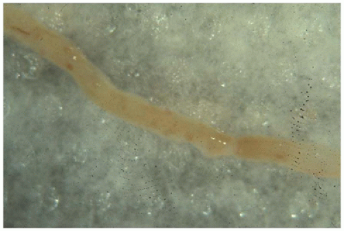
FIGURE 17-20 • Needle biopsy specimen of kidney viewed through a dissecting microscope. Glomeruli appear as red dots in the central region and vasa recta in the outer medulla as linear striations at either end. (Original magnification, 5×.)


TABLE 17-5 TERMS USED IN DESCRIBING GLOMERULAR LESIONS


Focal


Involvement of <50% of all glomeruli


Diffuse


Involvement of ≥50% of all glomeruli


Segmental


Involvement of <50% of a glomerulus


Global


Involvement of ≥50% of a glomerulus


Hyalinosis


Accumulation of eosinophilic, PAS-positive, silver-negative, structureless material that stains red with trichrome stains (glycoproteins and lipids)


Sclerosis


Accumulation of eosinophilic, PAS-positive, silver-positive structureless material that stains blue or green with trichrome stains (collagen IV)


Fibrosis


Accumulation of eosinophilic, PAS-negative, silver-negative fibrillar material that stains blue or green with trichrome stains (collagen I, III)


Mesangial proliferation


More than three mesangial cells per peripheral mesangial area


Mesangiocapillary (membranoproliferative) glomerulonephritis


A combination of mesangial proliferation and capillary wall thickening


Adhesion (synechiae)


Attachment of part or the entire circumference of a glomerular tuft to Bowman capsule. Adhesions may be fibrous or fibrinous


Crescent


A proliferation of glomerular epithelial cells and inflammatory cells that fills part (segmental) or all (circumferential) of Bowman space. Crescents may be cellular, fibrocellular, or fibrous


PAS, periodic acid-Schiff stain.


Our understanding of pediatric renal pathology has been greatly facilitated by contributions from two multiinstitutional collaborative studies, the International Study of Kidney Disease in Children (ISKDC) and the Southwest Pediatric Nephrology Study Group (SPNSG). The terms most commonly used to describe the lesions encountered in renal biopsy specimens are listed in Table 17-5. Children with renal disease usually present with proteinuria or hematuria, alone or in combination, with or without associated systemic disease. Less commonly, patients present with a nephritic syndrome that includes proteinuria, hematuria, red blood cell and white blood cell casts, and decreased plasma levels of complement components, or with acute renal failure, renal concentration defects, or chronic renal failure without known antecedent disease. Isolated proteinuria and hematuria do not usually warrant biopsy study, and most children with nephrotic syndrome responsive to steroid therapy or acute glomerulonephritis attributable to streptococcal disease do not undergo biopsy unless the course is atypical or the response to therapy is suboptimal. Typically, the glomeruli in patients with isolated proteinuria or hematuria are optically normal or show focal and segmental glomerulosclerosis (Figure 17-21A) or mesangial hypercellularity (Figure 17-21B). Diffuse and global mesangial hypercellularity with thickening of capillary walls and obliteration of capillary loops resulting in accentuation of the lobular architecture of the glomerulus (Figure 17-21C) or the presence of crescents and proliferations of parietal epithelial cells and inflammatory cells in Bowman space (Figure 17-21D) are usually associated with a nephritic syndrome or acute renal failure. IF and electron microscopic studies are usually necessary to arrive at a more precise diagnosis. A granular pattern of immunofluorescence— along capillary loops (Figure 17-22A),
within mesangia (Figure 17-22B), or both (Figure 17-22C)—indicates immune complex (IC) deposition, and the site and the composition of the immunoreactant(s) depend on the disease. Crescents often stain brightly for fibrinogen. Linear staining along the capillary wall may indicate antiglomerular basement membrane disease (usually only IgG). Dense deposit disease, or so-called C3 glomerulopathy, will usually have C3 only or at least C3 predominance (Figure 17-22D). The histologic, IF, and ultrastructural lesions for specific diseases are detailed below, but a careful inventory of the lesions in all renal compartments—glomeruli, tubules, interstitium, and vessels—and correlation of the morphologic findings with the clinical history and the results of renal function tests and serologic studies are necessary for the proper clinicopathologic interpretation of renal biopsy specimens from patients of any age.


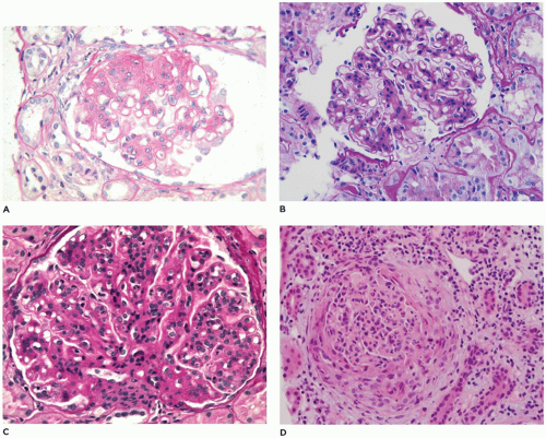
FIGURE 17-21 • Glomerular lesions observed in pediatric renal biopsy specimens. A: FSGS with the sclerotic tuft in the 11 o’clock position adherent to Bowman capsule and segmental proliferation of visceral epithelial cells at the 2 o’clock to 4 o’clock position. B: Mesangial proliferation is defined as more than three mesangial cell nuclei per peripheral mesangial focus. C: Mesangiocapillary or MPGN shows both mesangial proliferation and thickening of capillary loops. D: In CGN, a segmental, or in this case, circumferential proliferation of epithelial and inflammatory cells in Bowman space compresses the underlying glomerular tuft. (D: hematoxylineosin, A-C periodic acid-Schiff stain, original magnifications ×400.)


Glomerulopathies that Usually Present with Proteinuria or Nephrotic Syndrome

The incidence of idiopathic nephrotic syndrome is two to seven per 100,000 children, 95% of whom respond to steroid therapy, although 60% to 80% of these will experience one or more relapses (125). The distribution of lesions in multiple series of untreated children with nephrotic syndrome was minimal change disease (MCD), 34% to 53%; membranoproliferative glomerulonephritis (MPGN), 2% to 16%; FSGS,
9% to 40%; membranous glomerulonephritis (MGN), 1.5% to 9%; and chronic or unclassified glomerulonephritis, 6% to 17% (126). The incidence of MCD is higher in children under 6 years old at diagnosis (71% versus 24%), more frequent in males (male to female ratios of 60:40), and less often associated with hypertension or hematuria than other causes of nephrotic syndrome. A response to prednisone at 8 weeks was seen in 93% of patients with MCD, 75% with focal global glomerulosclerosis, 30% with FSGS, 56% with diffuse mesangial hypercellularity, 7% with MPGN, and none with MGN. MCD is underrepresented in more recent series, reflecting the current practice of not performing biopsies in children with nephrotic syndrome unless unresponsive or resistant to steroid therapy. However, even allowing for the changes in biopsy practice, the incidence of FSGS in children appears to be increasing in all ethnic groups, becomes more apparent in patients over 6 years of age at presentation, and appears to be more common and more aggressive in African American and possibly Japanese children (127). Familial FSGS has been attributed to mutations that alter slit-diaphragm proteins (nephrin [NPHS1], podocin [NPHS2], CD2-associated protein [CD2AP], short transient receptor potential channel 6 [TRPC6]), actin-regulating proteins (alpha-actinin-4 [ACTN4], inverted formin-2 [INF2], Rho GTPase-activating proteins 24 [ARHGAP24], Rho GDP-dissociation inhibitor 1 [ARHGDIA]), transcription factors (Wilms tumor protein [WT1], LIM homeobox transcription factor 1β [LMX1B], WSI/SNF-related matrix-associated actin-dependent regulator of chromatin subfamily A-like protein [SMARCAL1]), glomerular basement membrane proteins (laminin subunit β2 [LAMB2], integrin β4 [ITGB4]), and various mitochondrial and lysosomal proteins. Many of these patients do not
present until adulthood, and there is considerable variability in the clinical severity and response to therapy, especially among heterozygotes, implying that other genes or nongenetic triggers may be involved (127,128). Altered expression or distribution of these and other podocyte proteins has been found in studies of nonfamilial FSGS suggesting that reorganization of podocyte proteins in response to injury is a key step in the pathogenesis of proteinuria (129). Gene expression profiling of isolated glomeruli from patients with FSGS, MCD, and normal controls demonstrated numerous significantly differentially regulated genes including some known to be part of the slit-diaphragm complex and previously described in dysregulated podocyte phenotype (130). FSGS, often with lesser (nonnephrotic) levels of proteinuria, is also the lesion seen in cyanotic congenital heart disease, sickle cell anemia, massive obesity, HIV, and other viral infections (parvovirus, SV40, and some cases of hepatitis C). Nephrotic syndrome in the first year of life may be associated with lesions that occur in older children but is more often caused by one of two lesions unique to this age group—congenital nephrotic syndrome (CNS) of the Finnish type (CNF) and diffuse mesangial sclerosis (DMS) that are discussed later. Medical complications of nephrotic syndrome include acute infections and thromboembolic disease related to the nephrotic state and long-term effects on bones, growth, and the cardiovascular system related to the disease and its treatment (125).


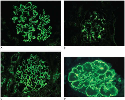
FIGURE 17-22 • Immunofluorescence patterns observed in pediatric renal biopsy specimens. A: Granular staining along the capillary loops. B: Confluent granular staining in mesangia. C: Combination of capillary and mesangial granular staining. D: Linear staining for C3 along the capillary wall and bright rings with mesangia in dense deposit disease (type II MPGN). [Fluorescein isothiocyanate-conjugated anti-IgG (A-C) or anti-C3 (D), original magnifications 400×].


Minimal Change Disease, IgM and C1q Nephropathy

Glomerulopathy with minimal change is the most common cause of nephrotic syndrome in children and referred to as minimal change nephrotic syndrome (MCNS) or MCD. The pathogenesis of MCD remains unknown, although it is likely the result of complex interactions among multiple systems including lymphokines that increase glomerular permeability, regulatory T-cell dysfunction, upregulation of CD80 and angiopoietin-like-4 (Ang-4) and Ang-3 by podocytes, or various epigenetic modifications triggered by environmental or immune factors (131,132).

By definition, MCD shows no significant abnormalities by LM. Slight segmental increases in mesangial matrix and cellularity (three or more mesangial cells in most tufts of most glomeruli [Figure 17-21B]) and focal interstitial fibrosis are within the spectrum of “minimal change.” The historically termed “diffuse mesangial hypercellularity” is also now considered within the range of MCD (126); some have noted signs of glomerular immaturity in association with hypercellularity (133). The long-term prognosis in these patients is good although there is a trend to initial steroid nonresponsiveness.

The classic light microscopic appearance in MCD includes patent glomerular capillaries that are surrounded by regular walls of expected thickness. Mesangial matrix may show a slight increase, but absent are focal or segmental scarring or collapse, adhesion to Bowman capsule, and endocapillary proliferation. Occasionally, immature glomeruli are seen. IF microscopy is usually negative or reveals only segmentally variable, mesangial staining for IgM with or without C3 or C1q. Ultrastructural findings in patients with the nephrotic syndrome include diffuse retraction of foot processes of visceral epithelial cells, microvillous transformation along the cell membrane, and vacuolization and lipid droplets within visceral epithelial cell cytoplasm. The GBM is usually normal.

Most authors consider any segmental glomerulosclerosis significant, but rare globally sclerotic or hyalinized glomeruli are occasionally seen in otherwise normal infant kidneys. Emery and MacDonald found hyalinized glomeruli in the kidneys of 75 of 200 (38%) infants and children up to 15 years of age (0.5% to 30% of glomeruli in affected kidneys, but in most cases, the range was 1% to 2%) and noted that rare sclerotic glomeruli were present in many of the kidneys that had no such glomeruli in the selected field (134). Global glomerulosclerosis may occur as a part of normal aging and repair in the absence of renal disease. Less than 5% globally sclerosed glomeruli is the expectation up to mid-adulthood. Thus, a rare globally sclerotic glomerulus might be within normal limits but should initiate a search of serial sections through the block for a segmentally sclerotic glomerulus. Examination of serial sections is also recommended if focal tubular atrophy, interstitial fibrosis, enlarged glomeruli, segmental hyalinosis, segmentally positive immunofluorescence, collagen in glomeruli by EM, or an incomplete therapeutic response is found.

The differential diagnosis of MCD is IgM nephropathy, C1q nephropathy, FSGS, and CNS. The latter two are discussed in other sections. Controversy exists as to the definition and significance of IgM nephropathy, as IgM is frequently considered a nonspecific finding due to trapping. The typical light microscopic appearance is normal or mild mesangial hypercellularity with bright mesangial IgM on immunofluorescence; however, various authors have systematically required or excluded the presence of deposits on EM. Several have shown a poorer prognosis or variable responses to steroids, cyclophosphamide, or cyclosporine (135,136).

Jennette et al. described a proliferative glomerulonephritis with mesangial granular C1q as the dominant or codominant immunoreactant and no evidence of systemic lupus erythematosus (SLE) in 15 adolescents and young adults who presented with proteinuria or nephrotic syndrome (137). C1q nephropathy is noted most frequently in children and young adults (age range: 3 to 42 years, mean: 24.2 years) with a female and African American preponderance, and renal biopsies typically show minimal change, mesangial hypercellularity, or FSGS (138). Many authors concluded that C1q nephropathy falls within the spectrum of MCD and FSGS, but recent reports fail to demonstrate significant outcome differences between similar cases with or without C1q (136,139,140).

MCD is most often idiopathic in children and exquisitely sensitive to steroid therapy, such that treatment in
most patients is initiated without a biopsy and 90% to 95% of patients respond within 8 weeks, although approximately 60% relapse (126). Less frequent relapse is reported in those who receive extended initial therapy, and supplemental therapies have included calcineurin inhibitors, especially cyclosporine; alkylating agents, particularly cyclophosphamide; and more recent introduction of mycophenolate mofetil (MMF) and rituximab (141). Secondary causes of MCD include NSAIDs, lymphomas, infections, and allergic reaction, and the diagnosis is suspected by clinical history.


Focal Segmental Glomerulosclerosis

Although often considered a spectrum of one disease, with FSGS resulting from progression of MCD, contemporary evidence supports the notion that these two are distinct entities with different pathogeneses (142,143,144,145). Most children with primary FSGS present with nephrotic syndrome, and the proportion of pediatric patients with nephrotic syndrome due to FSGS increases with age.

Segmental proliferation of visceral epithelial cells (Figure 17-21A, 2 o’clock to 4 o’clock position) may be the earliest lesion of FSGS. D’Agati et al. (146) have subdivided FSGS into five categories: NOS, cellular, perihilar, tip, and collapsing variants. FSGS-NOS is the most common form seen in children and adults. The collapsing and tip lesion variants are most likely to present with full nephrotic syndrome, which have the lowest and highest rates of remission, respectively, and conversely, the highest and lowest rates of ESRD (147,148). While not all espouse this classification, it can be applied to primary and secondary FSGS and may provide clues to the underlying etiologies.

The classic appearance of FSGS includes segmental tuft sclerosis with adhesion to Bowman capsule in a minority of glomeruli (Figure 17-21A). Juxtamedullary glomeruli are more frequently affected than superficial glomeruli. Mild endocapillary or extracapillary cellular increase may be associated with matrix accumulation in the tuft. IF microscopy is typically negative although nonspecific trapping of IgM or C3 is often noted in sclerotic segments. Podocyte foot process effacement is expected by ultrastructural assessment as well as podocyte swelling, microvillous transformation, and increased organelles; podocytes may be detached from the GBM (148). Proximal tubules often contain resorption droplets, and tubular atrophy is accompanied by interstitial fibrosis. The pathology of primary (idiopathic) and familial FSGS is similar such that clinical and family history may be more significant than the histopathology in determining response to therapy. The treatment approach is similar as with MCD; however, the majority of patients in whom a sustained response is not achieved will eventually progress to ESRD. A recurrence rate after transplant as high as 50% has been reported with initial steroid responsiveness highly predictive of recurrence, while pathogenic genetic mutations are protective of recurrence (149).


Membranous Glomerulonephritis

MGN is seen in 1.5% to 9% of children and 18.5% of adolescents with nephrotic syndrome (126). In children, the age at onset is usually 8 to 16 years, and the sex ratio varies among studies from equal to a male predominance (150). Most patients have microscopic hematuria in addition to proteinuria, but macroscopic hematuria is uncommon. Thirty-five percent of cases of MGN in children are secondary to systemic diseases, whereas the incidence of secondary MGN in adults is 20% (151). The designation of “idiopathic” (primary) may be disappearing as most cases of MGN in adults appear to be mediated by autoantibodies to phospholipase A2 receptor (PLA2R) expressed on podocytes (152). The antibody seems to be relatively specific for primary MGN (153), although other antipodocyte antibodies may be circulating (154). Moreover, primary MGN in the pediatric population must have more etiologies as less than 45% show PLA2R autoantibodies (155). Most cases of secondary MGN are due to SLE with the proportion due to hepatitis B decreasing as vaccination has become routine in many places. Additional secondary causes in children include infection, neoplasia, and other autoimmune disorders.

Following the description in 2002 of a remarkable case of antenatal MGN due to maternal antibodies directed against neutral endopeptidase (NEP), a podocyte and tubular brush border protein, which was present in the fetus but not the mother (156), other cases of MGN in early life attributable to antenatal alloimmunization have been identified (157). This particular disease mechanism bears similarities to the autoimmune form of anti-PLA2R-associated MGN in adults, where autoantibodies bind in situ to endogenous podocyte proteins. More recently, the same investigators identified circulating antibodies directed against cationic bovine serum albumin (BSA) and BSA within subepithelial deposits (158). This phenomenon is more analogous to secondary MGN, such as hepatitis B, where planted antigens contribute to an immune complex, which stimulates complement activation and collateral damage to the podocyte.

Histologically, glomeruli in MGN appear large and have uniformly thickened capillary walls but patent capillary lumens (Figure 17-23A). The diagnostic “spikes” seen on silver stains (Figure 17-23B) represent notches along the outer aspect of the normally argyrophilic basement membrane due to immune complexes that do not take up the silver. Spikes cannot be detected when the deposits are small or sparse (Figure 17-23C) or when they have been fully incorporated into the basement membrane (Figure 17-23D). Mesangial hypercellularity, glomerular lobulation, and segmental inflammation, necrosis, or sclerosis are more common in secondary MGN (151). The degree of proteinuria or stage of disease does not correlate with disease course or outcome, although glomerulosclerosis and interstitial fibrosis may portend an unfavorable course (159).

IF microscopy reveals granular staining along capillary walls (Figure 17-22A) and occasionally also within the mesangia (Figure 17-22C). IgG and C3 are very commonly
present, but a “full house” of immunoreactants suggests lupus or another systemic disease. Mesangial deposits also suggest systemic disease. Four stages have been described in MGN: stage I, small subepithelial deposits (Figure 17-23C); stage II, larger and more numerous deposits bordered by projections of the lamina densa; stage III, incorporation of deposits into the lamina densa (Figure 17-23D); and stage IV, a thickened and irregular basement membrane without recognizable deposits. These observations provide much insight into the morphology of MGN, but they simplify the biologic complexity as the stages do not correlate with proteinuria, outcome, or clinical change and often overlap. Patients may present at any stage and may have deposits characteristic of more than one stage. Foot process retraction is typically extensive in all stages. Treatment is influenced by clinical severity; asymptomatic children with nonnephrotic proteinuria are often managed conservatively with angiotensin-converting enzyme inhibitors or angiotensin receptor blockers, while those with nephrotic syndrome receive various combinations of corticosteroids, alkylating agents, or other immunosuppressants such as calcineurin inhibitors, MMF, and rituximab (150,159).

Only gold members can continue reading. Log In or Register to continue

Stay updated, free articles. Join our Telegram channel

Sep 23, 2016 | Posted by in PATHOLOGY & LABORATORY MEDICINE | Comments Off on The Kidney and Lower Urinary Tract

Full access? Get Clinical Tree

Get Clinical Tree app for offline access