13 FIG. 13.1 Lip FIG. 13.2 Palatal mucosa, monkey FIG. 13.3 Lip and tooth FIG. 13.4 Tooth structure FIG. 13.5 Tooth development (illustrations opposite)
Oral tissues
Introduction
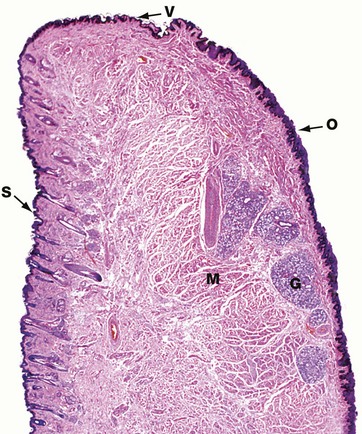
H&E (LP)
This micrograph illustrates a midline section through a human lower lip, the bulk of which is made up of bundles of circumoral skeletal muscle M seen in transverse section. The external surface of the lip is covered by hair-bearing skin S which passes through a transition zone to merge with the oral mucosa O of the inner surface. The transition zone constitutes the free vermilion border of the lip V, and derives its colour from the richly vascular dermis, which here has only a thin, lightly keratinised epidermal covering. The free border is highly sensitive due to its rich sensory innervation. Since the vermilion border is devoid of sweat and sebaceous glands, it requires continuous moistening by saliva to prevent cracking. The oral mucosa covering the inner surface of the lip has a thick stratified squamous epithelium and the underlying submucosa contains numerous accessory salivary glands G of serous, mucous and mixed seromucous types.
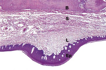
H&E (LP)
Like the rest of the mouth, the palate is covered by a thick stratified squamous epithelium Ep supported by a tough, densely collagenous lamina propria L. To assist mastication, the palatal mucosa is thrown up into transverse folds or rugae, one of which is shown in this micrograph. The mucosa of the hard palate is bound down to the underlying bone B by relatively dense submucosal tissue S containing a few accessory salivary glands.
In rodents and many other mammals with a coarse diet, the surface epithelium of particularly exposed areas is keratinised for extra protection, as in this specimen taken from a monkey.
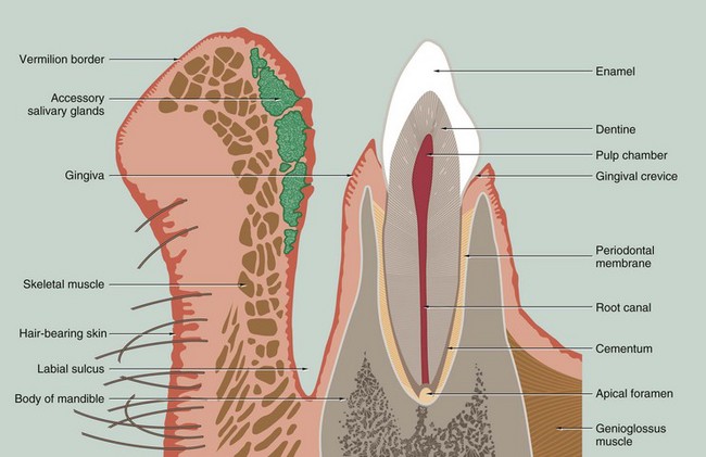
This drawing of a section through the lower jaw near the midline illustrates the general arrangement of the lip and a tooth with its supporting structures. Each tooth may be grossly divided into two segments, the crown and the root; the crown is that portion which projects into the oral cavity and is protected by a layer of highly mineralised enamel which covers it entirely. The bulk of the tooth is made up of dentine, a mineralised tissue which has a similar chemical composition to bone. The dentine has a central pulp cavity or chamber containing the dental pulp which consists of specialised supporting tissue containing many sensory nerve fibres. The tooth root is embedded in a bony ridge in the jaw called the alveolar ridge; the tooth socket is known as the alveolus. At the lip or cheek (buccal) aspect of the alveolus, the bony plate is generally thinner than at the tongue (palatal) aspect. The root of the tooth is invested by a thin layer of cementum which is connected to the bone of the socket by a thin fibrous layer called the periodontal ligament or periodontal membrane.
The oral mucosa covering the upper part of the alveolar ridge is called the gingiva and, at the junction of the crown and root of the tooth (the neck of the tooth), the gingiva forms a tight protective cuff around the tooth. The potential space between the gingival cuff and the enamel of the crown is called the gingival crevice. All of the tissues which surround and support the tooth are collectively known as the periodontium.
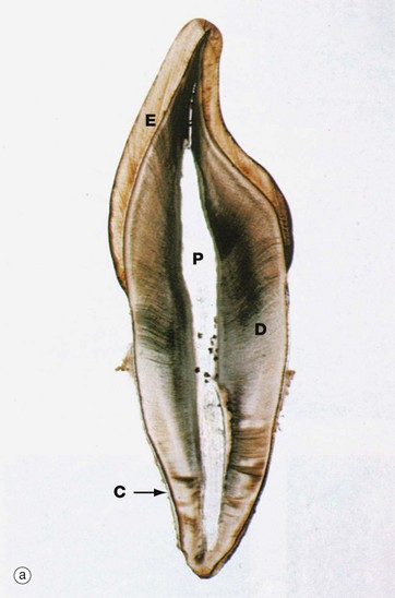
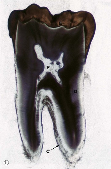
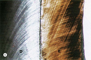
Undecalcified sections, unstained (a) LP (b) LP (c) (HP)
These undecalcified sections, cut with a diamond wheel, demonstrate the arrangement of the calcified tissues of an upper central incisor tooth, micrograph (a), and a lower molar tooth, micrograph (b). Micrograph (c) demonstrates the tissues of the crown at high magnification.
The dentine D, which forms the bulk of the crown and root, is composed of a calcified organic matrix similar to that of bone. The inorganic component constitutes a somewhat larger proportion of the matrix of dentine than that of bone and exists mainly in the form of hydroxyapatite crystals. Teeth are thus harder than bone. From the pulp cavity P, minute parallel tubules called dentine tubules radiate to the periphery of the dentine.
The crown is covered by enamel E, a translucent substance composed of parallel enamel rods or prisms of highly calcified material, cemented together by an almost equally calcified interprismatic material.
The root is invested by a thin layer of cementum C which is generally thicker towards the apex of the root. The cementum is an amorphous calcified tissue into which the fibres of the periodontal membrane are anchored.
The morphological form of the tooth crown and roots varies considerably in different parts of the mouth; nevertheless, the basic arrangement of the dental tissues is the same in all teeth.
In humans, the primary (deciduous) dentition consists of 20 teeth comprising two incisors, one canine and two molars in each quadrant. These begin to be formed at the age of 6 weeks during fetal development and they erupt between the ages of 6 and 30 months after birth. Between the ages of 6 and 12 years, the deciduous teeth are succeeded by permanent teeth, namely two incisors, one canine and two premolars in each quadrant. Distal to these will develop three permanent molars which have no primary precursors; the first permanent molar erupts at age 6, the second at age 12 and the third (wisdom tooth) at age 17 to 21 years. The sharp points found on the posterior teeth are known as cusps.
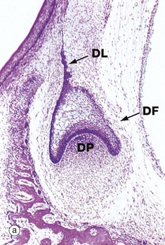
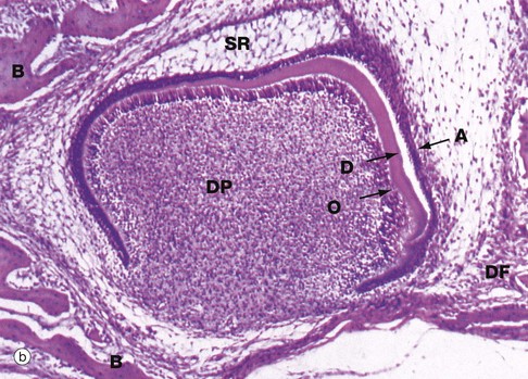
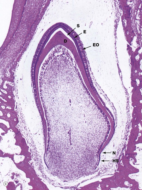
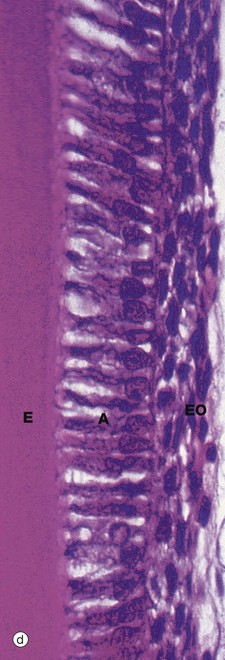
(a) H&E, cap stage (LP) (b) H&E, bell stage (MP) (c) H&E, onset of root development (LP) (d) H&E, ameloblasts (HP)
This series of micrographs illustrates the important stages of tooth development. The tissues of the teeth are derived from two embryological sources. The enamel is of epithelial (ectodermal) origin, while the dentine, cementum, pulp and periodontal ligament are of mesenchymal (mesodermal) origin. The first evidence of tooth development in humans occurs at 6 weeks of fetal life with the proliferation of a horseshoe-shaped epithelial ridge from the basal layer of the primitive oral epithelium into the underlying mesoderm in the position of the future jaws; this is known as the dental lamina. In each quadrant of the mouth, the lamina then develops four globular swellings which will become the enamel organs of the future deciduous central and lateral incisors, canines and first molar teeth. Subsequently, the dental lamina proliferates backwards in each arch, successively giving rise to the enamel organs of the future second deciduous molar and the three permanent molars. The permanent successors of the deciduous teeth will later develop from enamel organs which bud off from the inner aspect of the enamel organs of their deciduous predecessors.
The primitive mesenchyme immediately subjacent to the developing enamel organ proliferates to form a cellular mass, the dental papilla DP. At the same time, the enamel organ becomes progressively cap-shaped, as seen in micrograph (a), enveloping the dental papilla. During the cap stage, the cells lining the concave face of the enamel organ in contact with the dental papilla begin to differentiate into tall columnar cells, ameloblasts, which will be responsible for the production of enamel. This, in turn, induces the differentiation of a layer of columnar odontoblasts, the future dentine-producing cells, in the apical region of the dental papilla. The interface between the differentiating ameloblast and odontoblast layers marks the position and shape of the future junction between enamel and dentine.
As the enamel organ develops further, it assumes a characteristic bell shape as seen in micrograph (b), the free edge of the ‘bell’ proliferating so as to determine the eventual shape of the tooth crown. Meanwhile, the cells of the main bulk of the enamel organ become large and star-shaped, forming the stellate reticulum SR, the extracellular matrix of which is rich in glycosaminoglycans. Between the stellate reticulum and ameloblast layer, two or three layers of flattened cells form the stratum intermedium, while the outer surface of the enamel organ consists of a simple cuboidal epithelium called the external enamel epithelium. By the cap stage of development, the dental lamina DL connecting the enamel organ with the oral mucosa has become fragmented and, around the whole developing bud, a condensation of mesenchyme forms the dental follicle DF which will eventually become the periodontal ligament.
As ameloblasts and odontoblasts differentiate at the tip of the crown, a layer of dentine matrix is progressively laid down between the ameloblast and odontoblast layers. As the odontoblasts retreat, each leaves a long cytoplasmic extension, the odontoblastic process, embedded within the dentine matrix, thereby forming the dentine tubules. Dentine matrix has a similar biochemical composition to that of bone and undergoes calcification in a similar fashion. Deposition of dentine induces the production of enamel by the adjacent ameloblasts. Each retreating ameloblast lays down a column of enamel matrix which then undergoes mineralisation, resulting in the formation of a dense prismatic structure as described below. With the deposition of dentine and enamel, the overlying stellate reticulum atrophies and the enamel organ is much reduced in thickness. These changes are well demonstrated in micrograph (b). A thin layer of dentine D has been laid down by the underlying odontoblastic layer O of the highly cellular dental papilla DP. The ameloblastic layer A is about to lay down enamel in the space next to the dentine; note that in this area, the stellate reticulum has disappeared. Note also the surrounding dental follicle DF and early formation of cancellous bone B.
By the time dentine and enamel formation is well underway at the incisal edge or tips of the cusps (as the case may be), the enamel organ will have fully outlined the shape of the whole tooth crown. This is the case in micrograph (c), the neck of the tooth N marking the junction of crown and root. A thin, densely stained layer of poorly mineralised enamel E can be seen, covered at its external surface by the now much thinner enamel organ EO. The unstained space S between this and the underlying dentine D represents fully mineralised enamel laid down earlier but dissolved away during tissue preparation. Although enamel production is confined to the crown, the rim of the ‘bell’ of the enamel organ nevertheless continues to proliferate, inducing dentine formation and thereby determining the shape of the tooth root. This part of the enamel organ, known as the epithelial sheath of Hertwig HS, disintegrates once the outline of the root is completed. The cementum which later forms on the root surface is derived from the dental follicle. As the dentine of the crown and root are progressively laid down, the dental papilla shrinks and eventually becomes the dental pulp contained within the pulp chamber and root canals.
Growth of the tooth root is one of the principal mechanisms of tooth eruption and root formation is not completed until some time after the crown has fully erupted into the oral cavity.
Micrograph (d) illustrates the characteristic appearance of ameloblasts. Active ameloblasts A are tall columnar epithelial cells which form a single layer apposed to the forming surface of the enamel E. Each ameloblast elaborates a column of organic enamel matrix which undergoes progressive mineralisation by the deposition of calcium phosphate, mainly in the form of hydroxyapatite crystals. Fully formed enamel contains less than 1% organic material and is the hardest and most dense tissue in the body.
Mature enamel consists of highly calcified enamel prisms separated by interprismatic enamel, consisting of similar crystals orientated in a different direction. Each prism extends from the dentino-enamel junction to the enamel surface. The prisms are made up of groups of long, thin, parallel crystallites of hydroxyapatite, covered by a surface layer of organic material. Underlying the ameloblast layer are several layers of cells, also of epithelial origin, which constitute the remainder of the enamel organ EO. As enamel formation progresses, the enamel organ becomes much reduced in thickness compared with earlier stages of its development. At tooth eruption, the enamel organ, including the ameloblasts, degenerates leaving the enamel exposed to the hostile oral environment, completely incapable of regeneration.![]()
Stay updated, free articles. Join our Telegram channel

Full access? Get Clinical Tree


Basicmedical Key
Fastest Basicmedical Insight Engine
