15 • Converting carbohydrates and proteins into fatty acids and triglyceride • Regulation of blood glucose concentration by glycogenesis, glycogenolysis and gluconeogenesis • Synthesis of plasma proteins, including albumin and clotting factors • Synthesis of non-essential amino acids • Detoxification of metabolic waste products (e.g. deamination of amino acids and production of urea) FIG. 15.1 Liver (caption and illustration (b) opposite) FIG. 15.2 Hepatocytes FIG. 15.3 Portal tract FIG. 15.4 Liver FIG. 15.5 Hepatic vasculature and biliary system FIG. 15.6 Perfusion method FIG. 15.7 Liver architecture (illustrations opposite)
Liver and pancreas
Liver and Biliary System
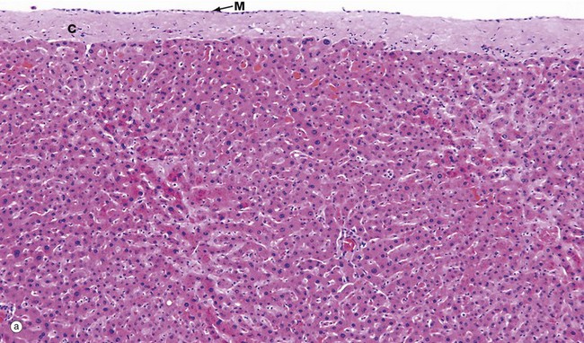
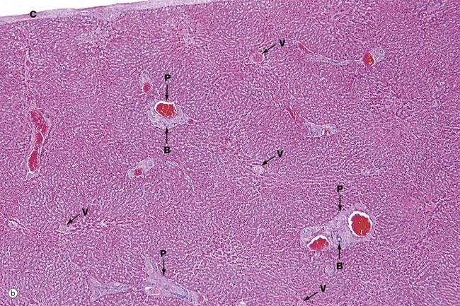
(a) Capsule and parenchyma, H&E (MP) (b) Architecture, H&E (LP)
Micrograph (a) shows the structure of the liver, which is a solid organ composed of tightly packed pink-staining plates of hepatocytes. The outer surface of the liver is covered by a capsule composed of collagenous tissue C called Glisson’s capsule, covered by a layer of mesothelial cells M from the peritoneum.
The sinusoids can just be seen as pale-stained spaces between the plates of liver cells. The hepatic sinusoids form a very low-resistance system of vascular channels that allows blood to come into contact with the hepatocytes over a huge surface area.
Micrograph (b) shows the overall architecture of the liver at a slightly lower magnification. The liver does not contain much in the way of connective tissue. Most of the collagenous connective tissue in the liver is found in the portal tracts P which contain the main blood vessels running into the liver. Larger vessels can be seen containing bright red blood, even at this low magnification. The other structures that run in the portal tracts are branches of the bile ducts B.
Less conspicuous than the portal tracts are the centrilobular venules (hepatic venules) V that drain the liver. These are tributaries of the hepatic vein and take blood away from the liver.
The very close association of the sinusoidal vasculature of the liver with the hepatocytes is essential for normal function. Certain diseases of the liver cause obliteration of the normal sinusoidal arrangement and this then causes impairment of liver function.
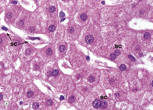
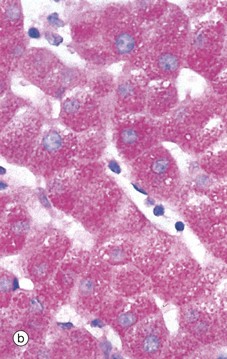
(a) H&E (HP) (b) PAS/haematoxylin (HP)
Hepatocytes are large polyhedral cells with round nuclei, peripherally dispersed chromatin and prominent nucleoli. The nuclei vary greatly in size, reflecting an unusual cellular feature; more than half of the hepatocytes contain twice the normal (diploid) complement of chromosomes within a single nucleus (i.e. they are tetraploid) and some contain four or even eight times this amount (polyploid). Binucleate cells BC are also common in normal liver.
The extensive cytoplasm has a variable appearance, depending on the nutritional status of the individual. When well-nourished, hepatocytes store significant quantities of glycogen and process large quantities of lipid. Both of these metabolites are partially removed during routine histological preparation, leaving irregular unstained areas within the cytoplasm. The cytoplasm is otherwise strongly eosinophilic due to numerous mitochondria, with a fine basophilic granularity due to extensive free ribosomes and rough endoplasmic reticulum. Fine brown granules of the ‘wear-and-tear’ pigment lipofuscin (see Fig. 1.25) are present in variable amounts, increasing with age. All of these features are seen in micrograph (a).
The sinusoids are lined by flat endothelial lining cells SC which are readily distinguishable from hepatocytes by their flattened condensed nuclei and attenuated poorly stained cytoplasm.
Micrograph (b) shows glycogen in hepatocytes which, being polysaccharide, is PAS-positive (i.e. stains magenta). In this preparation, the nuclei are counterstained blue with haematoxylin.
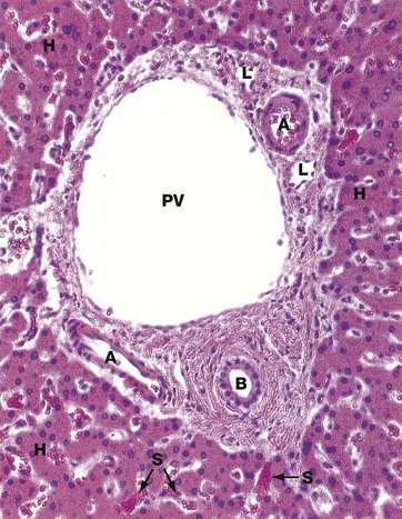
H&E (MP)
This micrograph shows a typical portal tract containing three main structures. The largest is a terminal branch of the hepatic portal vein PV (terminal portal venule) which has a thin wall lined by endothelial cells. Smaller-diameter thick-walled vessels are terminal branches of the hepatic artery A with the structure of arterioles.
A network of bile canaliculi is located within each plate of hepatocytes, but these are far too small to be seen at this magnification. These drain into bile collecting ducts lined by simple cuboidal or columnar epithelium, known as the canals of Hering, which in turn drain into the bile ductules B. The bile ductules are usually located at the periphery of the tract. The bile ductules merge to form larger, more centrally located trabecular ducts which drain via intrahepatic ducts into the right and left hepatic ducts, the common hepatic duct and then to the duodenum via the common bile duct.
Because these three structures are always found in the portal tracts, the tracts are often referred to as portal triads. Lymphatics L are also present in the portal tracts but, since their walls are delicate and often collapsed, they are less easily identified.
Surrounding the portal tract are anastomosing plates of hepatocytes H, between which are the hepatic sinusoids S. These receive blood from both the hepatic portal and hepatic arterial systems. The layer of hepatocytes immediately bordering the portal tract is known as the limiting plate.
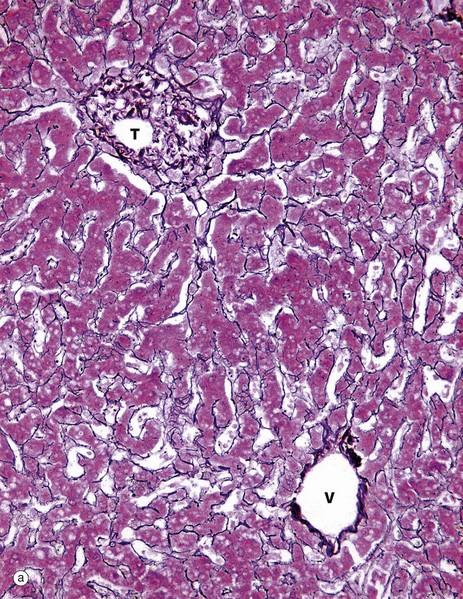
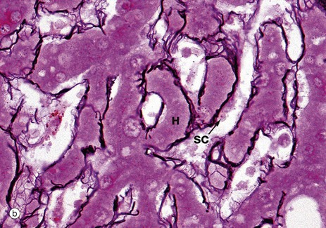
(a) Reticulin (MP) (b) Reticulin (HP)
The structural integrity of the liver is maintained by a delicate meshwork of extracellular matrix in the form of a fine meshwork of reticulin fibres (collagen type III). The reticulin meshwork supports both the hepatocytes and the sinusoidal lining cells (endothelial cells). These micrographs have both been stained by a silver method that shows reticulin as a black-stained material.
Micrograph (a) shows how reticulin is present on both sides of liver cell plates. The sinusoids are also bounded by the same reticulin framework. The reticulin merges with the sparse collagenous supporting tissue of the portal tract T and terminal hepatic venule V. At the periphery of the liver, the reticulin becomes continuous with Glisson’s capsule, which invests the external surface of the liver.
Micrograph (b) shows more detail of the reticulin scaffolding. Single layers of hepatocytes in the liver cell plates H lie immediately upon the reticulin framework. On the other side of the reticulin layer are the hepatic sinusoidal spaces. Some sinusoidal lining cells SC can just be seen.
The sinusoids are lined by a discontinuous fenestrated endothelium which has no basement membrane and is separated from the hepatocytes by a narrow space (the space of Disse) which drains into the lymphatics of the portal tracts.
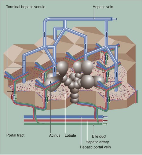
This diagram shows the hepatic vascular system and the bile collecting system. The hepatic portal vein and hepatic artery branch repeatedly within the liver. Their terminal branches run within the portal tracts and empty into the sinusoids. Blood from both systems percolates between plates of hepatocytes in the sinusoids, which converge to drain into a terminal hepatic (centrilobular) venule. These drain to intercalated veins and then to the hepatic vein, which drains into the inferior vena cava.
Bile is secreted into a network of minute bile canaliculi situated between the plasma membranes of adjacent hepatocytes. These canaliculi are too small to be represented in this diagram. The canalicular network drains into a system of bile ducts located in the portal tracts. Bile then flows through the extrahepatic biliary tree and is finally discharged into the second part of the duodenum. The hepatic lobule and acinus are explained in Fig. 15.7.
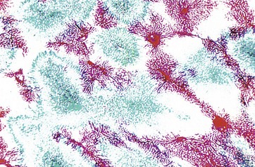
(LP)
This preparation shows one of the techniques used by early histologists in mapping hepatic blood flow. The hepatic portal vein (supplying the liver) has been perfused with a red dye, and the hepatic vein (draining the liver) has been back-perfused with a blue dye. Thus it can be seen how liver units can be defined by a number of portal tracts peripherally (stained red), with blood draining to a single terminal hepatic venule (stained blue) at the centre.
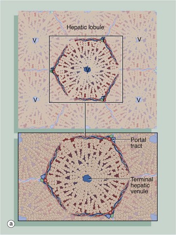
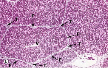
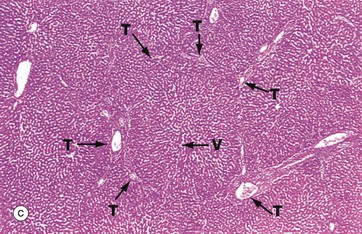
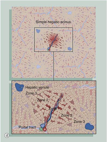
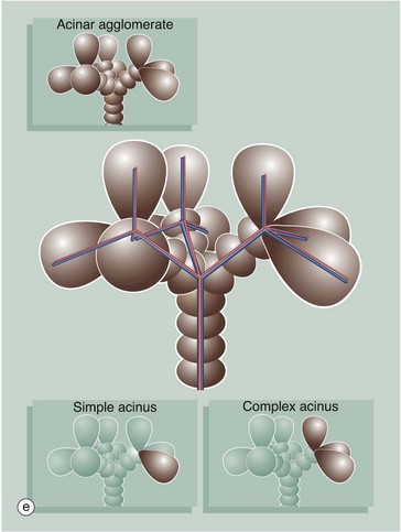
(a) Diagram of the liver lobule (b) Pig, H&E (LP) (c) Human, H&E (LP) (d) Diagram of the simple acinus (e) Diagram of acinar agglomerate
The structural unit of the liver can be considered as a conceptually simple hepatic lobule. However, the physiology of the liver is more accurately represented by a unit structure known as the hepatic acinus.
The hepatic lobule (a) is roughly hexagonal in shape and is centred on a terminal hepatic venule (centrilobular venule) V. The portal tracts T are positioned at the angles of the hexagon. The blood from the portal vein and hepatic artery branches flows away from the portal tract to the adjacent central veins. In some species, such as the pig (b), the lobule is outlined by bands of fibrous tissue F, giving a well-defined structural unit. In humans (c) and most other species, no such clear structural definition exists, although lobules can be roughly outlined as an hexagonal array of portal tracts T arranged around a terminal hepatic venule V.
The hepatic acinus (d) is a more physiologically useful model of liver anatomy, although more difficult to define histologically. The acinus is a roughly berry-shaped unit of liver parenchyma centered on a portal tract. The acinus lies between two or more terminal hepatic venules and blood flows from the portal tracts through the sinusoids to the venules. The acinus is divided into zones 1, 2 and 3 and the hepatocytes in these zones have different metabolic functions.
Zone 1 is closest to the portal tract and receives the most oxygenated blood, while zone 3 is furthest away and receives the least oxygen. Liver cells in zone 3 contain high levels of esterases and low levels of oxidative enzymes. Large branches of the portal vein and hepatic artery supply an agglomerate of acini, each of which is in turn composed of several complex acini which, at the lowest level, are made of simple acini, each supplied by terminal vascular branches. Although the structure looks on paper like a bunch of grapes, it must be remembered that this is a functional grouping and in reality the hepatic parenchyma is uniform and continuous.![]()
Stay updated, free articles. Join our Telegram channel

Full access? Get Clinical Tree


Basicmedical Key
Fastest Basicmedical Insight Engine