Xanthoma
Gregory Y. Lauwers, MD
Key Facts
Etiology/Pathogenesis
Helicobacter pylori
Common in operated stomach
Unknown etiology in many cases
Clinical Issues
Seen in 0.4-6% of endoscopies of nonoperated patients
Most commonly on lesser curvature
Yellowish nodule or plaque
May be multiple
10x more common after Billroth 2 resection
Microscopic Pathology
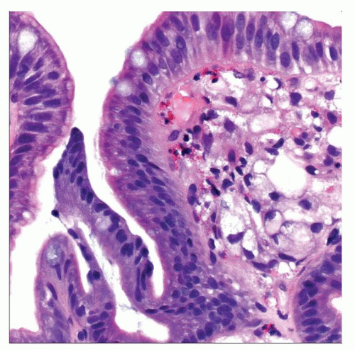
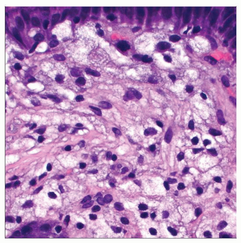
Aggregates of foamy lipid-laden histiocytes
Limited inflammation
Ancillary Tests
Positive histiocytic markers (e.g., CD68) and negative epithelial markers (e.g., AE1/AE3)
 Hematoxylin & eosin shows gastric xanthoma. Notice the subepithelial collection of bland foamy histiocytes underneath the surface epithelium. Several cells resemble signet-ring cells. |
TERMINOLOGY
Synonyms
Xanthelasma
Definitions
Collections of lipid-laden histiocytes within gastric mucosa
ETIOLOGY/PATHOGENESIS
Infectious Agents
Helicobacter pylori
Bile Reflux
Common in operated stomach
Metabolic
Hypercholesterolemia
Others
Cause is unknown in most cases
CLINICAL ISSUES
Epidemiology
Incidence
Relatively common
Seen in 0.4-6% of endoscopies of nonoperated stomachs
10x more common after Billroth 2 resection
Stay updated, free articles. Join our Telegram channel

Full access? Get Clinical Tree


