Wrist and distal forearm x-ray


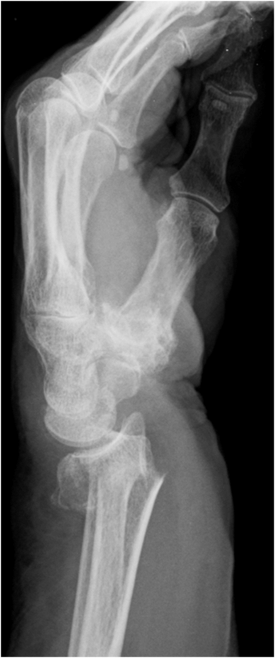
Radiograph of the wrist showing fracture of the distal radius with dorsal angulation (Colles’ fracture). The reverse would constitute a Smith’s fracture.


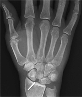
Radiograph of the right hand showing a widening of the scapholunate joint (Terry Thomas sign) (arrow).


Examination notes


What injuries should not be missed?


Subtle distal radius fracture: look for normal volar tilt of radius, radius distal to ulnar (AP view) and smooth cortical outline (lateral view).


Carpal fractures: normally waist of scaphoid intact.


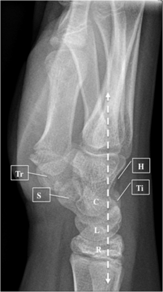
Lunate/perilunate dislocation – capitate normally sits in lunate (lateral view).

Only gold members can continue reading. Log In or Register to continue

Stay updated, free articles. Join our Telegram channel

Feb 18, 2017 | Posted by in GENERAL SURGERY | Comments Off on Wrist and distal forearm x-ray

Full access? Get Clinical Tree

Get Clinical Tree app for offline access