Water and Sodium


Water and Sodium


It is essential to understand the linked homeostatic mechanisms controlling water and sodium balance when interpreting the plasma sodium concentration and managing the clinically common disturbances of water and sodium balance. This is of major importance in deciding on the composition and amount, if any, of intravenous fluid to give. It must also be remembered that plasma results may be affected by such intravenous therapy, and can be dangerously misunderstood.

Water is an essential body constituent, and homeostatic processes are important to ensure that the total water balance is maintained within narrow limits, and the distribution of water among the vascular, interstitial and intracellular compartments is maintained. This depends on hydrostatic and osmotic forces acting across cell membranes.

Sodium is the most abundant extracellular cation and, with its associated anions, accounts for most of the osmotic activity of the extracellular fluid (ECF); it is important in determining water distribution across cell membranes.

Osmotic activity depends on concentration, and therefore on the relative amounts of sodium and water in the ECF compartment, rather than on the absolute quantity of either constituent. An imbalance may cause hyponatraemia (low plasma sodium concentration) or hypernatraemia (high plasma sodium concentration), and therefore changes in osmolality. If water and sodium are lost or gained in equivalent amounts, the plasma sodium, and therefore the osmolal concentration, is unchanged; symptoms are then due to extracellular volume depletion or overloading (Table 2.1). As the metabolism of sodium is so inextricably related to that of water, the two are discussed together in this chapter.


TOTAL SODIUM AND WATER BALANCE

In a 70-kg man, the total body water (TBW) is about 42 L and contributes about 60 per cent of the total body weight; there are approximately 3000 mmol of sodium, mainly in the ECF (Table 2.2). Water and electrolyte intake usually balance output in urine, faeces, sweat and expired air.


Water and sodium intake

The daily water and sodium intakes are variable, but in an adult amount to about 1.5-2 L and 60-150 mmol, respectively.


Water and sodium output


Kidneys and gastrointestinal tract

The kidneys and intestine deal with water and electrolytes in a similar way. Net loss through both organs depends on the balance between the volume filtered proximally
and that reabsorbed more distally. Any factor affecting either passive filtration or epithelial cellular function may disturb this balance.


Table 2.1 Approximate contributions of solutes to plasma osmolality


Osmolality (mmol/kg)


Total (%)


Sodium and its anions


270


92


Potassium and its anions


7


}8


Calcium (ionized) and its anions


3


Magnesium and its anions


1


Urea


5


Glucose


5


Protein


1


(approx.)


Total


292


(approx.)


Table 2.2 The approximate volumes in different body compartments through which water is distributed in a 70-kg adult


Volume (L)


Intracellular fluid compartment


24


Extracellular fluid compartment


18


Interstitial


( 13


)


Intravascular (blood volume)


( 5


)


Total body water


42


Approximately 200 L of water and 30 000 mmol of sodium are filtered by the kidneys each day; a further 10 L of water and 1500 mmol of sodium enter the intestinal lumen. The whole of the extracellular water and sodium could be lost by passive filtration in little more than an hour, but under normal circumstances about 99 per cent is reabsorbed. Consequently, the net daily losses amount to about 1.5-2 L of water and 100 mmol of sodium in the urine, and 100 mL and 15 mmol, respectively, in the faeces.

Fine adjustment of the relative amounts of water and sodium excretion occurs in the distal nephron and the large intestine, often under hormonal control. The effects of antidiuretic hormone (ADH) or vasopressin and the mineralocorticoid hormone aldosterone on the kidney are the most important physiologically, although natriuretic peptides are also important.


Sweat and expired air

About 1 L of water is lost daily in sweat and expired air, and less than 30 mmol of sodium a day is lost in sweat. The volume of sweat is primarily controlled by skin temperature, although ADH and aldosterone have some effect on its composition. Water loss in expired air depends on the respiratory rate. Normally, losses in sweat and expired air are rapidly corrected by changes in renal and intestinal loss. However, neither of these losses can be controlled to meet sodium and water requirements, and thus they may contribute considerably to abnormal balance when homeostatic mechanisms fail.


CONTROL OF WATER AND SODIUM BALANCE


Control of water balance

Both the intake and loss of water are controlled by osmotic gradients across cell membranes in the brain’s hypothalamic osmoreceptor centres. These centres, which are closely related anatomically, control thirst and the secretion of ADH.


Antidiuretic hormone (arginine vasopressin)

Antidiuretic hormone is a polypeptide with a half-life of about 20 min that is synthesized in the supraoptic and paraventricular nuclei of the hypothalamus and, after transport down the pituitary stalk, is secreted from the posterior pituitary gland (see Chapter 7).


Control of antidiuretic hormone secretion

The secretion of ADH is stimulated by the flow of water out of cerebral cells caused by a relatively high extracellular osmolality. If intracellular osmolality is unchanged, an extracellular increase of only 2 per cent quadruples ADH output; an equivalent fall almost completely inhibits it. This represents a change in plasma sodium concentration of only about 3 mmol/L. In more chronic changes, when the osmotic gradient has been minimized by solute redistribution, there may be little or no effect. In addition, stretch receptors in the left atrium and baroreceptors in the aortic arch and carotid sinus influence ADH secretion in response to the low intravascular pressure of severe hypovolaemia, stimulating ADH release. The stress due to, for example, nausea, vomiting and pain may also increase ADH secretion. Inhibition of ADH secretion occurs if the extracellular osmolality falls, for whatever reason.


Actions of antidiuretic hormone

Antidiuretic hormone, by regulating aquaporin 2, enhances water reabsorption in excess of solute from the collecting ducts of the kidney and so dilutes the extracellular osmolality. Aquaporins are cell membrane proteins acting as water channels that regulate water flow. When ADH secretion is a response to a high extracellular osmolality with the danger of cell dehydration, this is an appropriate response. However, if its secretion is in response to a low circulating volume alone, it is inappropriate to the osmolality. The retained water is then distributed throughout the TBW space, entering cells along the osmotic gradient; the correction of extracellular depletion with water alone is thus relatively inefficient in correcting hypovolaemia. Plasma osmolality normally varies by less than 1-2 per cent, despite great variation in water intake, which is largely due to the action of ADH.

In some circumstances, the action of ADH is opposed by other factors. For example, during an osmotic diuresis the urine, although not hypo-osmolal, contains more water than sodium. Patients with severe hyperglycaemia, as in poorly controlled diabetes mellitus, may show an osmotic diuresis.


Control of sodium balance

The major factors controlling sodium balance are renal blood flow and aldosterone. This hormone controls loss of sodium from the distal tubule and colon.


Aldosterone

Aldosterone, a mineralocorticoid hormone, is secreted by the zona glomerulosa of the adrenal cortex (see Chapter 8). It affects sodium-potassium and sodium- hydrogen ion exchange across all cell membranes. Its main effect is on renal tubular cells, but it also affects loss in faeces, sweat and saliva. Aldosterone stimulates sodium reabsorption from the lumen of the distal renal tubule in exchange for either potassium or hydrogen ions (Fig. 2.1). The net result is the retention of more sodium than water, and the loss of potassium and hydrogen ions. If the circulating aldosterone concentration is high and tubular function is normal, the urinary sodium concentration is low.

Many factors are involved in the feedback control of aldosterone secretion. These include local electrolyte concentrations, such as that of potassium in the adrenal gland, but they are probably of less physiological and clinical importance than the effect of the renin- angiotensin system.


The renin-angiotensin system

Renin is an aspartyl protease secreted by the juxtaglomerular apparatus, a cellular complex adjacent to the renal glomeruli, lying between the afferent arteriole and the distal convoluted tubule. Renin is derived from prorenin by proteolytic action, and secretion increases in response to a reduction in renal artery blood flow, possibly mediated by changes in the mean pressure in the afferent arterioles, and β-adrenergic stimulation. Renin splits a decapeptide (angiotensin I) from a circulating α2-globulin known as renin substrate. Another proteolytic enzyme, angiotensin-converting enzyme (ACE), which is located predominantly in the lungs but is also present in other tissues such as the kidneys, splits off a further two amino acid residues. This is the enzyme that ACE inhibitors (used to treat hypertension and congestive cardiac failure) act on. The remaining octapeptide, angiotensin II, has a number of important actions:


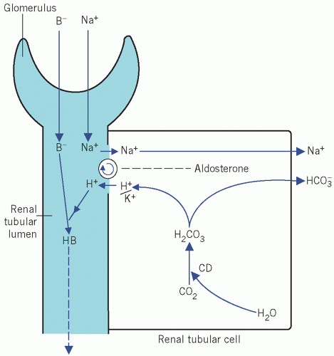
Figure 2.1 The action of aldosterone on the reabsorption of Na+ in exchange for either K+ or H+ from the distal renal tubules. See text for details. CD, carbonate dehydratase; B, associated anion.


  • It acts directly on capillary walls, causing vasoconstriction, and so probably helps to maintain blood pressure and alter the glomerular filtration rate (GFR). Vasoconstriction may raise the blood pressure before the circulating volume can be restored.


  • It stimulates the cells of the zona glomerulosa to synthesize and secrete aldosterone.


  • It stimulates the thirst centre and so promotes oral fluid intake.

Poor renal blood flow is often associated with an inadequate systemic blood pressure. The release of renin results in the production of angiotensin II, which tends to correct this by causing aldosterone release, which stimulates sodium and water retention and hence restores the circulating volume. Thus, aldosterone secretion responds, via renin, to a reduction in renal blood flow. Sodium excretion is not directly related to total body sodium content or to plasma sodium concentration.


Natriuretic peptides

A peptide hormone (or hormones) secreted from the right atrial or ventricular wall in response to the stimulation of stretch receptors may cause high sodium excretion (natriuresis) by increasing the GFR and by inhibiting renin and aldosterone secretion. However, the importance of this hormone (or hormones) in the physiological control of sodium excretion and in pathological states has not yet been fully elucidated, although it is important in the pathophysiology of congestive cardiac failure (see Chapter 22).


Monitoring fluid balance

The most important factor in assessing changes in day-to-day fluid balance is accurate records of fluid
intake and output; this is particularly pertinent for unconscious patients. ‘Insensible loss’ is usually assumed to be about 1 L/day, but there is endogenous water production of about 500 mL/day as a result of metabolic processes. Therefore the net daily ‘insensible loss’ is about 500 mL. The required daily intake may be calculated from the output during the previous day plus 500 mL to allow for ‘insensible loss’; this method is satisfactory if the patient is normally hydrated before day-to-day monitoring is started. Serial patient body weight determination can also be useful in the assessment of changes in fluid balance.

Pyrexial patients may lose 1 L or more of fluid in sweat and, if they are also hyperventilating, respiratory water loss may be considerable. In such cases an allowance of about 500 mL for ‘insensible loss’ may be totally inadequate. In addition, patients may be incontinent of urine, and having abnormal gastrointestinal losses makes the accurate assessment of fluid losses very difficult.

Inaccurate measurement and charting are useless and may be dangerous.

Keeping a cumulative fluid balance record is a useful way of detecting a trend, which may then be corrected before serious abnormalities develop.

In the example shown in Table 2.3, 500 mL has been allowed for as net ‘insensible daily loss’; calculated losses are therefore more likely to be underestimated than overestimated. This shows how insidiously a serious deficit can develop over a few days.

The volume of fluid infused should be based on the calculated cumulative balance and on clinical evidence of the state of hydration, and its composition adjusted to maintain normal plasma electrolyte concentrations.

Assessment of the state of hydration of a patient relies on clinical examination and on laboratory evidence of haemodilution or haemoconcentration.


  • Haemodilution Increasing plasma volume with protein-free fluid leads to a fall in the concentrations of proteins and haemoglobin. However, these findings may be affected by pre-existing abnormalities of protein or red cell concentrations.


    Table 2.3 Hypothetical cumulative fluid balance chart assuming an insensible daily loss of 500 mL


    Measured intake (mL)


    Measured output (mL)


    Total output (minimum mL)


    Daily balance (mL)


    Cumulative balance (mL)


    Day 1


    2000


    1900


    2400


    -400


    -400


    Day 2


    2000


    2000


    2500


    -500


    -900


    Day 3


    2100


    1900


    2400


    -300


    -1200


    Day 4


    2200


    2000


    2500


    -300


    -1500


  • Haemoconcentration ECF is usually lost from the vascular compartment first and, unless the fluid is whole blood, depletion of water and small molecules results in a rise in the concentration of large molecules, such as proteins and blood cells, with a rise in blood haemoglobin concentration and haematocrit, raised plasma urea concentration and reduced urine sodium concentration.

Table 2.4 shows various intravenous fluid regimens that can be used clinically. A summary of the British Consensus Guidelines on Intravenous Fluid Therapy for Adult Surgical Patients (GIFTASUP) can be found at www.bapen.org.uk/pdfs/bapen_pubs/giftasup.pdf.


DISTRIBUTION OF WATER AND SODIUM IN THE BODY

In mild disturbances of the balance of water and electrolytes, their total amounts in the body may be of less importance than their distribution between body compartments (see Table 2.2).

Water is distributed between the main body fluid compartments, in which different electrolytes contribute to the osmolality. These compartments are:


  • intracellular, in which potassium is the predominant cation,


  • extracellular, in which sodium is the predominant cation, and which can be subdivided into:


    • interstitial space, with very low protein concentration, and


    • intravascular (plasma) space, with a relatively high protein concentration.


Electrolyte distribution between cells and interstitial fluid

Sodium is the predominant extracellular cation, its intracellular concentration being less than one-tenth of that within the ECF. The intracellular potassium concentration is about 30 times that of the ECF. About 95 per cent of the osmotically active sodium is outside
cells, and about the same proportion of potassium is intracellular. Cell-surface energy-dependent sodium/potassium adenosine triphosphatase pumps maintain these differential concentrations.


Table 2.4 Some electrolyte-containing fluids for intravenous infusion


Na+ (mmol/L)


K+ (mmol/L)


Cl (mmol/L)


HCO3 (mmol/L)


Glucose (mmol/L)


Ca2+ (mmol/L)


Approximate osmolarity × plasma


Saline


‘Normal’ (physiological 0.9%)


154


154


× 1


Twice ‘normal’ (1.8%)


308


308


× 2


Half ‘normal’ (0.45%)


77


77


× 0.5


‘Dextrose’ saline


5%, 0.45%


77


77


278


× 1.5


Sodium bicarbonate


1.4%


167


167


× 1


8.4%a


1000


1000


× 6


Complex solutions


Ringer’s


147


4.2


156


2.2


× 1


Hartmann’s


131


5.4


112


29b


1.8


× 1


a Most commonly used bicarbonate solution. Note marked hyperosmolarity. Only used if strongly indicated.

b As lactate 29 mmol/L.


Other ions tend to move across cell membranes in association with sodium and potassium. (The movement of hydrogen ions is discussed in Chapter 4.) Magnesium and phosphate ions are predominantly intracellular, and chloride ions extracellular.


Distribution of electrolytes between plasma and interstitial fluid

The cell membranes of the capillary endothelium are more permeable to small ions than those of tissue cells. The plasma protein concentration is relatively high, but that of interstitial fluid is very low. The osmotic effect of the intravascular proteins is balanced by very slightly higher interstitial electrolyte concentrations (Gibbs-Donnan effect); this difference is small and, for practical purposes, plasma electrolyte concentrations can be assumed to be representative of those in the ECF as a whole.


Distribution of water

Over half the body water is intracellular (see Table 2.2). About 15-20 per cent of the extracellular water is intravascular; the remainder constitutes the interstitial fluid. The distribution of water across biological membranes depends on the balance between the hydrostatic pressure and the in vivo effective osmotic pressure differences on each side of the membrane.


Osmotic pressure

The net movement of water across a membrane that is permeable only to water depends on the concentration gradient of particles – either ions or molecules – across that membrane, and is known as the osmotic gradient. For any weight-to-volume ratio, the larger the particles, the fewer there are per unit volume, and therefore the lower the osmotic effect they exert. If the membranes were freely permeable to ions and smaller particles as well as to water, these diffusible particles would exert no osmotic effect across membranes, and therefore the larger ones would become more important in affecting water movement. This action gives rise to the effective colloid osmotic (oncotic) gradient. Water distribution in the body is thus dependent largely on three factors, namely:

1. the number of particles per unit volume,

2. particle size relative to membrane permeability,

3. concentration gradient across the membrane.


Units of measurement of osmotic pressure

Osmolar concentration can be expressed as:


  • the osmolarity (in mmol/L) of solution,


  • the osmolality (in mmol/kg) of solvent.

If solute is dissolved in pure water at concentrations such as those in body fluids, osmolarity and osmolality will hardly differ. However, as plasma is a complex solution containing large molecules such as proteins, the total volume of solution (water + protein) is
greater than the volume of solvent only (water) in which the small molecules are dissolved. At a protein concentration of 70 g/L, the volume of water is about 6 per cent less than the total volume of the solution (that is, the molarity should theoretically be about 6 per cent less than the molality). Most methods for measuring individual ions assess them in molarity (mmol/L). If the concentration of proteins in plasma is markedly increased, the volume of solvent is significantly reduced but the volume of solution remains unchanged. Therefore the molarity (in mmol/L) of certain ions such as sodium will be reduced but the molality will be unaltered. This apparently low sodium concentration is known as pseudohyponatraemia.


Measured plasma osmolality

Osmometers measure changes in the properties of a solution, such as freezing point depression or vapour pressure, which depend on the total osmolality of the solution – the osmotic effect that would be exerted by the sum of all the dissolved molecules and ions across a membrane permeable only to water. These properties are known as colligative properties. Sodium and its associated anions (mainly chloride) contribute 90 per cent or more to this measured plasma osmolality, the effect of protein being negligible. As the only major difference in composition between plasma and interstitial fluid is in protein content, the plasma osmolality is almost identical to the osmolality of the interstitial fluid surrounding cells.


Calculated plasma osmolarity

It is the osmolam, rather than the osmolar, concentration that exerts an effect across cell membranes and that is controlled by homeostatic mechanisms. However, as discussed below, the calculated plasma osmolarity is usually as informative as the measured plasma osmolality.

Although, because of the space-occupying effect of protein, the measured osmolality of plasma should be higher than the osmolarity, calculated from the sum of the molar concentrations of all the ions, there is usually little difference between the two figures. This is because there is incomplete ionization of, for example, NaCl to Na+ and Cl; this reduces the osmotic effect by almost the same amount as the volume occupied by protein raises it.

Consequently, the calculated plasma osmolarity is a valid approximation to the true measured osmolality. However, if there is gross hyperproteinaemia or hyperlipidaemia such that either protein or lipid contributes much more than 6 per cent to the measured plasma volume, the calculated osmolarity may be significantly lower than the true osmolality in the plasma water. A hypothetical example is shown in Figure 2.2.

Many formulae of varying complexity have been proposed to calculate plasma osmolarity. None of them can predict the osmotic effect, but the following formula (in which square brackets indicate concentration) gives a close approximation to plasma osmolality (although some equations omit the potassium, which may be preferable):


The factor of 2, which is applied to the sodium and potassium concentrations, allows for the associated anions and assumes complete ionization. This calculation is not valid if gross hyperproteinaemia or hyperlipidaemia is present or an unmeasured osmotically active solute, such as mannitol, methanol, ethanol or ethylene glycol, is circulating in plasma.

A significant difference between measured and calculated osmolality in the absence of hyperproteinaemia or hyperlipidaemia may suggest alcohol or other poisoning. For example, a plasma alcohol concentration of 100 mg/dL contributes about 20 mmol/kg to the osmolality. This osmotic difference is known as the osmolar gap and can be used to assess the presence in plasma of unmeasured osmotically active particles. In such cases the plasma sodium concentration may be misleading as a measure of the osmotic effect. It is not possible to calculate urinary osmolarity because of the considerable variation in the concentrations of different, sometimes unmeasured, solutes; the osmotic pressure of urine can be determined only by measuring the osmolality.


Distribution of water across cell membranes


Osmotic pressure gradient

Because the hydrostatic pressure difference across the cell membrane is negligible, cell hydration depends on the effective osmotic difference between intracellular and extracellular fluids. The cell membranes are freely permeable to water and to some solutes, but different solutes diffuse (or are actively transported) across cell membranes at different rates, although always more slowly than water. In a stable state, the total intracellular osmolality, due mostly to potassium and associated anions, equals that of the interstitial fluid, due mostly to sodium and associated anions; consequently, there is
no net movement of water into or out of cells. In some pathological states, rapid changes of extracellular solute concentration affect cell hydration; slower changes may allow time for the redistribution of solute and have little or no effect.


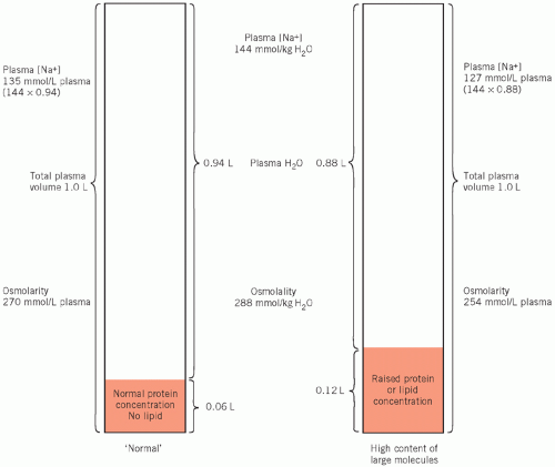
Figure 2.2 The consequence of gross hyperproteinaemia or hyperlipidaemia on the plasma water volume and its effect on the calculated plasma osmolarity and the true plasma osmolality.

Sodium In normal subjects sodium and its associated anions account for at least 90 per cent of extracellular osmolality. Rapid changes in their concentration therefore affect cellular hydration. If there is no significant change in the other solutes, a rise causes cellular dehydration and a fall causes cellular overhydration.

Urea Normal extracellular concentrations are so low as to contribute very little to the measured plasma osmolality. However, concentrations 15-fold or more above normal can occur in severe uraemia and can then make a significant contribution (see Chapter 3). However, urea does diffuse into cells very much more slowly than water. Consequently, in acute uraemia, the increased osmotic gradient alters cell hydration, but in chronic uraemia, although the measured plasma osmolality is often increased, the osmotic effect of urea is reduced as the concentrations gradually equalize on the two sides of the membrane.

Glucose Like urea, the normally low extracellular concentration of glucose does not contribute significantly to the osmolality. However, unlike urea, glucose is actively transported into many cells, but once there it is rapidly metabolized, even at high extracellular concentrations, and the intracellular concentration remains low. Severe hyperglycaemia, whether acute or chronic, causes a marked osmotic effect across cell membranes, with movement of water from cells into the extracellular compartment causing cellular dehydration.

Although hyperglycaemia and acute uraemia can cause cellular dehydration, the contribution of normal urea and glucose concentrations to plasma osmolality
is so small that reduced levels of these solutes, unlike those of sodium, do not cause cellular overhydration.

Solutes such as potassium, calcium and magnesium are present in the ECF at very low concentrations. Significant changes in these are lethal at much lower concentrations than those that would change osmolality.

Mannitol is an example of an exogenous substance that remains in the extracellular compartment because it is not transported into cells, and may be infused to reduce cerebral oedema. Ethanol is only slowly metabolized, and a high concentration in the ECF may lead to cerebral cellular dehydration; this may account for some of the symptoms of a hangover. High glucose concentrations account for the polyuria of severe diabetes mellitus.

Large rises in the osmotic gradient across cell membranes may result in the movement of enough water from the intracellular compartment to dilute extracellular constituents. Consequently, if the change in osmolality has not been caused by sodium and its associated anions, a fall in plasma sodium concentration is appropriate to the state of osmolality. If, under such circumstances, the plasma sodium concentration is not low, this indicates hyperosmolality.

Generally, plasma osmolarity calculated from sodium, potassium, urea and glucose concentrations is at least as clinically valuable as measured plasma osmolality. It has the advantage that the solute responsible, and therefore its likely osmotic effect, is often identified.


Distribution of water across capillary membranes

The maintenance of blood pressure depends on the retention of fluid within the intravascular compartment at a higher hydrostatic pressure than that of the interstitial space. Hydrostatic pressure in capillary lumina tends to force fluid into the extravascular space. In the absence of any effective opposing force, fluid would be lost rapidly from the vascular compartment. Unlike other cell membranes, those of the capillaries are permeable to small ions. Therefore sodium alone exerts almost no osmotic effect and the distribution of water across capillary membranes is little affected by changes in electrolyte concentration.


Colloid osmotic pressure

The very small osmotic effect of plasma protein molecules produces an effective osmotic gradient across capillary membranes; this is known as the colloid osmotic, or oncotic, pressure. It is the most important factor opposing the net outward hydrostatic pressure (Fig. 2.3). Albumin (molecular weight 65 kDa) is the most important protein contributing to the colloid osmotic pressure. It is present intravascularly at significant concentration but extravascularly only at a very low concentration because it cannot pass freely across the capillary wall.

The osmotic gradient across vascular walls cannot be estimated by simple means. The total plasma osmolality gives no information about this. Moreover, the plasma albumin concentration is a poor guide to the colloid osmotic pressure. Although other proteins, such as globulins, are present in the plasma at about the same concentration as albumin, their estimation for this purpose is even less useful: their higher molecular weights mean that they have even less effect than albumin.


Relation between sodium and water homeostasis

In normal subjects, the concentrations of sodium and its associated anions are the most important osmotic factors affecting ADH secretion. Plasma volume, by its effect on renal blood flow, controls aldosterone secretion and therefore sodium balance. The homeostatic mechanisms controlling sodium and water excretion are interdependent. (A simplified scheme is shown in Fig. 2.4.) Thirst depends on a rise in extracellular osmolality, whether due to water depletion or sodium excess, and also on a very large increase in the activity of the renin-angiotensin system.

A rise in extracellular osmolality reduces water loss by stimulating ADH release and increases intake by stimulating thirst; both these actions dilute the extracellular osmolality. Osmotic balance (and therefore cellular hydration) is rapidly corrected.


Assessment of sodium status

As already discussed, the plasma sodium concentration is important because of its osmotic effect on fluid distribution. Plasma sodium concentrations should be monitored while volume is being corrected to ensure that the distribution of fluid between the intracellular and extracellular compartments is optimal. The presence of other osmotically active solutes should be taken into account.


URINARY SODIUM ESTIMATION

Urinary sodium excretion is not related to body content but to renal blood flow.

Estimation of the urinary sodium concentration in a random specimen may be of value in the diagnosis of the syndrome of inappropriate antidiuretic hormone
secretion (SIADH) and may help to differentiate renal circulatory insufficiency (pre-renal) from intrinsic renal damage (see Chapter 3).


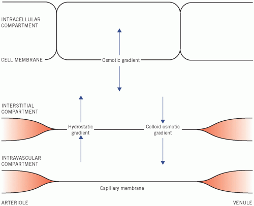
Figure 2.3 Osmotic factors that control the distribution of water between the fluid compartments of the body.


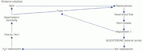
Figure 2.4 Control of water and sodium homeostasis. ADH, antidiuretic hormone.

The fractional excretion of sodium (FENa%) may also be useful in helping to assess renal blood flow and can be measured using a simultaneous blood sample and spot urine sample:


A value of less than 1 per cent may be found in poor renal perfusion, for example pre-renal failure, and of more than 1 per cent in intrinsic renal failure.


DISTURBANCES OF WATER AND SODIUM METABOLISM

The initial clinical consequences of primary sodium disturbances depend on changes of extracellular osmolality and hence of cellular hydration, and those of primary water disturbances depend on changes in extracellular volume.

Plasma sodium concentration is usually a substitute for measuring plasma osmolality. Plasma sodium concentrations per se are not important, but their effect on the osmotic gradient across cell membranes is, and it should be understood that the one does not always reflect the other.

If the concentration of plasma sodium alters rapidly, and the concentrations of other extracellular solutes remain the same, most of the clinical features are due to the consequence of the osmotic difference across cell membranes, with redistribution of fluid between cells and the ECF. However, gradual changes, which allow time for redistribution of diffusible solute such as urea, and therefore for equalization of osmolality without major shifts of water, may produce little effect on fluid distribution.


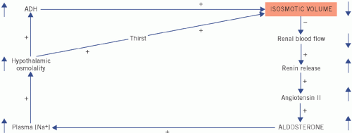
Figure 2.5 Homeostatic correction of isosmotic volume depletion. The reduced intravascular volume impairs renal blood flow and stimulates renin and therefore aldosterone secretion. There is selective sodium reabsorption from the distal tubules and a low urinary sodium concentration. (Shading indicates primary change.) ADH, antidiuretic hormone.

Only gold members can continue reading. Log In or Register to continue

Stay updated, free articles. Join our Telegram channel

Jun 30, 2016 | Posted by in BIOCHEMISTRY | Comments Off on Water and Sodium

Full access? Get Clinical Tree

Get Clinical Tree app for offline access