Viruses
Diagnosis of pulmonary viral infections can be made on cytologic or histologic specimens based on the virus-induced cytopathic changes. Confirmation by viral culture in equivocal cases is essential. Viral infections are common in immunocompromised subjects and often require expedited diagnosis so that antiviral treatment can be started. Immunohistochemistry is invaluable in the early and prompt diagnosis of viral infections. Commercially available immunostains that work well in our laboratory, and most other laboratories, include those for cytomegalovirus (CMV), herpes simplex virus (HSV) I and II, adenovirus, parvovirus, respiratory syncytial virus (RSV), Epstein-Barr virus (EBV), human papilloma virus, and varicella-zoster virus (VZV). In situ hybridization and polymerase chain reaction techniques for diagnosis of some viral infections are available in most laboratories and are more specific than immunostains.
Part 1 Cytomegalovirus
Sajid A. Haque
Abida K. Haque
Mary L. Ostrowski
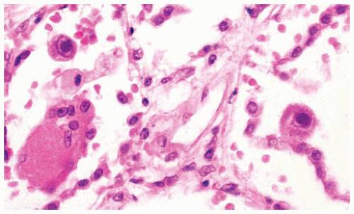
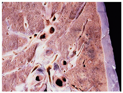
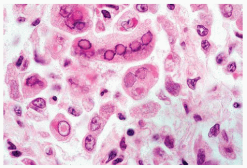
Cytomegalovirus (CMV), a member of the Herpesviridae family, is one of the most common viruses infecting infants and immunocompromised patients. The diagnostic hallmark of active CMV infection is cytomegaly with both nuclear and cytoplasmic enlargement and intranuclear and cytoplasmic inclusions. The lungs show interstitial pneumonitis with sparse mononuclear inflammation and characteristic nuclear and cytoplasmic inclusions. With severe infection, neutrophil infiltrates, necrosis, hemorrhage, hyaline membranes, and diffuse alveolar damage (DAD) are seen.
Cytologic Features
Marked enlargement of infected cells (cytomegaly).
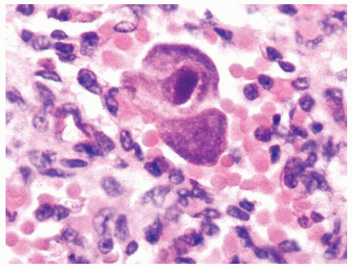
Enlarged nucleus with single, round to oval inclusion surrounded by a clear halo (Cowdry type A inclusion).
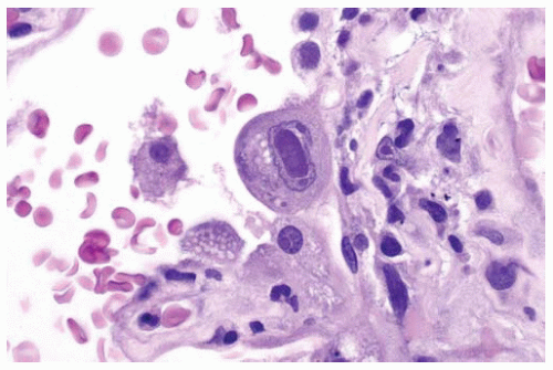
Cytoplasmic granular basophilic inclusions may be seen near the cell membrane.
Histologic Features
CMV inclusions may be seen in the alveolar pneumocytes, airway epithelium, vascular endothelium, macrophages, and interstitial cells.
The infected cell shows enlarged nucleus with a single, round to oval inclusion surrounded by a clear halo (Cowdry type A) with margination of chromatin on the inner aspect of the nuclear membrane.
Fully developed inclusions are easily seen at low power, permitting quick diagnosis; however, in the early stages, the inclusions resemble those seen in other Herpesviridae families (HSV, VZV).
Masson trichrome stain highlights the inclusions.
Cytoplasmic inclusions are valuable in the diagnosis of CMV; these are small, granular, and basophilic in early stages but later become larger (up to 3 μm) and aggregate near the cell membrane.
Inclusions are periodic acid-Schiff (PAS) and Gomori methenamine silver positive.
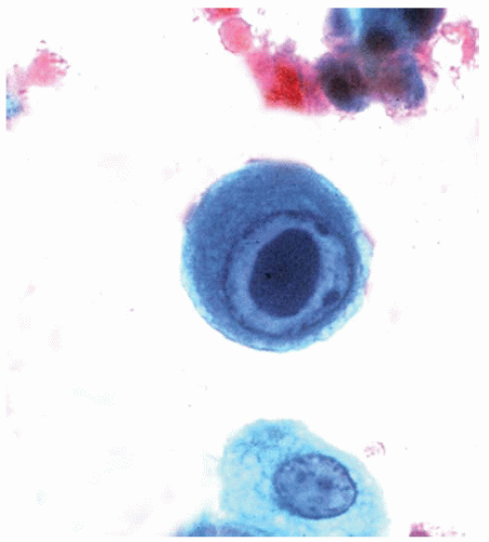 Figure 95.1 Cytology figure showing alveolar cell containing a large oval intranuclear inclusion (owl eye nucleus). |
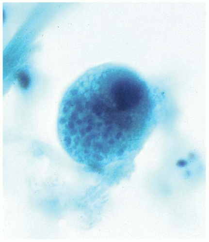 Figure 95.2 Cytology figure showing alveolar cell containing cytoplasmic inclusions consisting of small multiple round bodies. |
Part 2 Herpes Simplex Virus
Sajid A. Haque
Abida K. Haque
Mary L. Ostrowski
Herpes Simplex Virus (HSV) infections are usually asymptomatic in immunocompetent individuals. In immunocompromised adults, the infection is associated with necrotizing tracheobronchitis and bronchopneumonia. The airway mucosa and/or the lung parenchyma may show acute inflammation with necrosis, fibrinous exudates, and DAD in severe infection. The viral inclusions occur early in the course of infection but may be obscured by the inflammation. Both HSV I and II infections have been identified in the lungs of infants and immunocompromised adults.
Cytologic Features
Multinucleated cells with nuclear molding.
Chromatin pattern with opaque or ground-glass smudged appearance.
Eosinophilic nuclear inclusions.
Histologic Features
Infected cells are enlarged, often multinucleated, and may show two types of nuclear inclusions.
Characteristic amphophilic, homogenous, or “glassy” nuclear inclusions (3 to 8 μm in size) bordered by clumped chromatin (Cowdry type B), or deeply eosinophilic, homogenous, centrally placed, and surrounded by a clear halo (Cowdry type A).
No cytoplasmic inclusions.
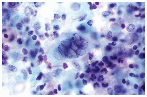 Figure 95.7 Cytologic figure showing a multinucleated giant cell with ground-glass nuclei diagnostic of HSV infection, surrounded by inflammatory cells. |
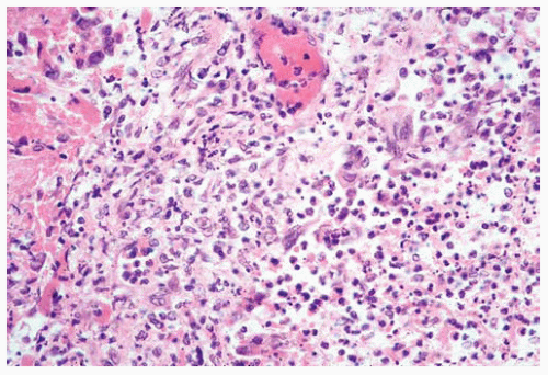 Figure 95.8 Low power showing an inflammatory infiltrate and a few multinucleated giant cells. Diagnosis of HSV pneumonia cannot be made at this magnification. |
Part 3 Influenza and Parainfluenza Virus Pneumonia
Sajid A. Haque
Abida K. Haque
The influenza and parainfluenza viruses primarily infect the respiratory epithelium. Infection in immunocompromised subjects and children may be severe, resulting in DAD. Secondary bacterial infection causes acute necrotizing tracheobronchitis and bronchopneumonia. Influenza virus A and B infections produce the influenza syndrome, whereas
parainfluenza infection predominantly causes upper respiratory infection. Squamous metaplasia of airway epithelium is often present in the healing phase. Diagnosis of influenza virus pneumonia requires viral culture.
parainfluenza infection predominantly causes upper respiratory infection. Squamous metaplasia of airway epithelium is often present in the healing phase. Diagnosis of influenza virus pneumonia requires viral culture.
Histologic Features
Classic pathologic finding in severe influenza infection is alveolar epithelial damage, either focal or diffuse, and associated with hyaline membranes.
Healing phase is characterized by interstitial lymphocytic infiltrate and bronchial epithelial squamous metaplasia.
Often no viral inclusions are seen with influenza virus infection.
Parainfluenza infection is characterized by formation of syncytial giant cells.
No intranuclear inclusions are present, but rarely small cytoplasmic inclusions may be seen.
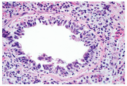 Figure 95.10 Small airway with characteristic pathologic changes of acute inflammation and damage to the lining of epithelial cells. |
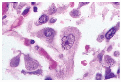 Figure 95.11 Small cytoplasmic inclusions are present in the centrally located large cell, probably a type 2 pneumocyte. |
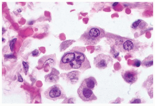 Figure 95.12 Centrally located multinucleated cell with viral cytopathic effect but no clear cytoplasmic inclusions.
Stay updated, free articles. Join our Telegram channel
Full access? Get Clinical Tree
 Get Clinical Tree app for offline access

|
