Chapter 10 Viruses
Viruses have dispensed with cell structure and metabolism but are capable of reproduction. Being unable to generate the metabolic energy required for their reproduction, they depend on a living cell to replicate their nucleic acid and synthesize their proteins. Thus viruses are villains not by choice but by necessity.
All viruses are obligatory intracellular parasites. They do nothing useful for the organism that harbors them. They are encountered mainly as the causes of viral diseases. These diseases are difficult to treat because viruses are so simple that they offer few targets for drug development. This chapter introduces the lifecycles of the major types of viruses.
Viruses can replicate only in a host cell
The viral genome can be formed from any kind of nucleic acid: double-stranded DNA, single-stranded DNA, single-stranded RNA, or double-stranded RNA. Viruses are genetic paupers, with anywhere between 3 and 250 genes, in comparison with 4435 genes in Escherichia coli and 25,000 to 30,000 in Homo sapiens.
Outside the cell, the virus exists as a particle with viral nucleic acid wrapped into a protective protein coat, or capsid. Viral genomes are too small to encode large numbers of structural proteins. Therefore capsids are formed from a few proteins that polymerize into a regular, crystalline structure. The protein coat protects the nucleic acid from physical insults and enzymatic attack, and it is required to recognize and invade the host cell.
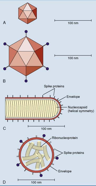
Many animal viruses are enclosed by an envelope, which is a piece of host cell membrane appropriated by the virus while it is budding out of its host cell. The envelope is studded with viral proteins, the spike proteins (Fig. 10.1).

Figure 10.1 Sizes and structures of some typical viruses. A, Papilloma (wart) virus: a nonenveloped DNA virus of icosahedral shape (spherical symmetry). B, Adenovirus: another nonenveloped DNA virus. C, Rabies virus: an enveloped RNA virus. D, Influenza virus: an enveloped RNA virus containing eight segments of ribonucleoprotein with helical symmetry.
The viral nucleic acid can be replicated only in the host cell, and host cell ribosomes are required for the synthesis of the viral proteins. Some viral proteins are enzymes for virus replication, and others form the capsid or appear as spike proteins in the viral envelope.
Bacteriophage T4 destroys its host cell
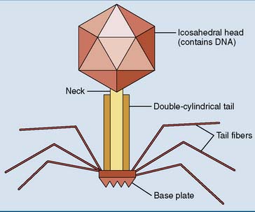
Viruses that infect bacteria are called bacteriophages (“bacteria eaters”) or simply “phages.” Bacteriophage T4 is a classic example. It is one of the most complex viruses known (Fig. 10.2), with a double-stranded DNA genome of about 150 genes tightly packed into the head portion of the virus particle. Attached to the head is a short neck followed by a cylindrical tail with two coaxial hollow tubes, a base plate, and six spidery tail fibers. This complex capsid consists of about 40 virus-encoded polypeptides, each present in many copies.

Figure 10.2 Structure of bacteriophage T4, one of the most complex DNA viruses known. Its capsid consists of approximately 40 different proteins.
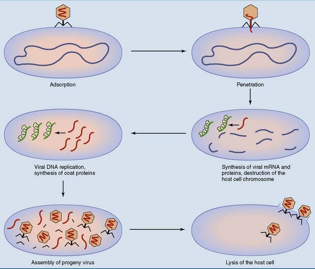
T4 is constructed like a syringe that injects its DNA into the host cell. First, the tail fibers bind to a component of the bacterial cell wall that serves as a virus receptor. Next, the sheath of the tail contracts, its inner core penetrates the cell wall, and the viral DNA is injected into the cell. Only the DNA enters the host cell. The protein coat remains outside (Fig. 10.3).
Some viral genes are transcribed immediately by the bacterial RNA polymerase. One of these “immediate-early” genes encodes a DNase that degrades the host cell chromosome. The viral DNA is not attacked by this DNase because it contains hydroxymethyl cytosine instead of cytosine.
During later stages of the infection, viral proteins substitute for the σ subunit of bacterial RNA polymerase and direct the transcription of the “delayed-early” and “late” viral genes. The promoters of these genes are not recognized by the bacterial σ subunit.
The early viral proteins include enzymes for nucleotide synthesis, DNA replication, and DNA modification. The viral coat proteins are synthesized late in the infectious cycle, and new virus particles are assembled from the replicated viral DNA and the newly synthesized coat proteins. Finally, virus-encoded phospholipase and lysozyme destroy the bacterial plasma membrane and cell wall.
This mode of virus replication is called the lytic pathway because it ends with the lysis (destruction) of the host cell. It takes approximately 20 minutes, and about 200 progeny viruses are released from the lysed host cell.
DNA viruses substitute their own DNA for the host cell DNA
Some but not all features of lytic infection by bacteriophage T4 are typical for viral infections in general:
Clinical example 10.1: Genetic AIDS Resistance
The human immunodeficiency virus (HIV) is an enveloped virus that causes acquired immunodeficiency syndrome (AIDS) by infecting helper T cells and macrophages. HIV can invade these cells because they express a glycoprotein called CD4 on their cell surface. Entry of HIV into the cell is facilitated by coreceptors in the membrane, which interact with the CD4-bound virus. One of these coreceptors is CCR5, a cytokine receptor expressed primarily on macrophages.
This cytokine receptor seems to be nonessential for immune responses, as about 15% to 20% of Europeans are heterozygous for a 32–base-pair deletion in the CCR5 gene (CCR5-Δ32), which prevents the synthesis of the receptor. Heterozygosity for this mutation delays the progression of HIV infection to clinical AIDS. Those lucky few who are homozygous for the mutation (up to 1 in 100 Europeans) are almost completely resistant to AIDS. The mutation is of recent origin. Whether it rose to its present frequency by chance or through darwinian selection by some infectious agent over the past one to five millennia is not known.
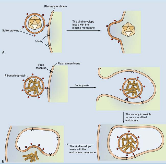
Some enveloped viruses fuse their envelope with the plasma membrane of the host cell, whereas others trigger their own endocytosis (Fig. 10.4).

Figure 10.4 Two strategies for uptake of an enveloped virus into its host cell. An initial noncovalent binding between a viral spike protein and the host cell membrane is essential in both cases. A, Uptake of human immunodeficiency virus (a retrovirus) is triggered by binding to the membrane glycoprotein CD4. The uptake of the nucleocapsid into the cytoplasm does not depend on endocytosis but is effected by direct fusion of the viral envelope with the plasma membrane. Only CD4-positive cells can be infected by this virus. B, Uptake of influenza virus, an enveloped RNA virus. Endocytosis is triggered by binding of the virus to the cell surface. The fusion of the viral envelope with the membrane of the endosome is facilitated by the low pH (5.0–6.0) of this organelle.
λ Phage can integrate its DNA into the host cell chromosome
Like T4 phage, λ phage is constructed as a syringe that injects its DNA into the host cell. Its genome, with about 50 genes, is a linear double-stranded DNA molecule of 48,502 base pairs with single-stranded ends of 12 nucleotides each. These single-stranded overhangs have complementary base sequences. They anneal (base pair) as soon as the viral DNA enters the host cell, and the viral genome is linked into a circle by bacterial DNA ligase (Fig. 10.5). Lytic infection can now proceed as described previously for T4 phage.

Figure 10.5 Circularization of λ phage DNA. This event takes place immediately after the entry of the viral DNA into the host cell and does not require virally encoded proteins. The circular DNA is then either replicated in the lytic pathway or integrated into the bacterial chromosome.

Stay updated, free articles. Join our Telegram channel

Full access? Get Clinical Tree

