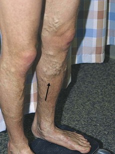
187 Venous ulcer
Salient features
History
• Varicose veins; ask about duration
• Past history of venous thromboembolism
• Duration of ulceration and whether this is the first episode or recurrent
• Pain (lack of pain suggests neuropathic aetiology)
• Rheumatoid arthritis (remember up to half the patients with rheumatoid arthritis have venous leg ulcers rather than the result of rheumatoid arthritis).
Examination
• Ulcer located on the medial aspect of leg (around the ankles) with pigmentation
• Surrounding dermatitis and excoriation from pruritus (stasis dermatitis)
• White atrophy with scars in the overlying skin
• The ulcer may be secondarily infected (cellulitis or thrombophlebitis)
• Look for varicose veins (Fig. 187.1)
• The range of hip, knee and ankle movement should be determined
• Sensation should be tested with a monofilament to exclude peripheral neuropathy (neuropathic arthritis or Charcot’s joints, see p. 496).
Advanced-level questions
What causes the surrounding pigmentation?
It results from extravasation of red blood cells and haemosiderin deposition.
What causes the white atrophy (atrophie blanche)?
It results from hyalization of skin vessels, leading to scars in the overlying skin.
How is the diagnosis confirmed?
Stay updated, free articles. Join our Telegram channel

Full access? Get Clinical Tree


