Venous Access: Saphenous Vein Cutdown
The greater saphenous vein is an anatomically constant vein that is easily cannulated for emergency venous access. The saphenous vein at the ankle is constant, although it may be involved by varicose vein disease in elderly patients. Although the interosseus route is faster in children, this remains a useful route of access, especially because it is somewhat removed from the central area and thus out of the way of resuscitative attempts. Bony landmarks render the vein easy to find.
The greater saphenous vein at the groin is sometimes used for introduction of an extremely large catheter, such as a sterile oxygen flow catheter, through which blood and intravenous fluids can be infused rapidly in a patient with traumatic injuries. The techniques of saphenous vein cutdown at the ankle and the groin are described in this chapter. Alternatives are discussed in the references at the end.
SCORE™, the Surgical Council on Resident Education, did not classify saphenous vein cutdown.
STEPS IN PROCEDURE
Cutdown at Ankle
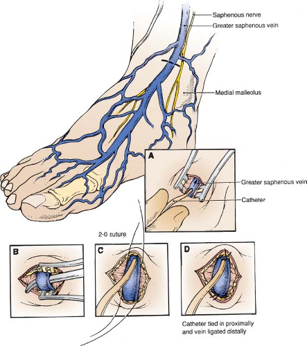
Local anesthesia two fingerbreadths above and two fingerbreadths medial to medial malleolus
Transverse skin incision
Spread tissues in longitudinal direction until vein is seen
Elevate the saphenous vein into field
Identify and protect saphenous nerve
Place ligatures proximally and distally
Cannulate vein and tie ligature around cannula
Ligate distal vein
Secure catheter and close incision
Cutdown at Groin
Moderate external rotation
Local anesthesia medial to femoral pulse, two fingerbreadths below inguinal crease
Incision parallel to inguinal crease
Dissect in subcutaneous fat
Identify the saphenous vein and elevate into incision
Cannulate as described above
Use Seldinger technique to avoid ligating vein, if desired
HALLMARK ANATOMIC COMPLICATIONS
Injury to saphenous nerve
Injury to femoral vein
LIST OF STRUCTURES
Common Femoral Vein
Greater saphenous vein
Saphenofemoral junction
Superficial epigastric vein
Superficial circumflex iliac vein
Superficial external pudendal vein
Medial malleolus
Patella
Inguinal ligament
Superficial fascia
Fascia Lata
Saphenous hiatus (fossa ovalis)
Femoral Nerve
Saphenous nerve
Medial femoral cutaneous nerve
Anterior femoral cutaneous nerve
 Figure 131.1 Saphenous vein cutdown at the ankle. A: Introduction of catheter into vein without ligation of vein. B: Isolation of vein. C: Insertion of catheter with vascular control of vein. D: Vein ligated distally and catheter tied in proximally.
Stay updated, free articles. Join our Telegram channel
Full access? Get Clinical Tree
 Get Clinical Tree app for offline access

|