Patricia Wieczorek
Vascular Surgery
Atherosclerosis continues to be one of the leading causes of death and disability in the Western world. It is estimated that peripheral atherosclerosis, including carotid, mesenteric, renal, and peripheral arterial disease (PAD) (also referred to as peripheral arterial occlusive disease [PAOD]), affects about 8 million people in the United States. The prevalence of PAD is approximately 12% of the adult population, with men being affected slightly more than women (Ferreria and Macedo, 2010). This is particularly striking, given that by the year 2030 the percentage of the U.S. population older than 65 years will increase to 20% (Federal Interagency Forum on Aging-Related Statistics, 2012). Many of these people will require interventions for the syndromes of peripheral ischemia, aneurysm, and venous disease.
Interventional therapy for peripheral atherosclerosis has become common. Aortic procedures are now routinely performed with minimal mortality. Carotid endarterectomy has also proven to be safe and effective. Peripheral angioplasty and bypass have a high initial success rate, but restenosis, graft failure, and progression of distal disease still lead to limb loss in certain patients after several years. Thus emphasis has been placed on decreasing morbidity and hospital stay associated with revascularization. Minimally invasive methods and strategies, including endovascular aortic aneurysm repair (Schwarze et al, 2009), carotid artery stenting (Liu et al, 2009) (Research Highlight), stenting for lower-limb ischemia, and percutaneous transluminal angioplasty (Zeller et al, 2009), have been developed, and their application and popularity continue to increase. New technologies, such as miniature shavers and laser arthrectomy devices for arterial plaque, continue to be developed (Kolakowski et al, 2009). Perioperative nurses must be prepared for the demands of patient care in vascular surgery. This chapter reviews surgical anatomy, perioperative nursing considerations, and surgical interventions for a variety of vascular procedures.
Surgical Anatomy
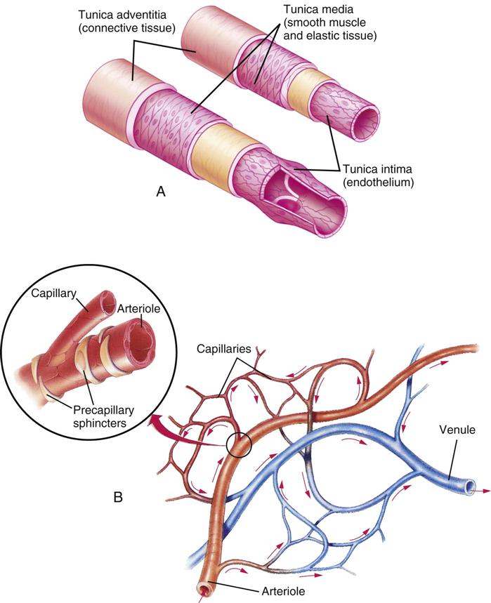
Basic knowledge of anatomy is essential when caring for perioperative patients with a vascular disorder. Figure 24-1 depicts the principal arteries and veins of the body. Arteries and veins have three layers:
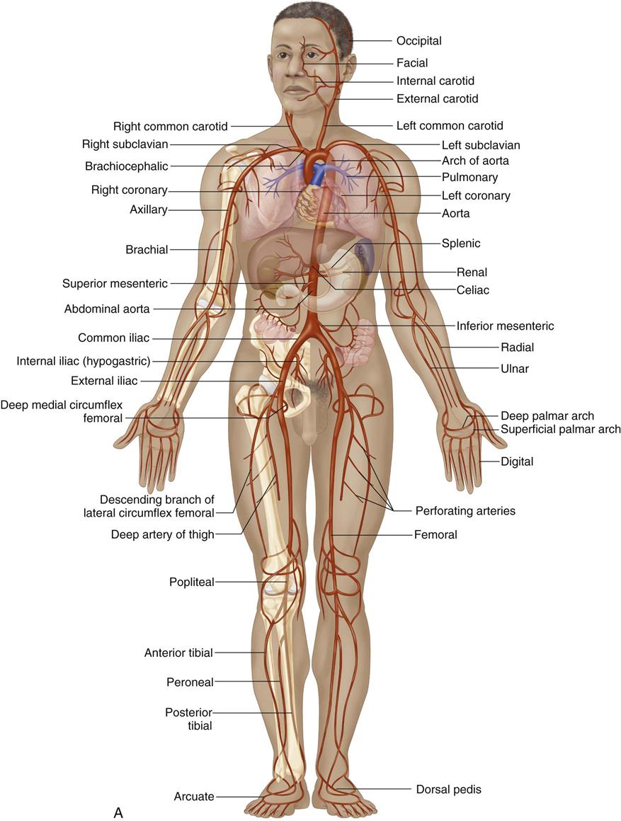
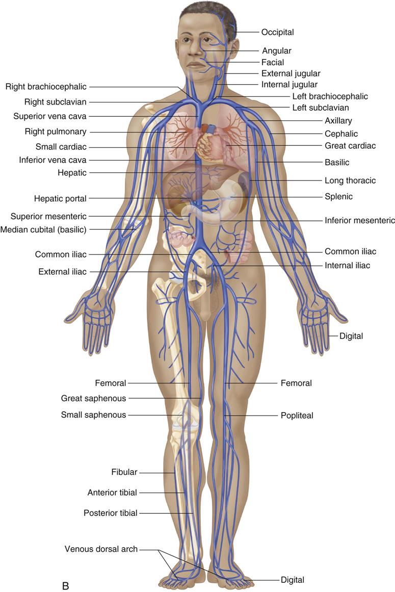
Arteries differ from veins in function and slightly in structure (Figure 24-2). Structurally, arteries have a thicker muscle layer and more elastic fibers than veins. The properties of elasticity and distensibility enable arteries to compensate for changes in blood pressure and volume. Because of the thicker muscle layer, severed arteries are capable of contracting and constricting enough to stop hemorrhage. In contrast, veins are more fragile than arteries, and whether its cause is traumatic or iatrogenic, venous bleeding may be difficult to control. Another difference is the presence of semilunar intimal folds, or valves, in veins that prevent backflow. Veins and arteries are nourished by a tiny network of vessels (the vasa vasorum), as well as from the intraluminal blood flow. Both are regulated by the autonomic nervous system, with veins having fewer nerve fibers than arteries. The two systems are connected (except for the pulmonary artery and pulmonary venous system): major arteries carry oxygenated blood, they branch into smaller arteries and arterioles, and then blood moves into capillaries to venules and to veins. The work of exchanging nutrients and metabolic waste is done at the capillary level.
Blood flow is a complex process that depends on many factors. Blood flows through arteries such that the blood in the center of the vessel moves faster than the blood at the periphery. Because the movement of the blood is in parallel lines, it is referred to as laminar. When flow is disrupted by an obstruction, stenosis, curve, or bifurcation, the particle motion is referred to as turbulent. Turbulence may be evidenced by the presence of a bruit (e.g., turbulent noise), detected by auscultation, or detected by a characteristic Doppler signal. Flow depends on blood viscosity, vessel wall resistance, and the peripheral resistance of the arterioles. There must be a difference in pressures, or a pressure gradient, to allow blood to flow. The gradient is provided by the contraction of the left ventricle. The negative pressure created by the relaxed right ventricle assists in venous return by creating a suctioning effect, and the skeletal and visceral muscles help propel venous return toward the heart.
Arterial Disease
Aneurysmal Disease
The most common cause of an arterial aneurysm is atherosclerotic degeneration of the arterial wall. The pathogenesis is a multifactorial process, involving atherosclerosis along with genetic predisposition, aging, inflammation, and localized activation of proteolytic enzymes (Sawabe, 2010). A true aneurysm is a dilation of all layers of the artery wall. A dissecting aneurysm results from a tear in the artery wall, allowing blood to dissect between the layers of the vessel wall. A false aneurysm, or pseudoaneurysm, is not an aneurysm but a disruption through all the layers of a vessel wall with the escaping blood being contained by the perivascular tissues. False aneurysms may result from trauma, infection, or disruption of an arterial suture line after surgery. True aneurysms are most frequently found in the abdominal aorta but are also found in the thoracic aorta and iliac, femoral, and popliteal arteries. More men than women are affected, and aneurysmal disease tends to be a disease of older adults. As early as 1977, a familial tendency was observed that subsequent research verified; as many as 18% of patients with abdominal aortic aneurysms have a first-degree relative with a similar diagnosis (Sawabe, 2010).
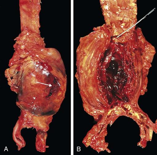
Abdominal aortic aneurysms (AAAs), which account for most aneurysms, occur primarily between the renal arteries and the aortic bifurcation (Little, 2013). An aneurysm involves intimal damage of the aorta and weakening of the tunica media (Sawabe, 2010) or elastic portion (collagen and elastin defects) of the arterial wall. Gradually the vessel wall in the damaged area expands and atheroma develops within the aneurysm sac (Figure 24-3). An AAA has minimal symptoms and is generally discovered on routine history and physical examination. In men, the diameter of the infrarenal aorta normally measures between 14 and 24 mm and in women it measures between 12 and 21 mm. An AAA is diagnosed if the diameter is 3 cm or larger for men or 2.6 cm or larger for females. Mortality is low with elective repair of the aneurysm. Dissection and rupture of the aneurysm (aortic dissection) dramatically increase operative mortality because of the abrupt and massive hemorrhagic shock that accompanies the rupture (Research Highlight). Aortic dissection is believed to arise from a sudden tear in the aortic intima, opening the way for blood to enter the aortic wall.
Acute Arterial Insufficiency
Arterial insufficiency may result from an acute occlusion, as in embolic disease, or from the rupture of an unstable atherosclerotic plaque, causing acute thrombosis of the vessel. Emboli usually arise from the heart as a result of atrial fibrillation but may occasionally result from a myocardial infarction (MI), where a clot forms on the endocardium (the lining of the heart) in an area of muscle damage. Atherosclerotic plaque can also break loose from other areas and result in an acute arterial blockage. Patients with acute arterial occlusion usually present with the onset of the six P’s: sudden severe pain, pulselessness, paresthesia, paralysis, pallor, and poikilothermia (coolness) of an extremity (Little, 2013). Heparin is the mainstay to prevent the enlargement of emboli while allowing time for collateral blood flow to develop. However, in the threatened limb there are basically two options: surgical removal of the clot (embolectomy) or chemical removal of the clot with the use of a thrombolytic medication. If the limb reaches the point where the muscle is rigid, the limb is not salvageable and amputation is a lifesaving procedure.
Chronic Arterial Insufficiency
Chronic arterial insufficiency occurs because of the deposition of calcium and cholesterol within the wall of the artery. Arteriosclerosis is a natural part of the aging process and occurs when the walls of the arterial vasculature undergo changes such as increased thickness and hardening, reducing the elasticity of the arteries. The decrease in elasticity should not be confused with atherosclerosis obliterans, which is a pathologic process that affects the intimal layer of the artery with the buildup of a fibrous plaque of lipids that can calcify and necrose. Atherosclerosis is the most common cause of PAD, the probable mechanism being initial damage to the intima and subsequent activation and aggregation of the body’s platelets. Inflammation follows, with the deposition of lipoproteins forming an atheroma. Calcification of this lesion leads to the development of an atherosclerotic plaque, resulting in inadequate muscle perfusion and ischemia (Zeller et al, 2009). The process is a gradual one, and a localized lesion usually indicates systemic disease. The body develops a network of collateral vessels as an adaptive mechanism to supply the tissues with oxygenated blood. Many theories have been postulated to explain the process of atherogenesis. The inflammatory process of intimal injury, as just described, seems to be the current and most widely accepted hypothesis. Box 24-1 presents risk factors for atherosclerosis. A large number of vascular surgical procedures revolve around the results of chronic arterial insufficiency.
Arterial Insufficiency: Cerebrovascular Disease and Stroke
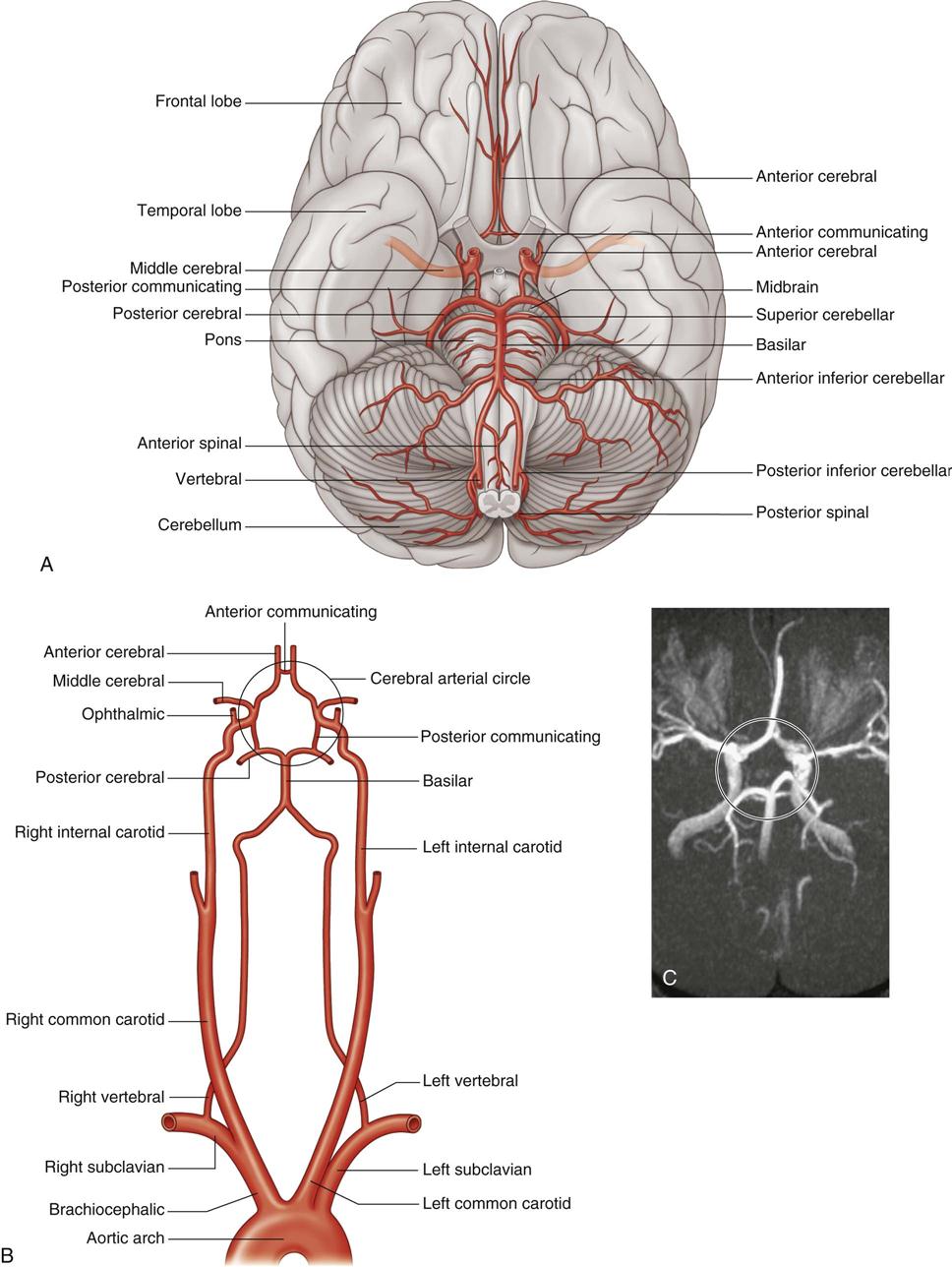
Cerebrovascular accident (CVA or stroke) is a leading cause of death in the United States and most industrialized countries. In the United States, approximately 795,000 people experience a new or recurrent stroke each year (Roger et al, 2012). Cerebrovascular disease may manifest itself as a transient ischemic attack (TIA) or as a major or minor stroke. A TIA is an episode of neurologic dysfunction that resolves in 24 hours. It may be caused by atheromatous debris or a thromboembolism from a carotid artery or the vertebral basilar system. Vascular lesions in the carotid artery occur primarily at the bifurcation of the common carotid artery into the internal and the external carotid arteries. The internal carotid artery supplies the brain with needed oxygenated blood. Obstruction in this arterial vessel leads to cerebrovascular insufficiency.
The right and the left carotid and vertebral arteries supply the brain (Figure 24-4). The first major branch of the internal carotid artery is the ophthalmic artery. Thromboembolic events that affect this artery may result in visual disturbances, ocular TIAs, or “amaurosis fugax” (complete or partial loss of vision). Patients often describe amaurosis fugax as a curtain over a partial field of vision, usually the top. Clinical conditions that generally indicate the need for a carotid endarterectomy (CEA) are transient cerebral ischemia, asymptomatic severe stenosis, and stable strokes. Carotid disease may recur after a CEA. Redo surgery for restenosis poses the same complication risks as the original procedure.
Arterial Insufficiency: Peripheral Vascular Disease
The initial and most important symptom of vascular disease in the aortoiliac vessels and distal arteries is intermittent claudication. The term claudication is derived from the Latin word claudicare, which means “to limp.” This is the most common symptom of lower-extremity PAD and occurs distal to the arterial obstruction while the patient is exercising. Many patients are asymptomatic and do not experience pain. When this symptom does occur, it is typically located in the working muscle, occurs with the same amount of exercise each time, and is relieved with rest. This is referred to as functional ischemia; blood flow is adequate at rest but inadequate to sustain exercise. The increased muscle demand for oxygen with exercise cannot be met distal to the arterial obstruction. Anaerobic metabolism occurs, and muscle cramping develops. Walking difficulties are often seen in the elderly patient with PAD (Patient-Centered Care). Surgery is not usually performed for claudication unless it is unusually disabling.
The second symptom—rest pain, which is located in the foot—develops as the vascular disease progresses (Oka et al, 2012). At this stage the ischemia is termed critical. Rest pain occurs without exercise and is a constant discomfort, often aggravated at night. The body is now unable to meet the oxygen needs of distal tissues even at rest. Rest pain may be somewhat relieved by analgesics or by lowering the legs off the bed. Gravity assists in increasing the tissue perfusion and oxygen supply to decrease the pain. Unless the vascular disease is corrected, nonhealing ulcers and gangrene can develop. Gangrene occurs when the arterial vessels are unable to meet the oxygen needs of distal tissues even at complete rest.
Venous Disease
Acute Venous Insufficiency
Acute venous insufficiency is caused by a clot in the deep venous system, or deep vein thrombosis (DVT). Such venous insufficiency can be a diagnosis of DVT, phlebitis, thrombophlebitis, or phlebothrombosis, which merely indicates that there is a clot, usually in the lower extremity. Virchow, a pathologist, identified the three elements that trigger venous thrombosis. Referred to as Virchow’s triad, these elements, or risk factors, are endothelial injury, venostasis, and hypercoagulability (Little, 2013). The cause of hypercoagulability is sometimes unknown but is seen in patients with tissue trauma (e.g., surgery, burns, or stroke), malignancy, sepsis, pregnancy or estrogen use, and diabetes mellitus. The patient may be asymptomatic or present with limb swelling, pain, and a skin color change. The danger lies in the potential emboli migrating to the right ventricle and proceeding to the lungs. A pulmonary embolus (PE) can be fatal. The majority of pulmonary emboli cases are caused by DVT. The majority of these originate in the lower extremities. The use of heparin and bed rest is the usual medical treatment. In cases that preclude the use of systemic heparin or in which heparin is ineffective, surgical insertion of a vena cava filter may be indicated.
Chronic Venous Insufficiency
Patients with chronic venous insufficiency (CVI) have not been treated surgically as often as patients with arterial disease for several reasons. CVI is generally not life- or limb-threatening. Improved imaging techniques (e.g., duplex ultrasonography) allow better diagnoses of the precise problem. The treatment of the majority of venous disorders is nonsurgical and aimed at increasing venous return and decreasing edema. CVI, which presents with stasis ulcers from postphlebitic syndrome, usually occurs in one leg. The leg is usually very swollen with a cyclic edema, which does not change visibly after overnight leg elevation. Stasis ulcers and hyperpigmentation usually are found in the “gaiter” area (e.g., midcalf to about 1 inch above the medial malleolus) on the leg. The condition is caused by incompetent perforator valves. The perforating veins connect the superficial and deep venous systems. The usual management is to apply 20 to 30 mm Hg of external pressure by means of special pressure stockings. Surgical interventions such as valvuloplasty (direct repair of the valve), valve transposition or transplantation (moving a valve from the arm to the leg), or perforator interruptions are occasionally performed but have had limited success. Patient selection is critical, and long-term results are mixed.
Diabetic and Vascular Foot Conditions
The severity of these conditions is conveyed by diagnoses and sequelae (e.g., necrotizing fasciitis, gas gangrene, ascending cellulitis, and infection with systemic toxicity or metabolic instability). Because patients with diabetes mellitus may have peripheral neuropathies, they may not feel and therefore not notice any tenderness or early signs of infection. These foot infections can lead to hospitalization and eventual lower extremity amputation for patients with preexisting ulcerations; surgical management is often required for the severe infections. The experienced surgeon determines when and how to intervene. Main principles for management include stabilization of the patient; adequate debridement along with antibiotic therapy; vascular evaluation and revascularization as necessary; delayed soft tissue reconstruction; and postoperative information and intervention for medical and surgical issues. In addressing all these aspects, the surgeon optimizes the possibility for limb salvage. This overview serves as a reminder to emphasize the importance of overall adherence in managing diabetes mellitus to prevent further complications (Aragón-Sánchez et al, 2011).
These complex patients are best handled with multidisciplinary consultations to coordinate care and determine optimal timing for soft tissue reconstruction. Primary closure may not be an option and secondary healing is not necessarily reliable. The proactive surgical approach aims to salvage the diabetic limb by improving overall health and to provide a stable, mechanically sound limb that will resist further breakdown once the patient is able to walk again. Patients need ongoing support and validation to prevent complications. Hospital care focuses on dressing changes, wound care, physical therapy, limb position, and laboratory testing. Postoperative nursing care should provide instruction for follow-up and frequency of monitoring of the surgical results, ambulatory status, work and social restrictions, and bathing. Especially important for the perioperative nurse to review with the patient are identification of infection and ways of contacting the surgical team if problems arise (Aragón-Sánchez et al, 2011).
Perioperative Nursing Considerations
Assessment
Nursing Responsibilities.
A preoperative assessment is necessary for an adequate understanding of the patient’s disease, the patient’s response, and the proposed surgical procedure. Knowledge of vascular disease and its progression assists the perioperative nurse in performing a critical review of the comprehensive assessment and developing a plan of care for patients undergoing vascular surgical procedures.
The perioperative nurse should assess the patient, reviewing an already completed, current nursing assessment for the development and extent of vascular symptoms. The nurse must assess medical conditions—including cardiac, renal, and pulmonary disease; coagulation status; and allergies (Box 24-2)—to ensure that the patient can tolerate a possible angiogram, because the contrast medium is toxic and increased fluid volumes are needed if angioscopy or intraoperative angiography is planned. This is a shared responsibility of the nursing, surgical, and anesthesia providers. The patient’s nutritional status, the patient’s use of alcohol and tobacco, and the existence of any skin lesions should also be noted. Preoperative location, grading, and marking of distal peripheral pulses assist the perioperative nurse with intraoperative and postoperative assessment of tissue perfusion. The nurse shares pertinent information gathered during the assessment phase with the rest of the surgical team, including the scrub person (who may be a surgical technologist or registered nurse), as appropriate.
The nurse greets the patient by name and verifies identification using two unique identifiers. When possible, the patient should confirm such identification. Identification must also be confirmed by the name band, medical record, surgical consent, operating room (OR) schedule, and marked surgical site. Safety is always the first priority. The surgical procedure must be confirmed and the patient’s understanding of the procedure and associated risks assessed; inaccuracies or misconceptions must be clarified before proceeding with surgery. Written consent is designed to protect the patient by providing written clarification of the proposed procedure. The National Quality Forum (NQF) identified Safe Practice 10 for informed consent, recommending that each patient (or legal surrogate) be asked to “teach-back” what he or she has been told during the informed consent discussion (Fink et al, 2010). Consents that are valid and obtained freely from a competent person remain in effect for as long as the person still agrees to the surgery or procedure. This may vary by institutional modification.
The nurse assesses patient comfort before continuing the interview and assessment. This reflects caring and will promote nurse-patient rapport and facilitate obtaining accurate and complete information. The acuity of the patient’s condition will also affect the order of priority of the assessments and interactions. After reviewing the results of the patient’s physical examination, the perioperative nurse should verify signs and symptoms of vascular disease that need to be considered during intraoperative care. Muscle and skin atrophy, the presence of tissue ulceration or necrosis, pain, neurovascular status, skin color and temperature, and other integumentary changes should be noted. Elderly, cachectic, or obese patients are at increased risk for pressure injuries.
The perioperative nurse should assess the patient’s mental status and determine the level of understanding and emotional response to the surgery. Patients with vascular disorders usually have systemic disease, and the fear of a stroke (if the patient is having a carotid procedure), amputation (if the patient has an ischemic limb), or other complications may be a realistic concern. A skin assessment should include notation of color, integrity, pain, and pulses. Any musculoskeletal problems that preclude patients moving themselves to the OR bed and any weaknesses that may have resulted from a stroke or that would modify the positioning for surgery should be noted. Correction of misunderstandings is possible only if the perioperative nurse identifies the patient’s current level of knowledge. Identification of the patient’s fears and concerns helps with planning supportive nursing interventions.
Vascular surgery can be lengthy. The nurse must consider the effects of hypothermia during long procedures. The patient’s extremities should be assessed for color, temperature, and strength of pedal pulses before the surgical procedure so that baseline data will be available for comparison with perioperative assessment data. This assessment evaluates tissue perfusion distal to the arterial obstruction.
Preoperative Tests.
A variety of tests may be required to plan for surgical interventions. Segmental pressure measurements give partial anatomic information in that they assist in locating lesions. Hemodynamic tests provide information on the flow of blood, such as that to the brain or an extremity, and the effects on flow caused by a vascular lesion.
Angiography.
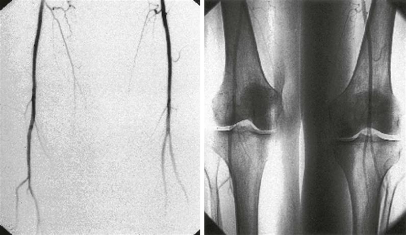
Invasive diagnostic tests may be performed preoperatively to identify the extent and location of the patient’s peripheral vascular disease (Oka et al, 2012). The introduction of a contrast medium through a catheter into the arterial or venous system of the patient facilitates this visualization. Angiography also involves injecting a contrast medium into the patient’s arterial system and taking serial radiographs of the movement of the dye through the arteries. Digital subtraction angiography (DSA) is one such technique that uses a computer along with contrast medium injection to make the image (Figure 24-5). Usually one side of the film shows the bone for orientation, and the other side subtracts the density of the bone and soft tissue to allow a clearer view of the vessels. Arteriography provides information on arterial anatomy and the location of stenotic or occluded vessels and assists the surgeon in planning bypass procedures. A venogram (contrast venography) is performed to show venous abnormalities in extremities, the vena cava, and the hepatic and renal systems. Ascending venography can differentiate between acute and chronic thrombosis and can define anatomy. Descending venography assesses valve competence of the lower extremities.
Doppler Scanning.
The Doppler effect is the change in the frequency of echo signals that occurs whenever there is a change in the distance between the sources of a sound and the receiving object. The probe, or transducer, is aimed toward the blood vessel at an angle of 45 to 60 degrees. This directs an ultrasound beam that is reflected back to the probe by moving red blood cells (RBCs). The velocity of the flow of cells is converted into an audible signal heard through a speaker. The signal is described as a swishing sound. The sound is called a signal, not a pulse. The tip of the probe is made of an element called a ceramic piezoelectric crystal, which can send, receive, and convert signals when an electric current is applied. The element becomes thicker and thinner, thus resulting in a pressure wave converted to an audible signal. The simplest form is the continuous wave (CW) Doppler probe. It has two elements: one sends a high-frequency wave, and the other receives it. In a pulsed Doppler probe, the same element sends and receives signals. The pulsed Doppler probe has the advantage of being able to differentiate among vessels of different depths. A normal arterial Doppler signal is either biphasic or triphasic. The first sound corresponds to systolic flow and is forward-moving and of high velocity. The second sound is related to early diastole and has a lesser reversal of flow. The third sound is later diastole and is smaller, forward-flowing, and of a lower velocity. The pitch is described as rising quickly in systole and dropping quickly in early diastole. An abnormal signal, indicating stenosis or occlusion is heard as low-pitched and monophasic. These abnormal arterial signals may sound like venous signals.
The Doppler probe can provide information in three forms: the audible signal, a visible graph printout similar to an electrocardiogram (ECG) tracing, and a spectral analysis that appears on a screen and may be recorded on paper as well. The Doppler transducer is the most widely used instrument for vascular study. It has the advantages of being readily available, inexpensive, and easy to use. A small, portable battery unit is durable and can be transported and stored easily. When the probe is used on intact skin, a water-soluble gel is needed to conduct a signal. Probes can be used directly on a vessel intraoperatively. The probes are heat sensitive and must either be sterilized according to manufacturer’s instructions or be inserted into a sterile sleeve or probe cover. If they are handled gently, the probes have a reasonable life span. Care must be taken to protect the sensitive tip from being dropped or crushed. The biggest drawback of the Doppler probe is a negative finding in the presence of a stenotic lesion pronounced enough to produce a flow disturbance resulting in an altered signal.
A bruit is a sound disturbance that is sometimes described as a low-pitched, blowing sound. It can be heard through a stethoscope over an area of blood flow turbulence that occurs at points of vessel stenoses. Bruits do not provide information on the extent of a lesion—only that an abnormal flow may exist. They occur at points of significant stenosis and are not heard when severe flow restriction or total flow occlusion occurs. The Doppler probe is noninvasive and painless for patients.
Ultrasonography.
Ultrasonography is done to obtain information about structures through the emission of high-frequency sound waves. These sound waves are reflected, or bounced back, to the probe or transducer that emits them and are electronically transformed into an image.
B-Mode Ultrasonography.
B-mode is brightness modulation, a technique in ultrasound imaging that projects a two-dimensional image on an oscilloscope screen. The image appears as dots from the echoes of the signal. The strength of the echo is shown by the intensity and brightness of the dots on the screen.
Duplex Ultrasonography.
A duplex ultrasound machine is a combination of the pulsed Doppler image and the so-called real-time B-mode image ultrasonogram. “Real time” simply refers to the image projecting current, undelayed information. B-mode image is best when the probe is perpendicular to the vessel, but the Doppler probe does not pick up signals at a perpendicular angle. Some manipulation of the probe angle is required to obtain the best results. Color duplex imaging converts the detected signals caused by blood flow into a color, depending on the direction of flow. Flow toward the probe may be displayed as red, away from the probe as blue, and turbulence as multiple colors. This imaging provides both hemodynamic and anatomic information. The technology is also used in transesophageal echocardiography (TEE) and is the diagnostic method of choice for venous insufficiency.
Pulse Volume Recording (PVR) or Sequential Volume Plethysmography.
A plethysmograph measures and records the changes in the sizes and volumes in extremities by measuring the blood volumes at blood pressure cuffs placed at intervals along the extremity. The methods include electrical impedance, mercury in Silastic strain gauges, and air or fluid displacement. This test, which is used to determine the location of an arterial lesion and estimate the severity of the disease, requires careful limb positioning and a cooperative patient. A negative study is a good predictor of low risk for PE.
This test is inexpensive, has good predictive value, and is accurate in detecting thrombosis. It has the disadvantage of a high rate of false-positive results in the presence of old DVT, congestive heart failure (CHF), and external compression.
Magnetic Resonance Imaging.
Magnetic resonance imaging (MRI) measures the behavior of atoms in a strong magnetic field. This test provides detailed and three-dimensional images of anatomy for evaluation of carotid, aortic, and lower-extremity disease (Oka et al, 2012). An MRI provides more detail than ultrasonography or computed tomography (CT) scan and avoids the complications of contrast medium injection and exposure to x-rays. MRI is contraindicated for patients with preexisting implantable devices.
Nursing Diagnosis
Nursing diagnoses related to the care of patients undergoing vascular surgery might include the following:
• Anxiety related to the surgical intervention and its outcomes
• Hypothermia related to surgical exposure and anesthesia
• Risk for Deficient Fluid Volume related to loss of body fluids
Based on the perioperative nurse’s assessment, identification and prioritization of nursing diagnoses aid in the development of an individualized plan of care.
Outcome Identification
Outcomes identified for the selected nursing diagnoses could be stated as follows:
Planning
Before the patient is transported to the OR, the perioperative nurse should procure the necessary supplies and equipment for the intended surgical intervention. Because the need for intraoperative arteriography or fluoroscopy for endovascular procedures is a possibility, the patient with a vascular disorder should be positioned on an appropriate OR bed with x-ray capabilities. The perioperative nurse needs to coordinate the availability of x-ray department personnel. Appropriate contrast media, catheters, and impermeable sterile x-ray covers must be available. Radiation-protection devices, such as lead aprons and shields, should be used for the patient when possible and for surgical team members. A typical plan of care for a patient undergoing vascular surgery, using the suggested nursing diagnoses, is seen in the Sample Plan of Care below.
Arterial procedures, especially those that involve the aorta, may place the patient at risk for significant blood loss. The perioperative nurse should confirm the availability of ordered blood-replacement products. The use of rapid infusion systems or blood salvage equipment should be determined and planned.
Implementation
Site Verification: Time-Out.
Patient safety is of utmost importance. It is the entire surgical team’s responsibility to verify that the correct patient is receiving the correct procedure on the correct site immediately before the start of any surgical procedure. The surgical site needs to be marked and visible after draping if laterality is involved. The time-out is a requirement of The Joint Commission. During the time-out, the patient’s name, procedure, site verification, and laterality are reviewed. Other items that may be discussed include the consent, anesthesia plan/concerns, the patient’s allergies, antibiotics ordered, the patient’s position, required instruments and special equipment, availability of blood, and anticipated length of the procedure. Such briefings improve team communication and intraoperative patient care (Johnson and Kimsey, 2012).
Intraoperative Monitoring.
Intraoperative monitoring for patients with vascular disorders includes the use of the basic ECG, pulse oximeter, and blood pressure cuff. For patients undergoing saphenous vein stripping or amputation, these are usually adequate. For lengthy procedures, such as arterial bypass or reconstruction, the anesthesia provider usually places an arterial line into the radial artery. This line is kept open by a pressurized heparin drip line attached to a transducer, and a waveform monitor reads the systolic and diastolic pressures. The monitor calculates the mean arterial pressure (MAP), which aids in the evaluation of the perfusion of systemic and cardiac circulation. This arterial line also allows easy access for collecting specimens for arterial blood gas (ABG) analysis. Continuous assessment of the patient’s arterial pressure is a critical part of the surgical procedure. Pulmonary capillary wedge pressure, as an index of left atrial pressure (LAP), may be monitored depending on the patient’s physiologic status. A general anesthetic may be administered and the patient intubated; local or regional anesthesia may also be used, depending on the surgical intervention. Epidural catheters may be placed to provide intraoperative anesthesia that can be augmented to accommodate increased surgical time, as opposed to a spinal anesthetic, which provides a finite period of anesthesia. Epidural catheters may be left in place postoperatively for pain management as well. Because many patients undergoing vascular surgery have generalized atherosclerotic disease, the perioperative nurse should be constantly alert for cardiac dysrhythmias and blood pressure changes. Acid-base balance and pulmonary gas exchange are assessed from the ABG analysis.
The anesthesia provider may also place a central venous pressure (CVP) catheter or pulmonary artery (PA) catheter, usually by way of the right internal jugular vein. The CVP line allows assessment of blood volume and vascular tone. The PA catheter monitors cardiac output, fluid balance, and the cardiac response to medications. PA catheters are commonly used for patients undergoing aortic surgery or for patients with cardiac disease.
TEE may be used to monitor the heart noninvasively during aortic surgery. The device looks similar to a bronchoscope and can be passed down the esophagus to provide an ultrasonic image. The cardiac structures, blood flow, wall motion, and great vessels can be observed. Use of TEE requires highly skilled personnel and may not be available in all surgical settings.
Electroencephalographic (EEG) monitoring is used for patients undergoing a carotid endarterectomy and allows for immediate observation of the slowing of brain waves caused by cerebral ischemia or reduced perfusion. The surgeon may elect to place a temporary shunt in the artery if this occurs during clamping, potentially reducing the chances of perioperative stroke.
The perioperative nurse inserts a urinary catheter for many procedures, including the following: if the proposed procedure involves the renal arteries or clamping of the aorta above the renal arteries; if considerable blood loss is anticipated; if the planned procedure time is lengthy; or whenever spinal or epidural anesthesia is used, because they delay the patient’s ability to void voluntarily. Urinary catheterization facilitates accurate hourly measurements of urine during and after the surgical procedure and assists in the assessment of renal perfusion and fluid status.
Positioning.
Positioning of the patient undergoing vascular surgery is of particular importance because of restricted circulation distal to the area of arterial obstruction and a generalized state of poor circulation. Particular care must be exercised in positioning elderly patients (see Chapter 27). Awareness of joint range-of-motion limitations attributable to immobility or joint surgery is critical even for a procedure as routine as urinary catheter insertion. Again, preoperative assessment can prevent injury and decrease OR time. Whenever possible, the perioperative nurse should have the patient demonstrate the ability to assume the position for the proposed procedure and provide feedback. A footboard may be applied to the OR bed to prevent the weight of drapes resting on the patient’s lower extremities. A head support may be used to position the head. A roll may be placed between the scapulae. For surgical procedures involving a lower extremity, the patient’s thigh may be externally rotated and abducted with the knee flexed. A small bolster may be used under the knee to support the patient’s leg. Proper skeletal alignment during surgery prevents injury to the neuromuscular system. The nurse pays close attention to the skin overlying bony prominences, especially the heels, sacrum, and elbows, and uses the proper supports and pads to prevent pressure and potential positioning injury to the patient. If the procedure will be lengthy, a pressure-reducing mattress or pad can be placed on the OR bed to help prevent patient injury. Arms are placed on padded armboards with the palms up and fingers extended. Armboards are maintained at less than a 90-degree angle to prevent brachial plexus stretch. If there are surgical reasons to tuck the arms at the side, the elbows are padded to protect the ulnar nerve, the palms face inward, and the wrist is maintained in a neutral position (Denholm, 2009). A drape secures the arms. It should be tucked snuggly under the patient, not under the mattress. This prevents the arm from shifting downward intraoperatively and resting against the OR bed rail. During the procedure the perioperative nurse and scrub person continually monitor the sterile field to ensure heavy instruments and drapes do not rest on the patient’s body and cause pressure injuries.
Skin Preparation and Draping.
Skin preparation for vascular surgery may be extensive. The patient’s hair should be removed preoperatively only if it interferes with the procedure; if hair removal is necessary, it should be done immediately before the surgical procedure using clippers (Evidence for Practice), not a razor. For carotid surgery, the patient is prepped from the ear and chin on the affected side to below the clavicle (Figure 24-6, A). For abdominal aortic surgery, the patient’s skin is prepped from the nipple line to the midthigh area (see Figure 24-6, B). For peripheral vascular surgery on the lower extremities, the patient is prepped from the umbilicus to the feet. The patient’s legs are prepped circumferentially. It is important that alcohol-based prep solutions are allowed to dry completely before applying the surgical drapes and starting the surgical procedure.
Stay updated, free articles. Join our Telegram channel

Full access? Get Clinical Tree


