Vascular and Other Mesenchymal Tumors and Lesions


Vascular and Other Mesenchymal Tumors and Lesions


Vascular Lesions

Vascular lesions of the skeleton occupy a tenuous niche with regard to nomenclature and unpredictable biologic behavior. They range from benign localized processes, some of which are arteriovenous malformations, to highly malignant neoplasms. They are often multicentric. Concomitant skin, soft tissue, and even visceral involvement are reported.

Vascular anomalies have been characterized by their speed of flow with fast-flow arterial lesions contrasting with slow-flow capillary, lymphatic, or venous lesions. Combinations of these also occur.

Vascular abnormalities in the soft tissue are common and have been associated with bone involvement in as much as 20 percent of cases (1). Most common in the pediatric age group, lesions may be associated with pain and disparity in leg length. Venous malformations are the most common. Present at birth, they grow proportionally with the patient and generally do not regress (2). The following is a review of relatively well-recognized clinicopathologic vascular entities (Tables 11.1 and 11.2).


Vascular Malformations

Vascular malformations have been characterized by both flow dynamics (high vs. low flow), and the histology of the vessels (lymphatic, capillary, venous, arteriovenous, arterial, or combinations of these) (3).

They are, in general, most common in the lower extremity soft tissues and may cause symptoms and imaging findings such as a localized soft tissue mass. Lesions can cause periosteal reactions and pressure defects in the local bone. Classic signs of a vascular abnormality, such as a bruit, thrill, disturbance in growth, skin changes, cardiac symptomatology, hemolytic anemias, or thrombocytopenia, may be noted.

The majority of A.V. malformations are isolated lesions in otherwise healthy individuals. Intraosseous lesions are rare and can be confused with osseous hemangiomas on histopathology (4).

Varices can cause defects in adjacent bones.


TABLE 11.1 Classification of Benign Vascular Lesions of Bone (and Soft Tissue)


Glomus tumor


Hemangiomas and arteriovenous malformations


Hemangiomatosis and Gorham syndrome


Hemangioma-associated hypophosphatemic rickets (oncogenic osteomalacia)


Epithelioid hemangioma, histiocytoid hemangioma, and Kimura disease


AIDS-related vascular lesions


Kaposi sarcoma


Bacillary angiomatosis


Lymphangioma


Lymphangiomatosis and Gorham syndrome


AIDS, acquired immunodeficiency syndrome.


Vascular Tumors and Tumorlike Lesions


Benign Tumors


Glomus Tumor (Glomangioma)

Glomus tumors present as painful, bluish red nodules in the dermis or subcutaneous tissue of the extremities, with a predilection for a subungual location in the fingers (5) (Fig. 11.1). Symptoms have usually been present for years. Episodes of acute pain radiating from the lesion are suggestive of the diagnosis, the pain often elicited by changes in temperature, as on exposure to cold, or gentle tactile stimulation. Hypesthesia, osteoporosis, and muscle atrophy have been associated symptoms and findings.

Glomus tumors are specialized arteriovenous anastomoses consisting of modified perivascular smooth muscle cells arranged in sheets and nests. They arise from the neuromyoarterial glomus bodies, which are connections between preterminal arterioles and efferent veins. Glomus bodies are specialized complexes of vascular anastomoses that regulate body temperature. Common sites include the fingertips and nail beds, but other than the finger include
the ankle, foot, and knee. The presence of glomera elsewhere as anatomic curiosities—for example, at the tip of the coccyx (glomus coccygeum, originally described by Luschka in 1859) causing coccydynia—is well documented (6).


TABLE 11.2 Clinical, Radiological, Histological, and Genetic Features of Vascular Bone Tumors


Entry


Clinical Features


Mean Age (y)


Radiological Features


MRI


Histological Features


Mitoses


Genetic Features


Bones Involved


Percent Multifocal


Immuno-Histochemistry


Mortality Rate


Prognosis


Treatment


Hemangioma


Benign Incidental finding/rarely painful Spine/skull


40


Well demarcated Lytic Honey-combing Trabeculation Striations “Polka dots”


↓ T1


↑ T2


Cavernous/capillary/venous


Well demarcated


Vascular spaces


Endothelial cells


No atypia


None


Aberrant activation of the Notch pathway


Spine>> Craniofacial> long bones


Frequent


+ Factor VIII (vWF) CD31 CD34 +/- FLI1 and ERG


0


Excellent


Observation usually; If symptomatic sclerotherapy, kyphoplasty, or excisions


Epithelioid hemangioma (histiocytoid hemangioma, angiofollicular hyperplasia with eosinophilia, spindle cell hemangioma)


Benign Painful Rare metastatic


35


Well-defined Lytic lesion Rare focal cortical destruction with reactive periosteal new bone formation


T1


T2


Well circumscribed Lobular growth pattern Vasoformative with “mature” vessels Vacuolated cells Abundant cytoplasm “Tombstone” appearance of cells Inflammatory cells including eosinophils No atypia


<1/10HPF None atypical


None


Long tubular bones>> especially lower extremity Flat bones Vertebrae hands, feet


Up to 25%


Regional distribution


+Factor VIII (vWF) CD31,CD34 FLI1, ERG +/- keratins (AE1AE3,CAM 5.2,CK8,CK1 8,CK7,CK19), EMA


0


Excellent


Curettage or marginal en bloc excision (?) Radiation if inaccessible


Epithelioid hemangio-endothelioma


Low grade Malignancy Painful Metastatic Good prognosis


20 (wide range)


Poorly demarcated Expansive and infiltrative lytic lesion Small oval cortical-based lucencies


Intermediate T1 High T2


Infiltrative Strands/cords of solid cells Myxochondroid or sclerotic stroma Mild degree of nuclear atypia


Present


T(1:3) (p36.23:q25.1) resulting in WWTR1-CAMTA1 fusion


Long tubular bones, especially lower extremity>> pelvis > ribs > spine


50%-60% usually regionally


+ Factor VIII (vWF) CD31 CD34 FLI1, D240, ERG +/-keratin EMA


20%


Variable clinical course


Wide resection


Angiosarcoma


High-grade malignancy Painful Tumor mass Metastatic Poor prognosis


62


Lytic lesion Well or poorly defined margins Cortical destruction usually no periosteal rx Invades soft tissue


T1


T2


Infiltrative Atypical endothelial cells Vasoformative Nuclear atypia Atypical mitoses Inflammatory cells Necrosis


++


MYC Amplification Rare t(1;14) (p21;q24)


Long and short tubular bones Femur> pelvis >axial skeleton


33% contiguous or distant


++ CD31, Factor VIII (vWF) + CD34, ERG, FLI1 +/- EMA, D240 keratin, EMA


33% at 5 y


Unpredictable, but generally poor with up to 50%-80% developing metastases and dying of disease.


Wide resection ?radiation ?chemotherapy


FIGURE 11.1. Radiograph of a glomus tumor. Well-defined lucency is seen in the tuft of the distal phalanx. Differential diagnosis should include epidermoid inclusion cyst and, less likely, infection and metastasis. Enchondroma should not occur at the tip of the distal phalanges.

In a review of 37 coccygeal bones removed during rectal resection for carcinoma, Gatalica et al. identified, in 50 percent of the cases, sharply circumscribed glomus bodies of various sizes but without atypia, pleomorphism, expansile growth, or infiltration into the surrounding soft tissue or bone (6). These “glomus coccygeum” lesions can pose a diagnostic dilemma to pathologists unfamiliar with the entity. The cells are smooth muscle actin and neuron-specific enolase positive.

Glomus tumors usually appear as a well-defined radiolucency. On magnetic resonance imaging (MRI), they have a high signal on T2, an intermediate signal on T1, and are typically uniformly enhanced after gadolinium administration (7).

Histologically, glomus tumors are composed of an admixture of glomus cells, proliferating vascular spaces, and various stromal components, and smooth muscle cells. The term “glomangioma” has been used when the smooth muscle cell component predominates (Fig. 11.2). The sheets of glomus cells typically envelop small-caliber vessels. A fibrous pseudocapsule is seen.

Glomus cells are considered a derivative of smooth muscle, staining positively for vimentin and muscle-specific actin. However, they are negative for another smooth muscle marker, desmin, as well as for protein S100, factor VIII-related antigen, and myoglobin.

Primary intraosseous glomus tumors are rare, but have been documented, most commonly in the distal phalanx. The middle phalanx, ulna, and fibula are other locations that have been documented (8).

Malignant soft tissue glomus tumors have been reported (9).


FIGURE 11.2. Histology of glomus tumor. (A) Uniform dark round cells surround blood vessels. (B) Stroma may be inconspicuous or more prominent and consist of fibrovascular, myxoid, or chondroid tissue. The predominant “glomus” cell is a monomorphic bland cell with round nuclei.


Hemangiomas

Intraosseous hemangiomas are usually asymptomatic, solitary vascular hamartomatous, or congenital vascular malformations involving the vertebrae, calvaria, and facial bones in the middle years of life. Other sites, such as flat bones (pelvis, ribs, and scapulae) and long bones, may be affected. Vertebral hemangiomas are particularly well documented. They have been found in 10.7 percent of spines at autopsy and in 14.2 percent of people older than 60 years (10). However, symptoms related to vertebral hemangiomas (epidural compression of spinal cord, epidural hemorrhage, or compression fracture) are far less common (<1 percent).

Roentgenographically, hemangiomas of vertebrae are seen typically as lesions with coarse internal linear trabeculations, best seen on computed tomography (CT) (Fig. 11.3). This feature should not be confused with Paget disease (characterized by enlargement of the bone and peripherally increased density, “window framing”) or lymphomas (which are usually a mixture of lucency and sclerosis).

On imaging, hemangiomas may show a honeycomb or “polka dot” appearance and either well-defined or poorly defined lobular lytic appearances on routine radiographs, and high signal intensity on T2 and STIR MRI. T1 signals are typically low (11).

In sclerotic hemangiomas, the sclerosis is the result of reactive, thickened bone surrounding the hemangioma.

Hemangiomas of soft tissues are seen as abnormal soft tissue masses. Calcifications, especially those resembling phleboliths, can be demonstrated on plain radiographs (Fig. 11.4). MRI of a soft tissue hemangioma demonstrates, in most cases, increased signal intensity in the T2-weighted images, corresponding to regions of accumulation of slowly flowing blood in the hemangioma.

Soft tissue hemangiomas are at times associated with deformity of the adjacent bones, probably secondary to extrinsic pressure. Arteriovenous malformations, and, to a lesser extent, hemangiomas, may be associated with localized gigantism.

Anatomically, hemangiomas may be in soft tissue, within bone, or both. Devaney et al. (12) have suggested that the term skeletal-extraskeletal angiomatosis be used in patients who have lesions, regardless of size, that involve both the intramedullary cavity of bone and soft tissue and/or viscera. Skeletal-extraskeletal involvement may be unifocal, or several bones may be affected. In bone, hemangiomas may be confined to the medullary cavity or cortical bone. Intracortical hemangiomas roentgenographically mimic Brodie abscess or osteoid osteoma (13). Periosteal (surface) hemangiomas involve primarily the extremity (tibia, 45 percent; fibula, 36 percent) (14) (Fig. 7.4). These patients have pain (2 months to 5 years), and one-third have a mass-like effect. Bone scans may be hot and, like intracortical hemangiomas, lesions may be lucent with or without sclerosis. Differential diagnosis includes osteoid osteoma. No recurrences are anticipated after en bloc resection (14).

In the classic intraosseous hemangioma, grossly one notes a cystic, dark red cavity, which histopathologically consists of thin-walled cavernous blood vessels or proliferating capillaries lined by thin, flattened banal endothelium (Fig. 11.5).

Hemangiomas are most likely congenital malformations and, as such, can appear as either capillary (small) or cavernous (large) proliferating and anastomosing channels.

Treatment is most tested with vertebral hemangiomas. Surgical treatment can be associated with extensive hemorrhage, incomplete resection, and lengthy recovery periods. Radiotherapy may be moderately effective, but the effect is delayed, and radionecrosis of the spinal cord has been reported. Transarterial particulate embolization has only transient efficacy (15). Recent use of percutaneous vertebroplasty with injection of polymethylmethacrylate has been successful in some cases (16).

Ethanol sclerotherapy has been used to reduce pain (17). Algorithms have been developed to address treatment options. In suspected hemangiomas that are asymptomatic, observation suffices unless the diagnosis is unclear, in which case a needle biopsy is done. In symptomatic lesions with mild pain or intermittent pain, observation and medical management should be considered. For those with symptoms (nerve palsy, marked pain, functional deficit) or unclear diagnosis, confirmation of diagnosis by open biopsy and surgical excision has been used.

Lesions of long bones have been followed conservatively owing to an expected indolent course, but they may be complicated by fracture or extraosseous extension.

There is no familial tendency in solitary lesions, and malignant change has not been reported unless the lesion has been irradiated.


FIGURE 11.3. Intraosseous hemangioma. Roentgenographs of vertebra (A) and humerus (B) show typical coarsened, vertical trabeculation.


Hemangiomas versus Arteriovenous Malformations

Distinctions between hemangiomas and arteriovenous malformations are often tenuous at best, and they may be considered related entities. Either may be circumscribed or less well defined, and authors have described lesions with both soft tissue and osseous components.

One distinction between classic hemangiomas and classic arteriovenous malformations is that arteriovenous shunting does not occur in hemangiomas. Congenital arteriovenous malformations, lacking capillaries, act as low-resistance arteriovenous shunts (10).


FIGURE 11.4. Soft tissue hemangiomas. Roentgenograph of hand shows soft tissue masses, calcified phleboliths, and thinning of the tubular bones of the fourth and fifth rays of the hand.

Heiss et al. (10) has drawn a distinction in the spine. In the spine, hemangiomas generally originate in the vertebrae, and they may grow in the epidural space to compress the spinal cord and produce a slowly progressive myelopathy. Arteriovenous malformations generally originate intradurally and produce either slowly progressive myelopathy from spinal cord ischemia, venous
congestion or thrombosis, or acute myelopathy from intramedullary hemorrhage (10).


FIGURE 11.5. Hemangioma. Vertebral body with well-circumscribed hemangioma. (A) The lesional trabecular bone is coarse and thickened compared with adjacent normal bone. (B) Histology reveals large vascular channels filled with blood. Lining endothelial cells are bland and inconspicuous.


Miscellaneous Vascular Lesions

Other vascular lesions that may involve the bone include intravascular papillary endothelial hyperplasia (IPEH) and Kimura disease. IPEH, also known as “Masson tumor” is characterized by intravascular papillary proliferation of endothelial cells. Most often subcutaneously located, it may be large and adjacent to bone (18). Kimura disease is a chronic angiolymphoid proliferation of unknown etiology. Most common in the head and neck in Asian men, it is associated with elevated IgE levels and may involve bone (19).


Lymphangioma

Although lymphangiomas of soft tissue are relatively common, intraosseous lymphangiomas, excluding those seen in the larger and more systemic osteolysis associated with Gorham syndrome, are extremely rare (20).

Lymphangiomas of bone have been reported in the vertebra, frontal bone, femur, tibia and mandible (20), and elbow and distal phalanx (21). They are diaphyseal or metaphyseal in location.

Patients complain of pain and may have pathologic fracture.

Radiographically, lesions are osteolytic and may be multiloculated and expansile (Fig. 11.6).

Pathologically, the distinction between hemangioma and lymphangioma may be difficult and is best made by gross clinical observation. Lymphangiomas appear milk-like with a chylous effusion. Histologically, lymphangiomas may have septa with
lymphoid tissue (Fig. 11.7). In practice, congenital vascular lesions are often admixed complications of arterial, venous, and lymphatic tissue. Both hemangiomas and lymphangiomas stain for factor VIII endothelial-related antigen. Subtle ultrastructural differences have been noted (22).


FIGURE 11.6. Lymphangioma. Roentgenogram shows circumscribed lytic lesion in the proximal humerus.


FIGURE 11.7 Lymphangioma. Histology reveals dilated membranes containing inconspicuous cellularity, but grossly containing chylous fluid. Occasional lymphocytes may be seen [(A), bone. Lymphocytes are more conspicuous in the soft tissue component of lymphangiomatosis that affects both bone and soft tissue; (B), soft tissue].

Intraosseous lymph channels have been demonstrated by lymphangiography, producing an anatomic basis for lymphatic malformations and neoplasms, and lymphangiography has been used clinically to demonstrate the lesion.

Bone lesions are discovered incidentally or after the complication of fracture. Usually, lymphangiomas of bone grow slowly and may even regress. Currently, symptomatic treatment and surgery, if necessary, are recommended. Adequate surgical excision produces good results, with no recurrences reported.


Acquired Immunodeficiency Syndrome-Related Vascular Lesions


Kaposi Sarcoma

Kaposi sarcoma (KS), although classically described as an indolent, asymptomatic, proliferating vascular tumor of the lower extremity in elderly men of Italian and Eastern European ancestry, is now most commonly encountered in acquired immunodeficiency syndrome (AIDS) (23). Kaposi sarcoma is a multicentric vascular lesion that has been reported in as many as 40 percent of patients with AIDS.

In resource-privileged settings, the incidence of KS has decreased substantially with the availability of HART (antiviral therapy), but in Sub-Saharan Africa, where the epidemic is still on the rise, the incidence of KS is accelerating significantly. The risk of KS in HIV-infected patients is 10,000 times as great as the risk in patients without HIV (24).

There is now compelling evidence that Kaposi is induced by the human herpes virus 8.

Lesions may involve multiple organs, including skin, viscera, and bone marrow. Typical skin lesions have a purplish hue and vary from well circumscribed to irregular macular or papular nodules (Fig. 11.8).

Bone lesions that are rare show a predilection for the axial bones and have been described in the pelvis, hips, spine, and ribs (25). In one patient, the lesion extended to the shoulder joint from humeral involvement (26). Both contiguous spread from skin lesions, and, less commonly, dissemination has been documented (27).

Kaposi sarcoma in AIDS is thought to be the result of local hyperplasia of cells of endothelial origin, the net result of profound depression of cell-mediated immunity. Dysfunctional cell-mediated immunity combined with exposure to endothelial cell growth factors would explain the multifocal distribution.


Bacillary Angiomatosis

Bacillary angiomatosis is a newly reported infectious disease, first described by Stoler in 1983, seen mainly in advanced AIDS (28). It is caused by a small, slow-growing, fastidious, gram-negative bacillus of the Rochalimaea genus (R. henselae or R. quintana) currently referred to as genus Bartonella (Chapter 5). Unlike KS in AIDS, bacillary angiomatosis is frequently associated with bone lesions. They are usually osteolytic lesions but may be poorly defined and may or may not be associated with dermatologic changes. The tibia is most often affected, followed by the fibula, femur, humerus, radius, and flat bones. Results of conventional cultures may be negative. Warthin-Starry stains may reveal gram-negative, round-ended bacilli (Fig. 5.18). Organisms can be identified by electron microscopy or cultured in special media by lysis centrifugation blood cultures. PCR has been used and serological assays are available at the CDC.

Histologically, the organisms are enveloped in an admixed inflammatory infiltrate of polymorphonuclear leukocytes and lymphocytes, with areas of necrosis and proliferating capillaries lined by polygonal endothelial cells.

The two AIDS-related angioproliferative diseases have distinct clinical and histologic features (Table 11.3).

It has been estimated that bacillary angiomatosis exists in 1.2/1,000 patients infected with AIDS, and most bacillary angiomatosis/AIDS cases have a CD4 cell count of less than 100/µL. Patient symptoms include fever, malaise, anorexia, and headache. It is
important to recognize bacillary angiomatosis because bone lesions do not need to be biopsied.


FIGURE 11.8. Kaposi sarcoma. (A) Gross skin (B) and intraoral lesions. (C) Histology consists of poorly circumscribed (D) benign endothelial cell proliferation with slit-like vascular spaces and extravasated red blood cells. (Gross photos courtesy of Dr. Ernest Baden.)


TABLE 11.3 Bacillary angiomatosis versus Kaposi sarcoma


Bacillary Angiomatosis


Kaposi Sarcoma


Agent


Bartonella henselae


or


Bartonella quintana


Human Herpes Virus 8 (HHV8)


Imaging


Lytic bone lesions


Rare to see bone lesions


Histology


Vascular proliferation with endothelial and histiocytoid cells


Little extravasation of red blood cells


Spindle cell proliferation with vascular cleft-like spaces; extravasated red blood cells


Treatment


Antibiotics:


Erythromycin


or


Doxycycline


HAART therapy (Highly active antiretroviral therapy)


Bacillary angiomatosis, unlike KS, is treatable. Patients respond to antibiotic treatment with erythromycin, doxycycline, and rifampin.


Angiomatosis

The literature on angiomatosis of bone and soft tissue presents a plethora of terms and syndromes that are confusing to the clinician and best summarized by Devaney et al. (12):

The term angiomatosis has been applied to an array of disparate lesions. It has been used to describe—among other conditions—benign vascular proliferations that are restricted to the lung (diffuse pulmonary hemangiomatosis), infectious lesions (bacillary angiomatosis) (28), soft tissue lesions that may or may not involve bone as well (angiomatosis of soft tissue) (29), lesions restricted to the skeleton (cystic angiomatosis of bone) (30,31,32,33,34,35,36,37), unifocal or multifocal involvement of osseous structures by a benign vascular lesion (38), lesions involving both bone and extraosseous tissues (cystic or skeletal-extraskeletal angiomatosis) (30,31,32,33,34,35,36,37,38,39,40,41,42,43,44), lesions in adults involving just the viscera (cystic angiomatosis and nonsystemic
diffuse lymphangiomatosis) (45,46), vanishing bone disease (Gorham disease) (47,48,49,50,51,52,53,54,55,56,57,58,59,60), and a constellation of lesions in the neonatal period that may or may not involve the skeleton (diffuse neonatal hemangiomatosis) (61,62,63,64,65).


FIGURE 11.9. Angiomatous lesions of the skeleton: types and prognosis. (Modified after Devaney K, Vinh TN, Sweet DE. A clinicopathological study of 14 patients and nosologic considerations. J Bone Joint Surg Am. 1994;76:878-891. Copyright © 1994. Reprinted by permission of the Journal of Bone and Joint Surgery, Needham, MA.)

A functional classification scheme is presented in Fig. 11.9. Most entities are clinically significant orthopaedically in that, in general, they replace bone, causing osteolytic lesions that may mimic primary and metastatic malignancies. Skeletal hemangiomatosis/lymphangiomatosis and, in particular, its association at times with massive bone loss (Gorham syndrome) is a most compelling example.


Multicentric/Developmental Lesions


Skeletal Hemangiomatosis/Lymphangiomatosis (Cystic Angiomatosis of Bone; Lymphangiectasis of Bone)

Systemic hemangiomatosis/lymphangiomatosis is a hamartomatous malformation involving the skeleton and often the visceral organs. It is usually diagnosed incidentally on x-ray films or after complications, which may include pathologic fracture and rarely soft tissue masses or even chylous or hemorrhagic effusion (32,35). Patients are usually in the first three decades of life at the time of diagnosis (66). In childhood, the disorder may be asymptomatic until fracture occurs, in which case pain may be the presenting symptom. In adults, the symptoms may vary and include low back pain. Hemangiomas or lymphangiomas in the skeleton are often seen in association with visceral hemangiomas and lymphangiomas, which have a predilection for spleen, pleura, and skin.


FIGURE 11.10. Angiomatosis. Pelvis with multiple, well-circum-scribed osteolytic lesions, many with a fine rim of bone density.

Reid et al. (34) reviewed 12 cases spanning four generations, referring to the entity as familial diffuse cystic angiomatosis of bone. He proposed autosomal dominant transmission. Affected persons were clinically asymptomatic, with normal laboratory values. Serial radiographs showed healing by sclerosis. A greater prevalence of this entity than heretofore clinically appreciated is now apparent.

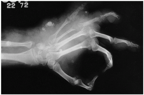
Roentgenographically, one notes multiple, cyst-like areas of bone destruction (
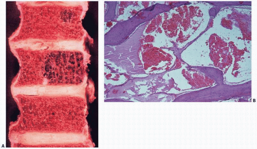
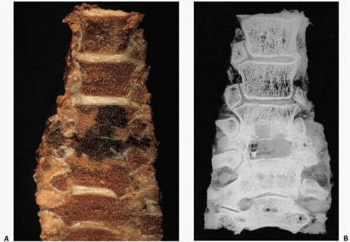
Fig. 11.10). There may be a fine peripheral rim of increased density. Isolated lesions may show the coarsened bone trabeculae of a solitary osseous hemangioma. Unlike the contour of bone involved by Paget disease, the contour of the exterior cortical bone is usually maintained in this condition, although vertebral collapse may occur (Fig. 11.11). In rare cases, a blastic appearance predominates, mimicking metastatic cancer (Fig. 11.12). Closer scrutiny, however, reveals central lucencies surrounded by dense, sclerotic bone. The lesions usually expand within the confines of the bone, but may involve the cortex, rarely breaking through the cortical shell.

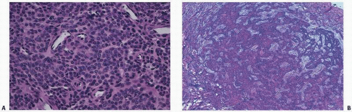
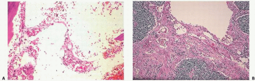
Laboratory findings may be unremarkable, although increases in alkaline phosphatase activity have been noted. On gross examination, lesions are cystic and filled with a reddish fluid consistent with blood, or a lymphangiomatous fluid consistent with lymph origin. Often, there may be combinations of both lesions. Support for a lymph origin is noted in many cases that present with chylothorax or chylopericardium, and in many cases with a thymic mass seen in communication with lymph channels. Lymphangiograms may also show dramatic findings. Microscopically, the lesions consist of thin-walled cavities lined by flattened endothelial cells separated by collagen septa, filled either with blood in the case of hemangiomas or a proteinaceous eosinophilic material. These lesions are usually surrounded by normal or thickened lamellar bone trabeculae.

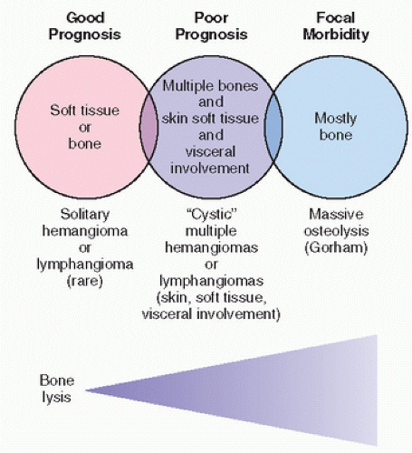
The prognosis in this disorder is variable, although usually it is self-limiting in its course. With extraskeletal involvement, however, the prognosis may be poor. The lesion is not hereditary, and most likely represents a maldeveloped vascular or lymphatic system.


FIGURE 11.11. Hemangiomatosis of spine. (A) Gross appearance, with both partial and nearly complete replacement of bone in several vertebrae. One vertebral body is completely collapsed. (B) The collapsed vertebrae and other coarsened trabeculae are noted in this specimen roentgenograph.


Gorham Syndrome (Massive Osteolysis; Disappearing Bone Disease; Phantom Bone Disease)

Massive osteolysis was first described in 1838 by Jackson (67), who documented a young man who had severe loss of bone of the upper extremity but lived to the age of 70 years, dying of unrelated causes. Numerous names have been used in the literature to describe this condition. In 1954, Gorham introduced the term massive osteolysis, and others have used the terms “disappearing” or “vanishing” bone disease and “phantom” bone (54). In 1955, Gorham and Stout (53) reviewed 24 cases, describing two additional cases, and concluded that the massive osteolysis was caused by a proliferation of vascular tissue, either vascular proliferation of blood or lymphatic tissue. Little has been added to the understanding or histologic assessment of this condition since that time. However, the identification of active osteoclastic resorption of bone and osteolysis in association with the proliferating vessels is now well documented. As reviewed by Choma et al. (48), Gorham syndrome is best described as a clinical, roentgenographic, and histologic entity characterized by a nonfamilial proliferating vascular condition, most often originating in bone and causing lysis of all or part of the bone. The disease is characteristic in that it usually begins in one bone and spreads in a direct fashion to involve adjacent skeletal tissue. It may affect soft tissue by extension or be part of a marked, most likely developmental, abnormality of the lymphatic system. Identification of abnormal proliferating lymphatic tissue in visceral organs such as the liver, spleen, and lymph nodes supports this concept. Osseous involvement in reported cases shows a predilection for the mandible, ribs, scapula, humerus, and femur (
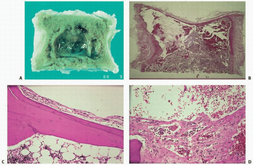
Fig. 11.13). Grossly, the bone is replaced by cyst-like vascular lesions (Figs. 11.14 and 11.15).

Clinical presentation varies from the incidental finding of regional osteolysis to massive bone loss. Severe physical deformities, disabilities, and life-threatening complications may occur (68). The onset is insidious, with dull pain in the affected region. Often, there is a history of preceding minor trauma or fracture in the remote past, but otherwise, the history is usually unremarkable. Localized pain as the chief complaint is frequent; however, a pathologic fracture or even deformity of the involved area with osteolysis may be found at initial presentation.

The age distribution of clinical presentation is varied. Presentation usually occurs before age 30 years, although the disease has been described in patients as young as 1 month to as old as 83 years. Gorham disease affects no particular sex or race, and does not appear to be associated with other disease entities or any pattern of genetic transmission. Bone lesions in Gorham disease appear to be equally distributed between the axial and appendicular skeleton; however, osteolysis has been described more frequently in the maxilla, mandible, clavicle, ribs, cervical vertebrae, pelvis, and femur. Osteolysis can be unifocal, with a single osteolytic focus affecting one bone area, or multifocal, with lesions occurring in more than one site simultaneously, particularly in a contiguous fashion.

The behavior of the osteolytic lesion is varied, ranging from slow, asymptomatic progression with spontaneous arrest after a number of years (69) to an extremely rapid and aggressive bone resorption that causes disability and even death when vital organs are involved.


FIGURE 11.12. Skeletal hemangiomatosis. Multiple osteolytic lesions appear osteoblastic because new bone has been produced encircling the osteolytic hemangiomas, mimicking osteoblastic metastatic cancer. Roentgenograph and computed tomography (CT) of pelvis (A, B). In gross spine (C), new bone appears solid and marble-like. Hemangiomas are cystic and sclerotic in corresponding roentgenograph (D).


FIGURE 11.13. Skeletal distribution in Gorham syndrome.

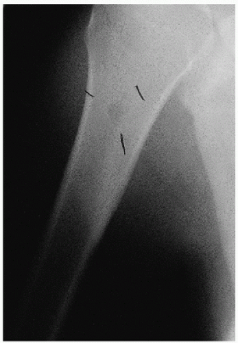
Radiographic changes occurring in massive osteolysis include initial intramedullary and subcortical radiolucent foci described as a “nondescript patchy osteoporosis” (57). Concentric shrinkage is characteristically seen in tubular bones, with tapering of the involved ends. Complete resorption of the involved bone follows, barring spontaneous arrest (Fig. 11.16). The osteolytic process may progress from intraosseous to extraosseous, with involvement of contiguous bones. Torg and Steel (59) illustrated the sequential radiographic changes occurring during a 10-year period in the hip of an 11-year-old with massive osteolysis. A diffuse lytic lesion within the confines of normal bone architecture was followed by increasing bone loss and deformity spreading to adjacent structures and across joints, and concentric shrinkage of involved long bone.

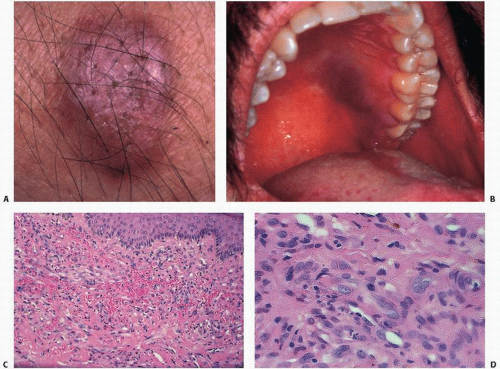
Arteriography, venography, and lymphangiography most commonly give only indirect evidence of Gorham disease and are not considered useful. Results of technetium pyrophosphate bone scans are routinely negative, or they demonstrate decreased uptake, confirming the absence of mineralization activity, or new bone formation or osteoblastic activity histopathologically. MRI has revealed a high signal intensity on T2-weighted images. There is no useful laboratory test to diagnose the disorder. The histopathology of Gorham disease is characterized by nonneoplastic, thin-walled lymphatic or blood vessels. There is usually little inflammation. One may observe mononucleated clear cells and multinucleated osteoclast-like cells.

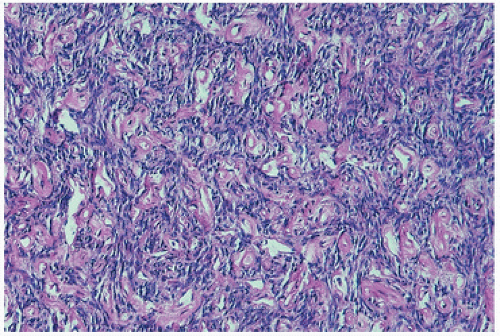
The replacement of the bone by proliferating vascular tissue is often seen in association with fibrous connective tissue. The vascular tissue varies from proliferating small capillary-type vascular tissue to that of proliferating or large hamartoma-like lymphatic- and/or endothelial-lined vascular channels (Fig. 11.14). Although the tissue is always histologically benign, the proliferation of vascular channels permeates the bone, cortex, and soft tissue in an aggressive fashion, similar to the uncontained proliferation of synovium in rheumatoid arthritis. There is usually little inflammation, although transient inflammation occurs, and fibrosis has been described in long-standing lesions. There are now several well-documented cases showing active mononuclear cells or phagocytic cells or even osteoclast-like cells resorbing bone at the membrane margin of the proliferating vascular channels (Fig. 11.15).

The prognosis in Gorham syndrome varies considerably. There is usually minimal disability, and the disease usually spontaneously regresses. However, with involvement of vital structures, such as the lung, death may ensue. Choma et al. (48) reported that 38 percent of patients with spinal and/or chest wall lesions died as a result of their disease, usually from extensive chylothorax. The development of chylothorax is particularly foreboding in that it may be very difficult to control the production of chyle.

Numerous therapeutic modalities have been described, including radiotherapy, bone grafts, prosthesis implantation, and (for chylothorax) irradiation and pleural adhesion therapies with minocycline and bleomycin (70). Although bone grafts may be resorbed in this condition, success with bone engraftment as well as prosthesis implantation has been reported. The condition often

spontaneously remits, and improvement and regrowth of bone may be seen, and in very rare instances, complete reconstitution of the bone.


FIGURE 11.14 Gorham Syndrome. (A) A vertebral body with almost complete replacement by a well-circumscribed cystic cavity (B) is histopathologically walled by vascular channels. (C) Lymphatic-type endothelial channels may be seen and may be membranous and flat (D) or proliferative and sponge-like.


FIGURE 11.15. (A) Membrane-associated resorption may be noted at low power by crenated bone surfaces. (B) At higher power, membrane-associated osteoclast resorption can be seen burrowing through bone.


FIGURE 11.16. Gorham syndrome. (A) Roentgenograph of lumbar spine shows complete disappearance of an L-4 pedicle. (B) Severe Gorham syndrome involving the femur and pelvis.

Radiotherapy has been used with reputed success for massive osteolysis of the chest with hemothorax (71). Radiation doses of less than 20 Gy are generally ineffective. Arrest of osteolysis has been reported with doses of 25 to 60 Gy.

Numerous etiologies have been suggested for Gorham disease, including posttraumatic hyperemia, similar to mechanisms suggested for bone resorption and reflex sympathetic dystrophy. The proliferation of fibrovascular tissue uncontrollably resorbing bone is similar to that seen in rheumatoid synovium. More than likely, the production and proliferation of vascular tissue are somehow directly linked to osteoclast activation or other bone-resorbing factors. Several mechanisms may be at work in this association. Endothelial cells may directly mediate bone resorption or modulate the extracellular matrix by producing enzymes that can degrade the matrix (72).

Because the regulation of cell growth most likely involves the interaction between growth factors and matrix proteins, the former (such as transforming growth factors) having great influence on cells of vascular lining tissue, and the latter (such as collagen I, III, IV, and V, elastin, and osteopontin) being abundant in the extracellular compartment of blood vessels, the potential for vascular lesions to remodel bone is great (72).

It has also been postulated that mesenchymal cells, which accompany blood vessels, may act as progenitor cells for lineage 2 osteoclasts. It has been observed that circulating monocytes in tissue culture, for example, are capable of inducing bone resorption.

Because no abnormality of the parathyroid glands is discernible in this condition, nor any systemic evidence of osteoclast-induced hypercalcemia via a parathormone mechanism, osteolysis appears to be caused by local factors and most probably is directly linked to the proliferating vascular channels.

A plausible explanation for the osteolysis in Gorham disease is that the proliferation of lymphatic endothelial cells (LECs) and blood endothelial cells (BECs) is stimulated by increased levels of vasoactive endothelial growth factors C and D (VEGF-C and VEGF-D) and macrophages-derived VEGF-A. In addition to the VEGFs, macrophages (progenitors of osteoclasts) can produce interleukin-6, all stimulators of osteoclast differentiation and all shown to be increased in the serum in different phases of the diseases (68). In fact, the endothelium of lymphatic vessels has been shown to harbor toll-like receptors that can upregulate osteoclast activation via tumor necrosis factor alpha and interleukin-6 (73). In addition, osteoblast progenitor cells may be inhibited by LECs and factors produced by osteocytes like sclerostin, Dickkopf- and soluble frizzled-related proteins.


The Lymphatic “Chylous” Variant of Gorham Disease

Gorham disease may also produce an indolent lymphatic proliferation that, over time, creates excessive chylous fluid (74).

Documented abnormalities of the lymphatic system including lymphangiomatous masses, lymphangiectatic dilations (small bowel, spleen, pancreas, thymus, and bones), and absence of a
portion of the thoracic duct have been shown. These findings are important because therapeutic attempts focusing on identification and ligation of the duct may be futile, as has been reported. Podevin et al. (75) recognized this futility by using the phrase “thoracic lymphatic dysplasia not further defined.”

Dilated lymphatic channels (lymphangiectasia) or accumulation of lymphangiomatous tissues in the pleural and peritoneal tissue, the diaphragm, mediastinum, and spleen correspond to known anatomic tracking of the thoracic duct.

There were several common findings during autopsies of five patients with Gorham disease (Table 11.4) (54,55,56,74,75,76,77). All had abnormal vascular proliferation that involved bone sites, some described as lymphatic and some admixed with fibrosis. Sites of involvement include pleural tissue, spleen, nodes, and vertebrae, as in our case. There is a predilection for osseous involvement of the clavicle, scapula, ribs, and vertebrae, as revealed by autopsy or premortem imaging. Numerous patients died from chylous effusions and pneumonia.

The lymphatic variant of Gorham disease may be a distinct variant of the disease. Some have suggested that Gorham is a generalized disorder of lymphangiogenesis and should be distinguished from cases that are a generalized lymphatic abnormality involving bone (78). Approximately 17 percent of patients with Gorham disease have chylothoraces develop, and approximately one-half die. Gorham disease generally affects younger patients and seems to have no racial or gender predilection, and most osseous involvement occurs in the shoulder region and vertebrae. In our patients and in reported cases, chylous effusions were frustrating to treat, with a poor prognosis.

A distinct variant of Gorham disease has been proposed on the basis of autopsy findings (Table 11.5). A maldeveloped (or totally absent) thoracic duct may lead to the development of an aberrant collateral lymphatic circulation including dilated lymphatic channels (lymphangiectasia) and lymph-rich soft tissue masses (lymphangiomas). Collateral lymphatic circulation develops and extends to the nascent intraosseous lymphatic system. This bone lymphatic tissue contains activated endothelial lymphatic cells leading to increased activity of osteoclasts, which lead to marked osteolysis.

Although there is no consensus regarding treatment of Gorham disease, therapeutic attempts must focus on the morbid and deadly chylous effusions. Surgical interventions in patients with Gorham disease complicated by chylous effusions have been mostly unsuccessful including pleurectomy, pleurodesis, thoracentesis, and thoracic duct embolization and ligation (76).

Thoracic duct ligation does not seem to be anatomically sound in all cases. The lymphatic dysplasia must be localized by imaging studies and addressed regionally. Although this is a challenging and elusive goal, patients with regionalized lymphatic dysplasia can be treated by an aggressive therapy such as radiotherapy or specific excision of the lesion. Osteoclastic resorption needs to be addressed in this condition, and may best be blocked by blunting local tissue factors or by using antiosteoclastic agents such as new-generation bisphosphonates.

Applying agents that interfere with osteoclast stimulation has been attempted in this condition. Agents previously utilized for antiosteoclastic activity include bisphosphonates, interferon alpha 2b, anti-VEGF-A antibody (Bevacizumab), propranolol, low molecular weight heparin, steroids, vitamin D, and calcitonin (68). Since the etiology of Gorham remains enigmatic, therapy is still frustrating.

Vascular tumors of bone are rare and vary from well-differentiated and locally aggressive tumors (epithelioid hemangiomas, previously called low-grade angiosarcomas) to highly anaplastic, poorly differentiated metastasizing neoplasms (angiosarcomas). Nomenclature is confusing, reflecting current debates over prognostic significance. Surgeons are often frustrated by the inability of pathologists to provide better guidance in the natural history of these lesions.

For those interested in the passion that nomenclature bias can invite, the letters published in the American Journal of Surgical Pathology regarding terminology of these lesions are worth reading (79,80,81,82). In general, there is a gradient of aggressive behavior with increasing pleomorphism and atypical mitotic activity, but lesions, especially multicentric lesions, may be hard to predict (83).

Peculiar features of vascular tumors of bone include concomitant lesions of soft tissue and multicentricity. The often-observed multifocality of these lesions remains enigmatic with regard to prognosis and therapy. Multiple primary tumors may be mistakenly confused with metastatic disease. The phenomenon of multicentricity in these lesions, often with both visceral and bone involvement, suggests corollaries with the KS phenomenon.

Concomitant histologic foci of solid spindle cell regions can be confused with other spindle cell lesions.


Intermediate and Malignant Tumors


Epithelioid Hemangioma of Bone (Histiocytoid Hemangioma)

Epithelioid hemangiomas are benign vascular tumors that can occur in bone, but more commonly involve soft tissue and skin. The older term histiocytoid hemangioma has given way to the term epithelioid hemangioma, thought to better reflect the cell of origin. Combined osseous and extraosseous lesions and multicentricity are documented. Indolent lesions once reported and regarded as low-grade epithelioid hemangioendotheliomas or low-grade epithelioid angiosarcomas may have been epithelioid hemangiomas.

The name of the tumor derives from the microscopic appearance of the predominant cell type: plump, often cuboidal cells with eosinophilic cytoplasm mimicking epithelial cells. In actuality, the cells mark for a battery of epithelial (epithelial membrane antigen [EMA]) and endothelial (factor VIII and the Ulex europaeus lectin) cells, justifying the term epithelioid hemangioma (84).

Radiographically, discrete radiolucency with or without sclerotic margins is characteristic.

Grossly, tumors are soft, lobular, hemorrhagic, and well circumscribed; there may be a shell of reactive subperiosteal bone.

Microscopically, the tumor is characterized by anastomosing cords of vascular channels lined by plump, eosinophilic, epithelial-like endothelial cells. Solid sheets of these polygonal cells may be seen obliterating identification of vascular lumina. The cells may appear to hobnail or fall off into the vascular spaces in a “tombstone” manner (84). The nuclei are large with vesicular nuclei and occasional nucleoli. Intracytoplasmic vacuoles are seen and even signet cell appearances. Extravasated red blood cells and scattered inflammatory cells are typical.

Immunohistochemical staining is characteristically positive for Factor VIII, CD31, CD34, FLI1, ERG, and may be positive for keratins (AE1/AE3. CAM52 [CK8/CK18] CK7 and CK19) and EMA.

Clinically, patients are in their middle years (ranging from 20s through 70s) and present with localized pain.

Treatment in the past has varied, but if properly classified, few patients with this lesion can be expected to do poorly.


TABLE 11.4 Autopsy Reports of Patients with Gorham Disease


Study


Age (years)/Gender


Bones Involved


Thoracic Duct


Vertebrae Involved


Histology


Osteoclasts


Cause of Death


Gorham et al.


16/male


Scapula, clavicle, vertebrae


Compressed, lost in inflamed tissue


Cervical and upper thoracic


Extreme vascularity with large thin-walled vessels


Present


Chylous effusions


Hambach et al.


16/male


Scapula, clavicle, humerus, vertebrae


No comment


Cervical


Dilated cavernous vessels


Not mentioned


Bronchopneumonia


Halliday et al.


27/male


Rib


No comment


Not mentioned


Thin-walled vessels


Not mentioned


Not mentioned


Fujiu et al.


15/male


Scapula, clavicle, ribs, humerus


Ligated in thorax, ineffective in stopping chylous effusions


Not mentioned


Vascular proliferation with lymphatic vessels


Present but not increased


Chylous effusions


Kawasaki et al.


44/male


Occipital bone, vertebrae


No comment


Cervical


Dilated vessels and fibrous tissue


Increased


Suicide


Vigorita et al.


35/male


Scapula, humerus, vertebrae, pelvis


Not identifiable


Cervical and lumbar


Dilated lymphatic vessels


Increased


Chylous effusions and pneumonia


Reprinted from Vigorita VJ, Magitsky S, Bryk E. Gorham’s disease. An autopsy report. Clin Orthop Rel Res. 2006;451:267-273.with permission. Autopsy reports are from references (54,55,56, 76, 78, and 79.)


TABLE 11.5 Gorham Syndrome, Lymphatic Variant Correlation of Proposed Pathogenesis with Autopsy Findings


Proposed Pathogenesis


Corroborative Autopsy Finding


Maldeveloped thoracic duct


Thoracic duct not identifiable at terminal end


Development of aberrant collateral circulation


Dysplastic lymph tissue (lymphangiectasis) in pleura, peritoneal cavities, small bowel, spleen, pancreas, and lymphangiomatous involvement in mediastinum


Compensatory development of nascent osseous lymphatic system


Dilated lymphatic channels in the vertebra


Activation of endothelial cells of intraosseous lymphatic channels to modulate bone remodeling


Proximity of vertebral osseous lymphatic channels to bone remodeling sites


Activation of local tissue such as interleukin-6


Increased osteoclast activity


Increased osteoclast activity


Marked osteolysis


Marked osteolysis of involved vertebra


In view of the rarity of epithelioid hemangiomas, it is problematic to be dogmatic about treatment. However, in one large report of 50 cases in which 36 were available for follow-up, treatment with curettage or marginal en bloc resection reported excellent results (85). Four patients recurred. There were no metastases.

Although surgical resection, curettage, or even radiation has been used, clinical and radiographic spontaneous remission has been observed, complicating a dogmatic approach to the treatment of this lesion (86).


Epithelioid Hemangioendothelioma (Myxoid Angioblastoma; Angioglomoid Tumor)

Epithelioid hemangioendotheliomas are considered borderline vascular tumors, the behavior of which is sometimes unpredictable (87,88). They have been reported throughout life (ages 7 to 76 years; mean, 30 years), particularly in the axial skeleton and lower extremities. Lesions are multifocal in approximately 50 percent of cases, with involvement especially of the lower extremity. Multifocal cases may be confined to one limb or contiguous bones, but tend to occur in the same geographic region.

Clinically, pain, swelling, and pathologic fracture are most frequently encountered.

Radiographically, tumors are usually osteolytic with or without bone expansion (Fig. 11.17). Occasional coarse trabeculation, peripheral sclerosis, or periosteal reaction occurs but is not characteristic. The most characteristic radiographic findings are small, oval, well-described cortical-based lucencies.

MRI studies have given variable results, but generally show an increased signal on T2-weighted images and intermediate T1.

The tumor is composed microscopically of anastomosing cords of often poorly defined vascular channels lined by plump endothelial cells having abundant, granular, eosinophilic, vacuolated cytoplasm. These lesions are often characterized by an abundance of vacuolated cells, so-called blister cells. Frank pleomorphism and significant mitotic activity are generally lacking, but are more commonly seen than in epithelioid hemangioma. Vascular channels are far more primitive. Solid foci or sheets of cells may be seen. The epithelioid appearance of the endothelial cells may lead to misdiagnosis as metastatic carcinoma. The stroma in EH may be very hyalinized.

Ultrastructure has revealed basal lamina with cell cytoplasmic processes interconnected by tight junctional complexes (88).

Peroxidase-antiperoxidase staining supports an endothelial origin, with positivity for factor VIII, CD31, CD34, the U. europaeus lectin, and vimentin (89). Immunohistochemical stains for
FLI1, ERG, and D240 are also positive. Keratin and EMA stains are less often positive.


FIGURE 11.17. Multicentric hemangioendothelioma with well-circumscribed, multifocal osteolytic lesions.

Sixty to 100 percent of patients with epithelioid hemangioendothelioma have a t(1;3) translocation (90) that appears to be unique to and specific to this tumor. Candidate genes thought to be transcriptional activators in oncogenesis include WWTR1 and CAMTA1. CAMTA1 belongs to the family of calmodulin-binding transcription activators and is a well-known tumor-suppressor gene lost in a subset of gliomas and neuroblastomas.

Although an indolent course is possible even in multicentric cases, aggressive behavior is possible; involvement of visceral organs carries a worse prognosis. Patients may die within 1 year of widespread pulmonary and skeletal metastases, but do so far less often than in high-grade angiosarcomas.

With surgery, there is a potential for intraoperative blood loss. Recurrences have been estimated at 13 percent (88), and metastases in primary soft tissue (not bone) hemangioendothelioma at 31 percent. Whereas surgery for a lesion with substantial involvement of a vertebra with imminent collapse may be warranted, a patient without compromise of spinal instability may benefit from radiation. Nonetheless, optimal treatment of this lesion continues to be debated and, therefore, needs to be individualized. Bisphosphonate therapy and antiangiogenic agents such as the VEGF inhibitor bevacizumab have been used.


Hemangiopericytoma

Hemangiopericytoma is a rare, low-grade vascular tumor of pericytes (91). The pericyte (peri, “around”; cyte, “cell”) is a cell of mesenchymal origin that contains branching cytoplasmic processes that encircle capillaries and postcapillary microvessels. Normal pericytes are contractile cells that may regulate blood flow. They share a common basement membrane or basal lamina with endothelial cells.


FIGURE 11.18. Low-grade hemangiopericytoma. The tumor cells are within the walls of the blood vessels.

Hemangiopericytomas are characterized microscopically by anastomosing vascular channels lined by bland, flattened endothelial cells and compressed by the intervascular stroma containing pericytes (92) (Fig. 11.18). The key to the diagnosis is the recognition that the neoplastic cells are not the endothelial lining cells but the intervascular proliferating pericyte. Reticulin stains, which show reticulin fibers enveloping the individual cells, have been used to identify a characteristic pattern.

There is a roughly 2:1 male predilection with a wide age range (12 to 90 years) peaking in the fourth and fifth decades.

Clinical symptoms are nonspecific, but lesions may be painful.

The most characteristic findings on roentgenograms are lytic destruction, cortical breakthrough, and soft tissue extension. Lesions may be poorly defined or well circumscribed.

On MRI, there is an intermediate signal on T1 hyperintense signal on gadolinium enhancement, and high intensity on T2 (93).

The most common locations are the pelvis and lower extremities and, in particular, the sacroiliac region, femora, and vertebrae.

Tang et al. (94) have recently proposed prognosis based on a histologic system that includes cellularity, cytologic features, and mitotic counts.

Prognosis is variable, depending on grade.

Surgical removal, when feasible, is the treatment of choice. Late recurrences and distant metastases are the rule, especially, as expected, in larger, axial, histologically high-grade tumors.


High-grade Malignancies


Angiosarcoma (Hemangioendothelial Sarcoma of Bone)

Angiosarcomas are fully malignant, metastasizing neoplasms of endothelial cells with no intratumoral cartilage or bone formation (95). They are characterized by rapid growth and extensive bone destruction with cortical erosion and soft tissue extension. The tumor consists grossly of soft reddish tissue with irregular borders and occurs at a mean age of 62 years (96).

Microscopically, irregular anastomosing vascular channels with prominent intravascular budding and striking cellular anaplasia with frequent mitoses are noted (Fig. 11.19). Foci of necrosis
as well as solid undifferentiated areas are common, the latter suggesting a poorly differentiated carcinoma or anaplastic lymphoma. Growth in solid sheets obliterating vascular lumina is characteristic.

Only gold members can continue reading. Log In or Register to continue

Stay updated, free articles. Join our Telegram channel

Jul 24, 2016 | Posted by in PATHOLOGY & LABORATORY MEDICINE | Comments Off on Vascular and Other Mesenchymal Tumors and Lesions

Full access? Get Clinical Tree

Get Clinical Tree app for offline access