Synovium


Synovium


Normal Synovium: Microanatomy and Function

The major function of the synovium is to provide joint tissue with lubrication and nutrient oxygen and proteins. Its complex structure leads to central roles in mediating the inflammatory response to injury and disease.

The synovium forms when a primitive mesenchymal tissue cavitates, forming a recognizable joint space at approximately 8 weeks of embryonic life (Fig. 15.1). Mature synovium appears pale pink in color and architecturally covers all the surfaces of the joint space, excluding the articular cartilage and most fibrocartilaginous structures (Fig. 15.2). Only in abnormal conditions does the synovium encroach on the surface of articular cartilage, a change classically seen in the reddish “pannus” or inflammatory synovial invasion of the articular cartilage in rheumatoid arthritis.

The synovial membrane, the most superficial layer of the synovium, lines the joint and forms the linings of tendon sheaths. Parietal and visceral synovium mimic their mesothelial-like counterparts in the thoracic and pericardial cavities (Fig. 15.3). The extensive synovial-like lining cells of tendon sheaths and ligaments explain a host of reactive synovitis and painful clinical tenosynovitis, bursitis, and enthesopathy syndromes (Table 15.1). The synovium is part of the biomechanically complex “joint” that is composed of articular cartilage coating the opposing ends of bone, the ligaments that hold the musculoskeletal components in check, and a fibrous capsule that defines and delimits the joint space from surrounding tissue. Joint capsule fibrous connective tissue is thought to add to the joint’s mechanical strength. The term joint capsule refers to the fibrofatty neurovascular tissue that envelops the nonarticular cartilaginous tissue of the joint space (1).

In pathologic states, synovial-like anatomic structures, not related to a joint space (“synovial metaplasia”), are known to form in a variety of circumstances, including (a) postsurgical states; (b) failed prostheses, including breast implants (2); (c) mechanical damage to connective tissue; and (d) experimental settings (injected air or oil in subcutaneous soft tissue) (3). These structures histologically resemble normal synovium and often have demonstrably similar secretory and phagocytic function.

Normally, the synovium appears smooth and transparent, but it turns thick, dull, and opaque with pathologic change. With hemorrhage, it becomes obviously bloody, but in chronic hemarthrosis it turns a reddish or rusty brown (Fig. 15.4) owing to hemosiderotic deposition and the release of iron from red blood cells. In cases of severe bleeding, a dark purple or rusty color may be noted. The appearance of reddish purple or rusty synovium indicates a hemangioma or bleeding and may be seen in trauma; bleeding disorders such as hemophilia, von Willebrand disease; and pigmented villonodular synovitis (PVNS). In ochronosis (alkaptonuria), the synovium may appear a dull gray, whereas fibrocartilage and articular cartilage are discolored black. Darkening or blackening also may be seen when there is extensive release of metallic debris. White foci in the synovium usually indicate gout (urate deposition), pseudogout (calcium pyrophosphate dihydrate [CPPD] deposition), or soft tissue calcifications (deposits caused by trauma or calcinosis syndromes). Cement debris also may lead to pallor. A yellow color
ensues with xanthoma cell accumulation, which may be a predominant feature of PVNS or xanthomatous disease.


FIGURE 15.1. Embryonic joint showing evolution of primitive mesenchyme (A) into a joint space (B) at about 8 weeks of intrauterine life.


FIGURE 15.2. Location and microanatomy of the synovium in the knee joint with relationship to articular cartilage, bone, fibrocartilage (meniscus), and soft tissues.


FIGURE 15.3. Location and microanatomy of the tenosynovial lining of a typical tendon of the finger.


Microanatomic Structure

The synovium consists of a thin layer of synovial cells or synoviocytes, the intimal layer, above a richly fibrovascular zone, the subintimal layer, which contains arterioles, fat, and other connective tissue cells, such as fibroblasts, histiocytes, and occasionally mast cells (Fig. 15.5). The intimal layer of lining cells is usually one to two cells thick with no discernible differences on light microscopy. The loose connective tissue subintimal zone layer becomes gradually more fibrous at capsular insertions.

The villous appearance of synovium is not abnormal, but rather nonspecific and may be seen in a broad range of conditions (Fig. 15.6). In general, traumatic synovitis and degenerative joint disease (DJD) (osteoarthritis) are attended by edematous change and mild villous hypertrophy. Inflammatory arthritis (classically rheumatoid arthritis) shows a dramatically reddish hyperplastic synovium with fibrinous exudation characterized by abundant tan fibrinous loose bodies, called rice bodies, and marked lymphoplasmacytic synovitis. In septic arthritis, leukocytes are seen in tissue.
Neuropathic joints and rapidly destructive joint processes are characterized by bone and cartilage debris.


TABLE 15.1 Etiologic Factors in the Development of Mechanically Induced Synovitis


Training environment


Poor preconditioning


Poor technique


Inappropriate equipment


Training errors


Personal characteristics of athlete


Leg-length discrepancy


Scoliosis


Pes planus; pes cavus


Malalignment


Excess weight


Muscle imbalance


Sport


Running (feet, ankles, knees, hips)


Swimming (shoulders, neck)


Tennis (shoulders, elbows)


Rowing (spine, ischial region, sacroiliac joints)


Modified after Moran ML, Arnold WJ. Mechanically-induced synovitis. In: Sports-Induced Inflammation. Rosemont, IL: American Academy of Orthopaedic Surgeons; 1989:331-336.


FIGURE 15.4. Gross appearance of the synovium in pathologic states, including bleeding (A), and reactions to foreign implant material such as cement and polyethylene (B) and metal (C). Bleeding induces a rusty brown appearance. Implant materials impart whitening, except metal, which induces darkening or blackening. Crystals such as gout and calcium pyrophosphate dihydrate (CPPD) can induce whitening (D).


Ultrastructure

The intimal zone consists of an admixture of cell types often conveniently classified as cells exhibiting macrophage function (synovial A cells) and cells more synthesizing in function (synovial B cells) (Fig. 15.7). Electron microscopy studies and immunophenotyping studies further characterized these cells. Ultrastructural studies show abundant mitochondria, Golgi apparatus, vacuoles, lysosomes, phagosomes, vesicles, and surface undulations (characteristics suited to macrophage activity) in type A cells and rough endoplasmic reticulum, free ribosomes, and smoother cytoplasmic profiles (characteristics suited to synthetic activity) in type B. As might be expected,


synoviocytes may be “intermediate” in nature, featuring organelle functions of types A and B. Some evidence supports the existence of other cells, such as antigen-type cells (HLA-DR, IA-like).


FIGURE 15.5. Composite histologic appearance of normal and hyperplastic synovium. (A, B) Normal synovium consists of a fine single or double layer of type A and type B synovial lining “intimal cells” on top of a subintimal zone of vascular and fatty tissue, fibroblasts, and rare histiocytes and mast cells. There is increasing collagen in the vicinity of dense connective tissue structures (B, higher power). (C-H) Hyperplastic synovium showing moderate hypertrophy of cells confined to the surface (C) with increasing hyperplasia (D), (Continued)


FIGURE 15.5. (Continued) hyperplastic cells migrating into the subintimal zone (E) with marked hypertrophy (F). Villous hypertrophy with giant cells (G) (H, higher power) of the Grimley-Sokoloff giant cell type in rheumatoid arthritis. (I) Mucinous hyperplasia illustrating secretory potential of synoviocytes. (J) Superficial zone foreign body giant cell reaction to failed prosthesis. (K) The zonal architecture may be distinct, as in this rheumatoid hyperplasia with both intimal and subintimal zonal change.


FIGURE 15.6. Synovium in degenerative joint disease (A), rheumatoid arthritis (B), traumatic synovitis (C), septic arthritis (D), and neuropathic joint (E). Degenerative joint disease is characterized by mild villous hyperplasia, rheumatoid arthritis by nodular lymphoplasmacytic inflammation, trauma by fibrinous change, septic arthritis by leukocytosis, and neuropathic joints by abundant bone and cartilage detritus.


FIGURE 15.7. Ultrastructure of synovial intimal cells. Type A cells (left) show prominent Golgi, vesicles, vacuoles, lysosomes, and mitochondria. Type B cells (right) show abundant rough endoplasmic reticulum. There are no basal lamina separating lining cells from subintimal connective tissue and unless inflamed, rarely cell junctions.

The use of special immunologic techniques, including monoclonal antibodies, against a range of antigens can confirm that synovial cells consist of many different cell types. Three distinct populations can be defined immunologically.

Arbitrarily designated type 1 (synovial “A” cells) is a group of cells that seem to be related to mononuclear phagocytes based on their expression of antigen and derivation from cells of the monocyte macrophage cell lineage (4). These cells exhibit phagocytosis and contain macrophage markers, abundant HLA-DR IA antigen, and Fc receptors. These cells constitute approximately one-third of the synovial cell population.

A second cell population, type 2, is characterized by nonphagocytic activity and a strong expression of IA antigen with the absence of IgG Fc receptors and antigens associated with the monocyte lineage, D and T lymphocytes, and fibroblasts. Type 2 cells include the IA antigen-positive dendritic cells. In rheumatoid arthritis, a considerable portion of the cells is type 2 cells.

A third cell population, type 3, is characterized by a nonlymphocytic population with characteristics typical of fibroblasts. These are the cells that usually predominate in tissue culture. They lack phagocytic activity and do not express monocyte antigens or IA antigens.

Cell types as defined by immunologic studies are consistent with previous morphologic observations. Type 1 cells expressing the monocyte macrophage antigens are similar to the cells historically described as characterized by phagocytosis type A and type C cells. Type 1 cells are most likely mononuclear phagocytes of bone marrow origin as ascertained by their antigenic marking with monocyte and IA antigen. Modifications in antigen expression suggest that they act more like tissue macrophages in the joint. The historically designated “type B” synovial cell that produces glycosaminoglycans is most likely of the type 3 cell variety, probably mesenchymal in origin. These cells lack IA antigens and most of the differentiated antigens of monocyte macrophage lineage (5).

The origin of normal synovial lining cells is controversial, but a dual-cell origin remains plausible with bone marrow derivation for the histiocytoid or type A cells, and local mesenchymal tissue for fibroblast or type B cells. Many clinicians believe, however, the heterogeneity of synovial cells is more an expression of functional activity resulting from various factors, and the cell types are possibly interconvertible.

Although the synovial cells lack desmosomes or tight junctions, characteristic of epithelial tissue, the complexity of this cell structure is evident in the changes seen in various pathologic states. Hyperplasia may be limited to a mild increase in intimal cell number, or there may be dramatic change, including large, bizarre cells, such as Grimley-Sokoloff giant cells or even striking mucin-producing cells. In this latter condition (mucinous hypertrophy of the synovium), the copious amount of material secreted testifies to the potential capacity of this membrane (Fig. 15.5I). Multipotential mesenchymal stem cells (MSCs) have been derived from the adult human synovial membrane and are inducible into chondrogenesis, myogenesis, osteogenesis, and adipogenesis (6).

Synovial MSCs, which are phenotypically similar to synovial type B cells, have been shown to be increased in number following minimal injuries to the knee and DJD (7). In addition, the synovium has been shown to be an excellent source of MSCs for cartilage regeneration. They may be a valuable source for chondrocytic regrowth in tissue-engineered menisci. In fact, gene expression profiles reveal that chondrogenic progenitor cells and synovial cells are closely related (8).

The ability of synovial MSCs to differentiate into chondrocytes, a chondroprogenitor potential greater than those originating from other tissues, is relevant in explaining the origins of synovial chondromatosis.


Functions of the Synovium

The functions of the synovium are best appreciated by understanding the characteristics of its cellular components and microarchitectural structure. The synovial A cells are suited to phagocytic (or macrophage) activity and ingest native or foreign material, such as hemosiderotic in chronic bleeding conditions (hemophilia) or iatrogenically introduced substances (gold in the treatment of rheumatoid arthritis). Most recently, synovial giant cell reactions to viscosupplementation products have been identified (vide infra). The phagocytic potential of the synovium is probably best illustrated by the marked foreign body giant cell and histiocytic reaction in some cases of loosened prostheses or in the resorption of bone and cartilage debris in rapidly destructive joint disease or neuropathic joints. Absorption of fluid material by synovial intimal cells is shown ultrastructurally by pinocytotic vesicles and vacuoles. Alternatively, the synovial B cell is suited to synthetic function and most characteristically secretes the hyaluronate protein of synovial fluid, hyaluronate contributing to the lubrication of joint structures. Type A and type B cells seem to have secretory and phagocytic potential, however. Other functions in conjunction with the vascular and lymphatic systems of the synovium include the regulation of movement of physiologically important proteins and electrolytes. The lack of a basal lamina, the presence of gaps between synovial cells, and the lack of a basement membrane in synovial blood vessels facilitate interchange between synovial fluid and blood vessels.


Synovial Fluid

Synovial fluid is a transudate or ultrafiltrate of plasma derived from synovial blood vessels supplemented by high molecular weight saccharide-rich molecules such as hyaluronans (9). This material gives synovial fluid its high viscosity. In addition, synovial fluid contains cells, mostly mononuclear phagocytes, and neutrophils. Synovial fluid content of glucose, uric acid, and lactate is similar to that in plasma, but there is less total protein. Albumin constitutes most of the total protein, whereas proteins of higher molecular weight, such as fibrinogen and other globulins, are relatively decreased compared with their plasma concentrations.

Arthrocentesis is the procedure whereby the synovial fluid is removed, a procedure that infrequently is complicated by infection. Arthrocentesis may be helpful in detecting crystal-induced synovitis, such as gout, CPPD disease, and hydroxyapatite (HA) crystal deposition disease. Blood is a nonspecific finding, which may be seen in a broad range of conditions, including trauma-induced hemarthrosis and PVNS, rheumatoid arthritis, and infection.

The proper collection and processing of synovial fluid is key to maximizing diagnostic information. Typically, normal synovial fluid has no clotting factors. With pathologies resulting in vascular leakage, high molecular weight clotting factors enter the joint, necessitating collection with anticoagulants. The anticoagulant lithium hepatin is preferred over EDTA since EDTA can sequester calcium ions, obscuring the diagnosis of hydroxyapatite and calcium pyrophosphate crystals and, therefore, DJD and CPPD. Viscosity can be measured by either the string test (dropping synovial fluid into a container from a pipette and assessing its stringiness, a sign of viscosity) or mixing the fluid with a 2 percent solution of acetic acid, which produces a white precipitate; the whiter the precipitate the more proteins and hyaluronans, and thus increased viscosity (9).

Heparinized tubes should be used for microbial cultures and Gram stain. For chemical analyses, observation of fresh synovial fluid in a clean test tube may be beneficial because normal synovial fluid does not clot (low fibrinogen concentration). Subsequent centrifugation can remove cells, fibrin, and other debris for further analysis for lactate, protein, uric acid, glucose, and other substances.


FIGURE 15.8. Synovial fluid composite. (A) Synovial fluid from patients with (from left to right) calcium pyrophosphate crystal deposition (cloudy), normal (clear amber), rheumatoid arthritis (cloudy yellow), and trauma-induced (blood stained). (B) Pigmented villonodular synovitis (PVNS), especially of the diffuse type, can give a bloody or rusty appearance. Septic arthritis yields a turbid yellowish-green.

The synovial fluid “wet prep” refers to the use of both thick and thin analyses of freshly removed fluid. In the “thick” prep technique, synovial fluid is removed and slowly returned to a container utilizing a pipette that will separate visible particles such as fibrin, cartilage, or crystals in the thin part of the pipette. Subsequently placed on a slide, the various components can be discerned against backgrounds that are both dark (for cartilage identification) and pale or white (to identify prosthetic debris). “Thin” preps drop fluid onto a slide that, when cover-slipped, will flatten cells for identification.

Synovial fluid analysis may be grouped for general classification of types of conditions producing synovial fluid (Fig. 15.8 and Table 15.2) (7). Noninflammatory conditions, such as DJD, trauma-induced arthritis, osteochondritis dissecans, and neuropathic arthropathies, or primary synovial metaplastic conditions, such as synovial chondromatosis, are characterized by a yellowish synovial fluid with high viscosity, low leukocyte counts, less than 10 mg/dL of glucose (i.e., below serum level), and low protein. Cultures are negative. This noninflammatory or class I type of synovial fluid group is distinct from the inflammatory or crystal-induced arthropathies, which are characterized by a synovial fluid with low viscosity, a yellow or whitish color, and a variable leukocyte count with a greater than 50 percent neutrophil population. Glucose usually is elevated greater than 25 mg/dL, with protein elevation greater than 3 g/dL.

Septic arthritis usually is characterized by a yellowish-green fluid of low viscosity with an elevated leukocyte count with greater than 75 percent neutrophils. Glucose is usually greater than 25 mg/dL with increased protein levels. A wide range of conditions can induce a hemorrhagic arthropathy, including idiopathic Hemosiderotic synovitis, trauma-induced arthritis, hemophilic arthropathy, and PVNS as a secondary change. These synovial disorders are characterized by a reddish brown color with decreased viscosity and a moderate elevation in white blood cell count with greater than

25 percent neutrophils. Glucose is in the normal range, usually 0 to 10 mg below serum level. Protein may be greater than 3 g/dL.


TABLE 15.2 Classification of Synovial Effusions


Gross Synovial Fluid


Gross Examination


Normal


Noninflammatory


Inflammatory


Septic


Hemorrhagic


Volume (mL) (knee)


<3.5


Often >3.5


Often >3.5


Often >3.5


Often >3.5


Viscosity


High


High


Low


Variable


Variable


Color


Colorless to straw


Straw to yellow


Yellow to white


Yellow/green


Rusty


Clarity


Transparent


Transparent


Cloudy


Opaque


Variable to rusty


WBC (mm3)


<200


200-2,000


2,000-75,000


Often 100,000


Variable


PMN leukocytes (%)


<25%


<25%


>50% often


>75%


Variable


Culture


Negative


Negative


Negative


Often positive


Negative


Mucin clot


Firm


Firm


Friable


Friable


Variable


Glucose (AM fasting)


Nearly equal to blood


Nearly equal to blood


<75 mg% than blood


<50 mg% than blood


Nearly equal to blood


Group I (Noninflammatory)


Group II (Inflammatory/crystal)


Group III (Septic)


Group IV (Hemorrhagic)


Osteoarthritis


Rheumatoid arthritis and variants


Bacterial, fungal (rare), and tuberculous infections


Hemorrhagic diatheses (e.g., Hemophilia) Trauma


Osteochondromatosis


Systemic lupus erythematosus


Anticoagulant therapy Sickle cell disease


Osteochondritis dissecans


Ankylosing spondylitis Lyme disease


PVNS Synovial hemangioma


Neuropathy


Gout Chondrocalcinosis (pseudogout)


Synovial sarcoma Joint Prostheses


Normal synovial fluid is usually less than 3.5 mL and clear or faintly yellow in color with clear transparency. It has high viscosity as a result of hyaluronic acid and does not clot because of low concentrations of fibrinogen and other clotting factors. In normal synovial fluid, there are fewer than 200 cells/µL, with a predominance of mononuclear phagocytes. There is a variable amount of lymphocytes, although lymphocytes and neutrophils increase dramatically in inflammatory arthropathies.

Eosinophilic synovitis has been described and may be associated with a mild-to-marked peripheral eosinophilia. Normal synovial fluid chemistries are typical. The differential diagnosis includes suppurative arthritis, tuberculosis and parasitic infections, allergies, and both degenerative and rheumatoid arthritis. The condition has been noted after arthrography and is usually accompanied by synovial fluid eosinophilia.

Crystal-induced arthropathy can be diagnosed with examination of synovial fluid using polarized light microscopy. Crystals other than gout (urate) and pseudogout (calcium pyrophosphate) may be seen. In chronic noninflammatory disorders, cholesterol crystals may be seen, which are square or rectangular with notched corners. Steroid injections may persist and be detectable as faintly birefringent.

Peculiar features of the joint on imaging would include the so-called “vacuum phenomenon.” In a synovial joint, a vacuum (a physical state of a space that is empty of matter) can be produced when the distraction of the articular surface creates a space where there is not enough synovial fluid to fill the space. Negative pressure attracts or extracts gas (typically nitrogen or CO2) from surrounding tissues into the joint space, essentially creating an intra-articular distended gas pouch (10).


Iron-Related Changes

Iron in tissue removed during orthopaedic or related conditions usually is seen in the form of hemosiderotic, typically identified as granular brown pigments in an intracytoplasmic localization. The presence of hemosiderotic can be seen in a broad variety of conditions (Table 15.3) (11). Traumatic hemarthrosis is seen in association with soft tissue injuries and fractures, including secondary fractures associated with other pathologic states. Iron can be a contributing factor to the pathophysiology of disorders such as hemophilic arthropathy and transfusional hemosiderosis of thalassemia. Deposition at the mineralization front, causing osteoporosis or even osteomalacia, can be seen in primary or secondary hemochromatosis. The term pigmented in PVNS refers to the brown pigmentation caused by coincidental iron bleeding in the synovial tissue surrounding the proliferating nodules of an essentially fibrous tumor. Yellow color may be seen as a result of the abundant presence of foamy histiocytes.


TABLE 15.3 Causes of Hemarthrosis


Trauma (with or without fractures)


Anticoagulant therapy


Pigmented villonodular synovitis


Myeloproliferative disease with thrombocytosis


Synovial Sarcoma


Thrombocytopenia


Hemangioma


Scurvy


Charcot joint


Ruptured aneurysm


Severe degenerative joint disease


Hemophilia or other bleeding disorders


Arteriovenous fistula


von Willebrand disease


Idiopathic


Incidental hemosiderotic deposition seen in association with microscopic or macroscopic hemorrhage is usually of little pathophysiologic consequence. Iron has been linked directly, however, to several important hemosiderotic-driven osteoarticular pathologies, such as trauma-related Hemosiderotic synovitis, hemophilic arthropathy, and osteoarticular iron osteopathy in hemochromatosis (Fig. 15.9).

The most commonly encountered iron-related injury in orthopaedics is that related to hemorrhage into the joint. Considering the rich vascularity of the subintimal layer of the synovium, microscopic bleeds from normal daily use of the joint may be expected. A few red blood cells are considered normal in joint fluid analysis. Trauma to the knee is often accompanied by significant
hemarthrosis. This is an important association because bleeding—or perhaps more specifically the release of iron from ruptured red blood cells—stimulates clinically significant synovial changes, characterized clinically by pain and swelling.


FIGURE 15.9. Etiologies and consequences of iron accumulation in the joint.

Acute hemarthrosis of the knee in athletes may result from a wide range of injuries to the meniscus and the cruciate ligaments, fracture of the bone, and tear of synovial tissue (12,13). These hemarthroses can lead to a mass-occupying lesion, sometimes black in color on cut section, resembling a hematoma (12), and may appear lytic in bone with extraosseous extension mimicking malignancy on imaging (14). In chronic hemarthrosis, iron accumulates in the synovium. Histopathologic localization includes the synovial intimal cells and the histiocytic cells of the subintimal zone (Fig. 15.10). Grossly the synovium may attain a “rusty” appearance (Fig. 15.2A).

Experimental evidence suggests that iron adversely affects synovial function. Chronic hemarthrosis may increase the synthetic function of the otherwise macrophagic synovial type A cell. Hemosiderotic synovitis without hemophilia is well described clinically (15). Hemophilia represents this situation in a clinical extreme.

Findings on magnetic resonance imaging (MRI) in iron or hemosiderotic accumulation are complex. In general, hemosiderotic has a so-called paramagnetic or ferromagnetic property, causing a signal dropout (16) in most cases.


FIGURE 15.10. Four synovial biopsies revealing hemosiderotic localization to bland synovial intimal lining cells (A), hyperplastic synoviocytes in intimal layer migrating to subintimal zone (B), marked hyperplasia and hemosiderotic accumulation in both intimal and subintimal zones (C), and hemosiderotic localized to subintimal cells alone (D).


Hemosiderotic Synovitis

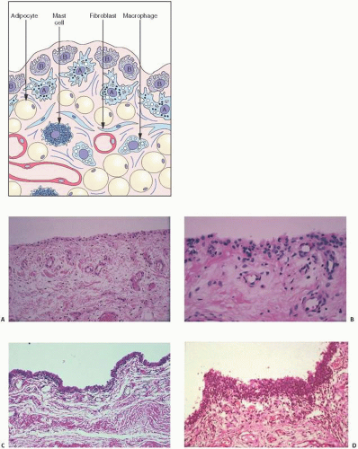
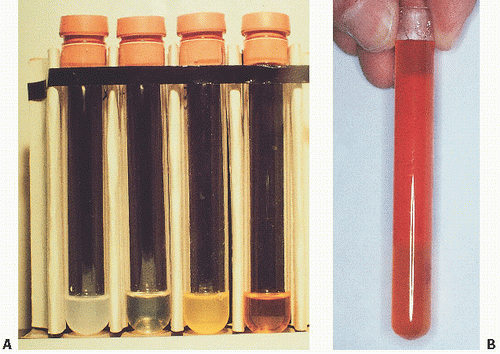
In hemorrhages that occur within the synovium, a highly vascularized tissue, red blood cells eventually disintegrate with phagocytosis occurring by histiocytes. Eventually, the hemoglobin is broken down and processed into hemosiderotic. Hemosiderotic characteristically is seen as a brown-pigmented granular substance in cells of the synovium. Hemosiderotic may occur as minute granules or accumulate into globules 25 µm in diameter. The intracellular deposition of hemosiderotic is observed mostly in macrophages or histiocytes, but also may be seen engulfed in various trauma-associated conditions by the hypertrophied synovial lining cells. In joints subjected to hemorrhage, hemosiderotic may be noted intracellularly or extracellularly. Cells within different compartments or layers of the synovium may be affected, ranging from the synovial lining cells to the subsynovial proliferating connective tissue cells. The gross appearance of the joint with chronic hemarthrosis is that of a rusty pigmentation, although more acute bleeding may be demonstrable as a blackish green discoloration.


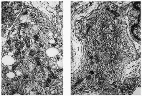
The response of the human joint to bleeding is denoted clinically by the formation of a hyperplastic vascular synovium within a few days. Examination of the tissue reveals a proliferation of the synovial cells and other subsynovial lining cell connective tissue elements, including often inflammatory cells. Under electron microscope, iron-containing, electron-dense particles that are membrane bound, called siderosomes, are noted within the synovial cells and subsynovial macrophages.

Clinically, Hemosiderotic synovitis may be due to a wide range of conditions, most notably trauma, particularly chronic trauma leading to chronic hemarthrosis. It may also be caused by the use of oral anticoagulant therapies, as a result of breakdown of synovial hemangiomas, or as a secondary phenomenon in conditions such as rheumatoid arthritis, PVNS, scurvy, and sickle cell anemia. The radiographic appearances may lead to joint space narrowing and may be confused with other ailments destructive to joints. Although most patients with chronic hemarthrosis or with an episode of hemarthrosis recover without any significant sequelae, the potential for joint destruction is present in any patient with bleeding into the joint.

With bleeding into the joint, two pathways may lead to damage. In one mechanism, the red blood cells may break down, causing macrophage activation. The ensuing inflammation may lead to destructive changes in and of itself, as seen in rheumatoid arthritis. Intracellular hemosiderotic precipitates the release of leukocyte-derived and synovium-derived chondrolytic enzymes (17). In excessive cases, the iron deposition in organs such as the meniscus may be severe enough to cause mechanical dysfunction and degenerative changes. In a second pathway, bleeding may lead directly to synovial proliferation, the release of an uncontrollable cascade of destructive substances such as proteases.


Hemophilia

Hemophilia A, the most common hereditary coagulation disorder, affects 20 live male infants per 100,000. It is caused by the deficiency, absence, or malfunction of coagulation factor VIII (18). In normal patients, a very low concentration of factor VIII (0.2 mg/mL plasma) is sufficient for adequate coagulation. Bleeding leading to clinically perceptible illness requires a reduction of at least 75 percent. The level of factor deficiency determines the severity of the disease: severe (factor VIII or IX baseline activity <1 percent), moderate (residual activity 1 to 5 percent), or mild (residual activity >5 percent).

Hemophilia A is a clinically heterogeneous disorder, ranging from mild (1 to 4 percent deficiency) to severe disease. Patients with mild or moderate disease may not be recognized unless a significant traumatic event precipitates abnormal bleeding. Excessive bleeding during surgery may be the first clue. Therapy centers around replacement of factor VIII (19,20). Traditionally done with shotgun frozen plasma, cryoprecipitate became available in the 1960s. More recently, purified concentrates have been used. In general, 1U of factor VIII increases plasma activity by 0.024 U/mL. Because 0.3 U/mL is usually needed to treat a mild episode of bleeding, clinical goals should strive for more than this amount. The development of inhibitory antibodies to the factor replacement products remains a clinical challenge (19). Therapy with recombinant concentrates have eliminated the risk of transmission of pathogens such as hepatitis and HIV viruses.

Clinically, abnormal hemorrhaging is key, especially into joints. Most commonly involved, in order of frequency, are the knees and elbows, followed by ankles, shoulders, and hips. Not usually evident in infancy, childhood signs include mild discomfort and limitation of joint motion. Pain and swelling follow. Numerous damaging microhemarthroses have transpired before clinical suspicion is initially aroused, so that the diagnosis is unfortunately delayed.

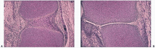
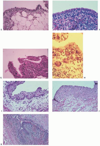
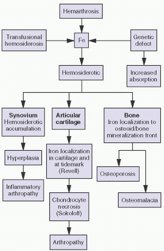
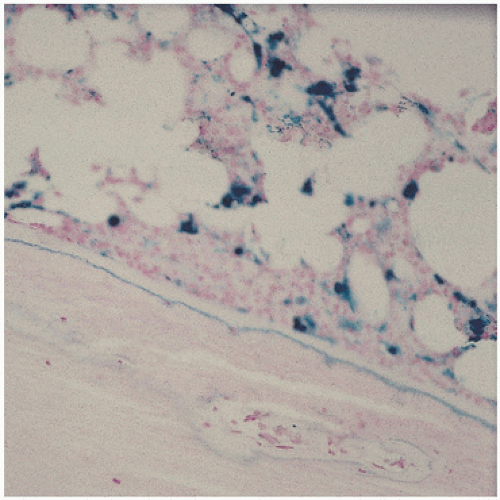
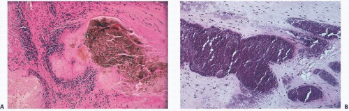
The pathology in the joint centers around iron-induced synovial inflammation (Fig 15.11), with both clinical and experimental observations documenting the damaging effects of hemosiderotic synovitis.

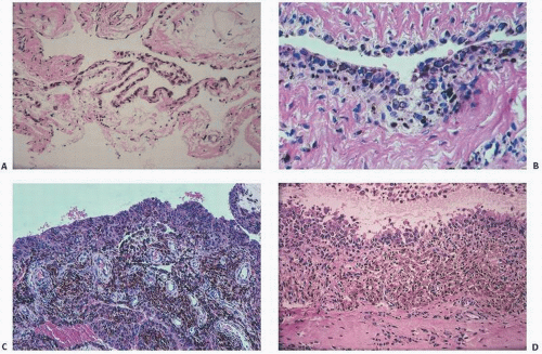
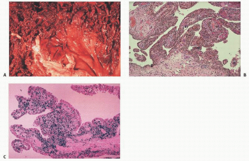
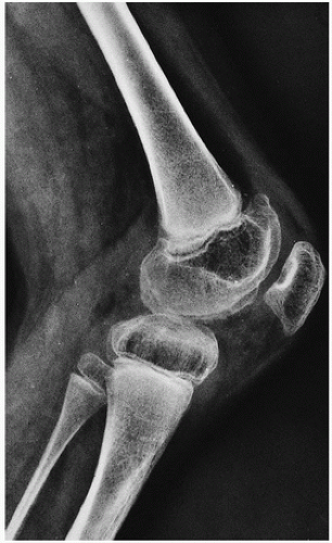
The adverse effect on synovium in patients with chronic hemorrhage has been studied most extensively in the joint changes of hemophilic arthropathy of the knee (Fig. 15.12). In tissue removed from hemophiliacs, the synovium shows marked villous hypertrophy with extensive hypertrophy and hyperplasia of the synovial lining cells and the subsynovial connective tissue components with abundant intracytoplasmic hemosiderotic granule accumulation (Fig. 15.11). In tissue cultures of synovium in hemophilia, pigment-laden fibroblast cells have been shown to proliferate, and explants of the synovial cells have been shown to secrete large amounts of latent collagenase and neutral proteinases, establishing the destructive potential of the synovitis in hemophilia. Synovium incited by hemophilia produces enzymes and may do so without coincident inflammation, the latter a significant component of the destructive arthropathy seen in rheumatoid arthritis.

Iron, a probable inducer of synovial inflammation, induces the expression of various oncogenes and proliferation of synovial fibroblasts. Hemosiderotic-laden tissue also leads to increased levels of proinflammatory cytokines such as interleukin 6, interleukin 1, and tissue necrosis factor alpha (21).

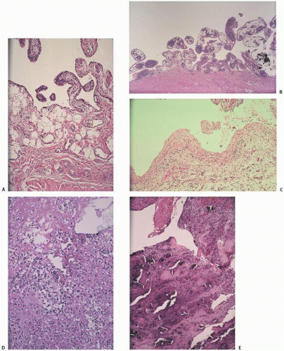
Iron accumulation may lead directly to chondrocyte destruction. In experiments aimed at developing a model for PVNS, a condition secondary to expanding nodules of proliferative fibroblasts and histiocytes, blood was experimentally injected into various animal models. The resultant proliferative synovitis was characterized by hyperplasia of fibroblasts, lipid-laden macrophages and giant cells, and extensive hemosiderotic accumulation similar to that seen in reactive Hemosiderotic synovitis or hemophilic arthropathy. The experimental bleeding model does not resemble PVNS.

Although less visible, iron is known to accumulate in cartilage as well. Extensive hemosiderotic deposition leading to grossly visivble brown menisci has been observed clinically. In studying hemophilic joints, iron has been localized to superficial chondrocytes, suggesting chondrocytic phagocytic uptake triggering a degradative enzyme release similar to that described in synovial cells. Iron has been localized histochemically to the tidemark in lupus and in hemochromatosis. Hemophilia A and hemophilia B are inherited X-linked deficiencies of factor VIII and factor X.

Roentgenographically, narrowing of joint spaces, loss of articular cartilage, cystic remodeling of bone (Fig. 12.8), and hemophilic pseudotumors characterize the illness (Fig. 15.13). However, scrupulous adherence to maintenance of factor VIII levels to prevent spontaneous hemorrhage has been associated with significantly decreased morbidity and has diminished, but not eliminated, the radiographic progression of disease.

Involvement of the musculoskeletal system is one of the major complications of hemophilia. The knee is the most commonly involved joint, followed by the elbow, ankle, shoulder, and hip.
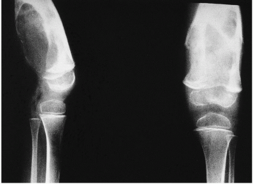
Hemophiliac arthropathy in the small joints of the hands and feet is rare, with few series of cases described in the literature. In decreasing order of frequency, the following are the imaging findings in hemophilic arthropathy: synovial hyperplasia, erosions, subchondral cysts, cartilage loss, and “pseudotumors.”


FIGURE 15.11. Hyperplastic hemosiderotic-laden rusty brown synovium from a hemophilic patient [(A) gross; (B, C) microscopic; (C) Prussian blue stain for iron].


FIGURE 15.12.Roentgenographic changes of hemophilic arthropathy.

The radiographic presentation of hemophilic arthropathy can be divided into stages for didactic purposes, but in real practice, there is frequent overlap between the different phases of the disease. Stage I is acute bleeding in the joint. Distension of the articular capsule is easily seen as a distended suprapatellar bursa in the case of the knee, or displacement of the humeral fat pads in the case of the elbow. Distension of the ankle joint capsule is usually easy to detect in the lateral projection. Bleeding into the shoulder or hip joints is difficult to determine in plain radiographs, but can be assessed by MRI and, to a lesser degree, also by computed tomography (CT).

Stage II is characterized by osteopenia in the subchondral surfaces of the affected joint. Overgrowth of the epiphyses is usually not noticeable. Joint contractures, such as fixed flexion of the knee, result in apparent enlargement of the ends of the bone on anteroposterior radiographs because of the increased distance between the knee and the x-ray cassette. This false enlargement is excluded if the lateral view is also evaluated. Enlargement of the ends of the bone is regarded to be secondary to hypervascularity, present in some cases of hemophilia. The deposition of hemosiderotic in the capsule of a joint renders it slightly more dense than the surrounding soft tissues. This is most easily detected in the elbow and ankle, but it can also be noticed in the knee.


FIGURE 15.13. Hemophilic pseudotumor (x-ray).

Changes in bone contour are noted in stage III. There is squaring of the patella, widening of the intercondylar notch, and subchondral cyst formation. Care should be exercised not to confuse a tunnel view of the knee (an anteroposterior view of the knee with flexion contracture is a tunnel view) with an intercondylar notch that is really widened. The subchondral cysts tend to be larger in hemophilia than in other inflammatory arthritides.

Erosion and destructive changes in the articular cartilage result in narrowing of the joint space. The changes in the articular cartilage are secondary to intra-articular liberation of digestive enzymes, as well as to prevention of the normal nourishment by synovial fluid, which cannot diffuse into cartilage coated by blood. These abnormalities are seen in stage IV.

The final stage, stage V, is defined by extensive bone destruction with marked deformity and rigidity of the joint.

The extra-articular manifestations of musculoskeletal hemophilia include muscle hemorrhage and pseudotumors. Hemorrhage in muscles can lead to neurovascular compromise. Such hemorrhage is most common in the gastrocnemius muscle and in the volar muscles of the forearm, where it can lead to a compartment-type syndrome. Bleeding in the iliacus muscle can result in femoral nerve palsy. MRI and CT are helpful in the diagnosis of episodes of bleeding into muscles.

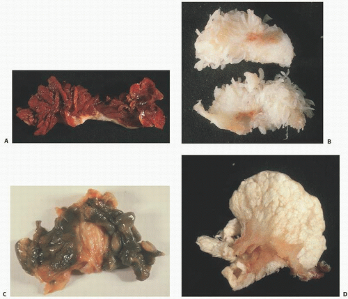
Pseudotumors are the result of the development of an encapsulated hematoma that slowly expands and may involve bone, the periosteum, or the soft tissue (22). This is a rare complication in which the hemorrhagic blood fails to be resorbed, accumulates, and slowly erodes the adjacent bones. It is usually an extremely painful condition and occurs in fewer than 1 percent of patients. Pseudotumors in adults are most common in the thigh, pelvis, and retroperitoneum, and most often in the vastus lateralis, soleus, or iliopsoas muscles.

In children, they can occur in the hands and feet. Ideally, pseudotumors should be surgically removed as they can continue to grow even after long dormant periods. However, surgical removal is not without complications including death, infection, fistulization, and pathologic fractures (23).

Surgical joint reconstruction in hemophiliacs has historically been necessary to preserve function. Although the mechanical survival of total knee replacement is quite good, the prevalence of infection as a complication is high, usually caused by Staphylococcus epidermidis (21).

Intra-articular, intrabursal, and soft tissue bleeding in hemophilia may result in painless masses clinically that roentgenographically mimic a tumor. These masses consist of spongy coagula of partially clotted blood encapsulated by thick, fibrous membranes. Complications of these so-called hemophilic pseudotumors include muscle and bone damage, infection, and neuropathies. Surgical removal is not without danger (22). They occur in 1 to 2 percent of hemophiliacs, mostly in the lower extremity and pelvis. Bleeding in the vicinity of the periosteum has been implicated in the peculiar juxtaosseous changes, which have included cyst-like bone changes and large soft tissue masses eroding bone. MRI findings depend on the stage of bleeding and associated effusion, erosions, cysts, cartilage damage, and degree of iron deposition in the tissue (24).

Before 1970, hemophilia A was still associated with significant severe disability and even death at a young age. However, the median life expectancy has increased significantly throughout the 20th century from 11.4 years to 68 years in carefully followed populations (25).

Nonetheless, the transfusion of blood products, including factor VIII concentrates, has led to one of the well-recognized modern complications of hemophilia-transfusion-related acquired immunodeficiency syndrome (AIDS). Life expectancy has now reversed after considerable gains. Serologically detectable antibodies to human immunodeficiency virus (HIV) developed in large numbers of hemophiliacs beginning around 1979. Approximately two-thirds of HIV-positive hemophiliacs have eventually died of AIDS.


Therapy more recently has been with factor VIII concentrates exposed to vigorous virus-killing heat treatment or solvent cleaning. Genetically engineered products are now available and are being tested for complications. Expense is a significant consideration.

In treating chronic hemophilic arthropathy, standard measures include factor replacement, physiotherapy, and operative synovectomy by conventional arthrotomy or arthroscopy. Nonoperative techniques, including the destruction of synovial tissue by intra-articular injection of radioactive agents such as colloidal 32P chromic phosphate, oxytetracycline chlorhydrate (Emicine), Yttrium-90 (for knees), or Rhenium-186 (for elbows and ankles) have been used (26).

In treating patients with HIV-positive hemophilia, the risk to the surgeon is minimal if proper precautions are taken (27).

Hemophilia B is due to deficiency of coagulation factor IX. Also known as “Christmas disease” after a 10-year-old patient named Stephen Christmas, hemophilia B has important royal associations. Queen Victoria was its most famous carrier, and Czar Nicholas II’s son Alexei Romanov, one of its most famous patients.

Recent advances in adenovirus-associated virus vector-mediated gene transfer offer great promise in treating and reducing the cost of the disease (28).


Iron Overload and Hemochromatosis

The most common iron overload disorders (IOD) are hemochromatosis and β-thalassemia, disorders that are typically clinically insidious. They cause progressive tissue damage including significant effects on the musculoskeletal system. Categories of iron overload and iron overload-related disease have been defined.

Screening laboratory tests include measurements of the serum ferritin level and transferrin saturation, with ferritin levels above 200 ng/mL (449 pmol/L) in women or 300 ng/mL (674 pmol/L) in men who have no signs of inflammatory disease, and transferrin saturation above 45 percent in women or above 50 percent in men warranting additional testing.

Four major cell types determine iron content and distribution (29):


  • Duodenal enterocytes by affecting dietary iron absorption;


  • Erythroid precursors by affecting iron utilization;


  • Reticuloendothelial macrophages by affecting iron storage and recycling; and


  • Hepatocytes by affecting iron storage and endocrine regulation.

Iron released from enterocytes (and macrophages) binds to plasma iron transport protein transferrin. Transferrin transport of iron to the hepatocyte and macrophage can be stored in those cells as ferritin. Erythroid precursors are the major site of iron utilization. Hepatocytes regulate the production of the hormone hepcidin, which downregulates the release of iron via ferroportin.

IODs can be categorized according to whether there is a defect in the hepcidin-ferroportin axis (“hemochromatosis”), impaired iron transport, or ineffective erythropoiesis (e.g., “thalassemia”).

The two major options to treat iron overload are phlebotomy for hemochromatosis and iron chelators (such as deferoxamine, deferasirox, or deferiprone) for β-thalassemia in which anemia coexists with iron overload.

The hepcidin-activating pathway has recently emerged as a target for treatments aimed at modulating iron status (30).

Much current knowledge about the effect of iron on tissue comes from studies of hemochromatosis, the systemic disorder in which iron deposition is associated with tissue dysfunction. In general, iron overload has been studied in clinical situations in which there has been hyperplastic refractory anemia, excessive blood transfusions for underlying hematopoietic abnormalities (thalassemia), and hereditary forms of hemochromatosis. In the United States and northern Europe, hereditary hemochromatosis is a common cause of iron overload; 15 percent of the population exhibits the gene in its heterozygous state. The abnormal gene, located on the short arm of chromosome 6, is closely linked to the HLAA locus.

In hereditary hemochromatosis, there is an increased absorption of dietary iron, resulting in excess iron deposition in parenchymal tissues of endocrine organs, liver, and heart; death often results from heart failure or chronic liver disease. In other IODs, the cause is ineffective erythropoiesis as seen in β-thalassemia and sideroblastic or aplastic anemia, for which the treatment may include repeated transfusions. IODs also may be caused by chronic liver disease, and in certain populations, such as within sub-Saharan Africa, hemochromatosis has been attributed to increased amounts of dietary iron intake, such as from large amounts of iron in beer brewed in steel drums. Individuals at greatest risk probably have a genetic predisposition (29).

The deposition of iron in bone, particularly at the osteoblast-osteoid interface or at the mineralization front or as hemosiderosis within the hematopoietic cells of the marrow, is a relatively common sequela to primary and transfusional hemosiderosis. Of import, iron directly inhibits osteoblast differentiation. Although the primary organs involved are the heart and the liver with eventual development of cirrhosis and hepatocellular carcinoma and heart failure, endocrine organs are particularly involved. Joint pain in the clinical presentation of arthritis has been noted in 11 percent in some studies, but its prevalence is as high as 72 percent. Bone pain is a less frequent cause for presentation (31) (Fig. 15.14, Table 15.4).


FIGURE 15.14. Iron localizing at the mineralization front interface between osteoid and bone in a patient with transfusional hemosiderosis.


TABLE 15.4 Presenting Clinical Features of Patients with Hemochromatosis


Presenting Symptoms


Percentage of Patients (%)


Incidental


30


Abdominal pain


16


Joint pain


11


Health examination


10


Weakness


9


Diabetes


8


Frequent infection


8


Contusion


5


Bone pain


4


Dyspnea


3


Impotence


2


After Adams PC, Kertesz AE, Valberg LS. Clinical presentation of hemochromatosis: a changing scene. Am J Med. 1991;90:445.


First recognized in 1964 as a feature of hemochromatosis, arthropathy is now generally believed to be a common clinical symptom. In hemochromatosis, the joint changes may mimic DJD. Less than 50 percent of patients seem to have clinically osteoarticular problems. The metacarpophalangeal joints and interphalangeal joints are most frequently reported. Wrists, elbows, shoulders, hips, and knees are less frequently affected. Iron may inhibit the pyrophosphatase activity in cartilage tissue, leading to the precipitation of calcium pyrophosphate crystals, an association between the two that is well recognized.

Bone changes in iron overload have been described in hemochromatosis as well as the iron-overloaded states of chronic renal dialysis. Osteoporosis is common, particularly if concomitant hypogonadism was present.

The osteopathy seen in iron overload has been associated with various concurrent metabolic abnormalities including hypogonadism, hyperparathyroidism, diabetes, and both vitamin C and vitamin D deficiency. Hypogonadism is a significant contributing factor to osteoporosis in this population, with both bone resorption parameters increased (osteoclast surfaces, osteoclast numbers, and urinary hydroxyproline levels), and bone formation parameters decreased. In hemochromatosis, iron may also infiltrate and adversely affect the parathyroid gland, with previous studies showing concurrent elevated parathyroid hormone.

Iron overload associated with hemodialysis can lead to a wide range of musculoskeletal problems. Proximal muscular weakness is well known and may be due to a number of problems including concomitant endocrinopathies, phosphate depletion, and osteomalacia. Although iron deposition at the mineralization front has been shown in numerous conditions including thalassemia, its histochemical detection in undecalcified bone biopsies may or may not be associated with osteomalacia (Fig. 15.14). In fact, as mentioned in hemochromatosis, the most common finding is osteoporosis, even with iron accumulation at the mineralization front. Tidemark iron accumulation has been demonstrated in articular cartilage, as well as in patients with hemochromatosis.

In the osteomalacia seen in association with renal disease, the disorder is usually the result of the toxic effects of aluminum, and, in fact, aluminum deposition at the mineralization front is now well established in the literature as a common cause of osteomalacia in this population. However, the presence of iron, obvious in dialysis patients by the significant hemosiderosis that complicates many cases, can also be the cause of osteomalacia (Fig. 4.4). Although iron may be the sole cause in rare cases of osteomalacia, in many cases it may also be deposited along with aluminum, causing peculiar staining properties at the mineralization front.

Although intestinal absorption of iron is normal in patients on hemodialysis, 1,25-vitamin D may stimulate iron absorption. However, it is more likely that iron deposition causing osteomalacia in the dialysis population is due to transfusion overload.


Lead Synovitis

The signs and symptoms of lead poisoning are subtle, and recognition of this entity may be difficult. Plumbism is caused most commonly by ingestion of lead-based paint by children; by occupational exposure, such as painting, lead mining, and working in battery factories; and by the consumption of contaminated beverages, such as moonshine (32).

Approximately 90 percent of lead is stored in bone with a half-life in bone of 20 to 30 years. It replaces calcium in the bone hydroxyapatite. Hypermetabolic conditions that cause increased bone turnover can lead to significant release of lead into the blood with the resultant lead toxic symptoms. Thus, increased bone metabolism induced by surgery, fractures, pregnancy, Paget, hyperparathyroidism, and medications can suddenly lead to lead symptoms unsuspected by the treating physician. Symptoms in these circumstances can be vague but include fatigue, malaise, headache, abdominal pain, or joint pain. Lab findings of lead toxicity include microcytic hypochromic anemia. Blood smears show a basophilic stippling.

A prototype of lead injury to the joint is the effect of retained bullet fragments (33). Lead poisoning secondary to retained projectiles is rare (Fig. 15.15), but has been reported since the ancient Roman wars. Lead toxicity may cause convulsions, somnolence, mania, delirium tremens, coma, neuritis, nausea, vomiting, abdominal cramps, anorexia, weight loss, renal insufficiency, general malaise, and death. Orthopaedic complications include bone cysts, localized arthropathy, pseudarthrosis, and gouty arthritis. Serum levels of lead of 920 µg/dL (44.40 mm/L) have been reported after retention of lead projectiles. The time between injury and onset of symptoms has ranged from 2 days to 40 years, although patients may be asymptomatic for long periods.

The location of the projectile in the body is a major factor in the likelihood of the development of lead intoxication. Exposure of lead to acidic synovial fluid results in greater dissolution than does exposure to human serum, water, or soft tissues. A second variable that may affect the duration of the symptomatic period is the surface area of the lead that is exposed to body tissue. Multiple small pellets have a greater surface area than one larger pellet, facilitating solubilization. Embedded lead particles usually are not absorbed systemically because they are encapsulated by dense, avascular fibrous tissue, inhibiting their dissolution in body fluids.


FIGURE 15.15. Lead synovitis. (A) Radiograph of the right knee showing multiple lead pellets. (B) Intraoperative photograph, showing the shotgun pellet in the intra-articular space (×10). (C) Intraoperative photograph, showing minute particulate matter engulfing the articular surface of the tibial plateau (×10). (D) Photomicrograph of hyperplastic and hypertrophied synovial tissue with irregular fragments of foreign material (black). A sparse and patchy chronic inflammation is present (hematoxylin-eosin, ×255). (E) Transmission electron micrograph of synovial tissue with cytoplasmic electron-dense particulate matter surrounding the nucleus (right). The scale marker represents 1 µm (×9,800).


The diagnosis of lead intoxication routinely has included analysis of the levels of lead in serum and urine and clinical findings. A blood level of 10 µg/dL or higher is considered elevated in adults. Studies in which electron microscopy was used suggested that lead is incorporated into cells to a level that causes death of cells, resulting in extracellular deposition of lead. This finding suggests a need for removing lead fragments from the intra-articular space as soon as possible to avoid localized arthropathy.

The treatment of lead intoxication has consisted primarily of chelation therapy and open techniques for removal of the fragments and cystic lead masses if present (34). Symptomatic lead poisoning can be treated with arthroscopic removal of lead. This technique allows for removal of the fragments, extensive synovectomy, and debridement without the need for an open arthrotomy. Frequently, chelation therapy is given preoperatively to avoid sudden exacerbation of lead intoxication caused by the stress of the operation. Theoretically, lead, which is stored mostly in bone, can be released during stresses, such as an operation, fever, immobilization, acidosis, and other conditions, and this would increase symptoms dramatically. The level of lead might not increase, however, with intervention such as arthroscopy.

Chelation therapy has been given intravenously and orally. Multiple courses of chelation therapy may be needed to deplete the body stores of lead. It should be combined with operative removal of lead from the intra-articular space. Arthroscopic excision may be an effective way to achieve this goal.

Reported clinical effects of lead on articular structures include extracellular subsynovial lead deposition, synovial hypertrophy and inflammation, and intracellular lead uptake. Lead can concentrate in articular cartilage where it can disrupt the normal chondrocyte phenotype by suppressing TGF-β signaling, leading to DJD (35). Radiographic effects reported in animals include synovial lead uptake and arthropathies. The presence of lead near the joint space can induce microscopic degenerative changes in the joint structures. Lead-implanted knees yield significantly greater degeneration compared with knees implanted with steel, knees undergoing simple arthrotomy, and control knees.

These joint changes constitute a lead arthritis or synovitis. Such changes are similar to those seen in DJD and may lead to chronic damage. Treatment considerations include surgical irrigation of all articulating surfaces. Exposure to lead via gunshot wounds or other trauma may present with arthropathies and even lead intoxication in humans. Irrigation may prevent not only lead arthropathy but also degeneration caused by other intra-articular debris such as cartilage or bone fragments found after gunshot wounds, thus avoiding or prolonging additional procedures such as synovectomy, fusion, or joint replacement.


Arthritis


Degenerative Joint Disease and Rheumatoid Arthritis

Although a broad range of disorders may give rise to arthritis, de novo arthritis may be readily classified into two groups: (1) DJD or osteoarthritis and (2) rheumatoid arthritis. They are distinct etiologically, clinically, radiographically, and pathologically (macroscopically and microscopically) (see Chapter 16).

There are significant differences in the primary component of the joint involved. Notwithstanding experimental interest in synovial tissue modulation of cartilage destruction by cytokines, such as catabolin (interleukin 1), the synovium in DJD seems, at least initially, to be an innocent bystander, with the brunt of damage initially involving the articular cartilage (fibrillation and eventual denudement) and bone (subchondral cyst formation and sclerosis with marginal new bone formation or osteophytosis). Biologically, osteoarthritis is associated with cartilage change, including reduction in the major proteoglycan, reduction in aggrecan, change in collagen fibril size and structure, and increased synthesis and degradation of matrix molecules. Although the synovium may show hyperplasia, this is usually minimal and nonspecific. Synovitis is limited if present at all (Fig. 15.6A). More active roles of the synovium in the pathogenesis of DJD have been proposed, including the reduction of the diffusion of synovial fluid and subchondral vascularization that accompanies high joint pressure, further deteriorating chondrocyte metabolism. Arthroscopic studies reveal that gross signs of inflammation in early DJD, in contrast to signs of rheumatoid arthritis, are limited anatomically to the points of the synovium in close proximity to articular cartilage. The overwhelming remaining synovium is essentially normal.

In rheumatoid arthritis, the synovium is the central inflammatory mediator. In acute rheumatoid arthritis, the synovium shows the most significant pathology (Fig. 15.6B). Infiltrated by lymphocytes and plasma cells, the synovium becomes hyperplastic, and the surface exudes a fibrinous exudate. Changes in the articular cartilage are truly secondary as the pannus, or inflammatory synovium, invades the surface of the joint, causing chondrolysis, eventual cartilage denudement, and, in chronic cases, the appearance of a secondary degenerative phenomenon. Chondrolysis and osteoporosis characterize rheumatoid arthritis, however, distinguishing it clearly from DJD. This distinction is evident in laboratory diagnosis and monitoring. The inflammatory changes in rheumatoid arthritis are discernible in elevated sedimentation rates and positive rheumatoid factors (an elevated immunoglobulin protein, usually IgM, circulating in the serum). There is no equivalent useful laboratory monitor for DJD.

Radiographic changes initially show joint space narrowing and subchondral sclerosis. Eventually, new bone forms at the margins of articular cartilage (osteophytosis), which may give rise to villous synovial hypertrophy and metaplasia, leading to chondro-osseous loose bodies.

Variants of DJD include an inflammatory type, characterized by more lymphocytic infiltration and hyperplasia of the synovium, and a rapidly destructive joint process that shows accelerated clinical and radiographic joint damage correlated pathologically with extensive cartilage and bone debris throughout the joint (24,36).

Rheumatoid arthritis is classically a chronic, symmetric, persistent arthritis that may be associated with systemic symptoms and rheumatoid nodules (classically subcutaneous). Its cause is obscure, but laboratory studies and familial history suggest immunologic and genetic factors in its expression. The rheumatoid synovium contains activated T cells, B cells, dendritic cells, and monocytes and macrophages. T cells are activated by antigen-specific (signal 1) and costimulatory receptors (signal 2); the former are modulated through major histocompatibility peptide complexes (37). Patients with class II major histocompatibility phenotypes, such as HLA-DR1 and HLA-DR4 (present in 80 percent of rheumatoid patients), are at risk because these molecules present antigens to CD4 and T cells. Proliferating T cells produce cytokines that can activate other inflammatory cells, such as macrophages, and cytokines such
as tumor necrosis factor-α and interleukin-1 have become therapeutic targets. Molecular mimicry involving these steps has long implicated synovial proteins and infectious agents in the etiology of rheumatoid arthritis. Most patients with rheumatoid arthritis have a circulating protein in their blood, usually IgM, which is the basis for the rheumatoid factor test, a nonspecific but often useful serologic test in corroborating the clinicopathologic diagnosis. Atypical infections may trigger an as yet undetermined genetic predisposition. The presence of rheumatoid factor and elevated sedimentation rates correlates well with the characteristic synovial changes of a hyperemic synovial tissue infiltrated by a pronounced lymphocyte and plasma cell infiltration, often producing a fibrinous exudate. The latter proteinaceous exudation may override the articular bone surfaces of the joint, often creating tan, friable bodies (rice bodies).


Variants

Other disorders have been associated with rheumatoid-like inflammatory joint disease, but these “rheumatoid variants,” such as psoriatic arthritis, Reiter syndrome, and the arthritides associated with colitis, show fewer inflammatory synovial changes, vary in clinical progression of disease, and usually are not associated with a positive rheumatoid factor.


Crystal-Induced Synovitis

Although numerous crystals may deposit in the joint and in the bone proper, two crystals are particularly synoviotropic and associated with clinical articular and osseous pathology and should be specifically contrasted. Gout, resulting from the deposition of sodium urate, and CPPD (pseudogout, chondrocalcinosis) are frequently encountered crystal deposition disorders presenting with rheumatologic or orthopaedic complications. In addition, HA crystals may be detected in synovial fluid in association with arthritis and degenerative joint changes. Other crystals, such as oxalosis and cystinosis, are rare and usually associated with renal disease.


FIGURE 15.16. Composite. Gout versus calcium pyrophosphate crystals (in tissue section). Gout (A) appears as brown accumulations if not dissolved in routine fixatives (right); otherwise amorphous whitish spaces remain (left). There is always an associated mononuclear and giant cell inflammation. Calcium pyrophosphate dihydrate (CPPD) (B) (Continued)


Gout

Gout refers to the painful clinical syndrome associated with the precipitation of sodium urate crystals in and around joint surfaces characterized by synovitis and juxta-articular destruction of articular bone. Because humans do not express the enzyme uricase, which degrades uric acid, and normal uric acid levels are close to the limits of urate solubility, hyperuricemia is an effective trigger to urate crystal deposition. Although gout often presents as a rheumatologic or orthopaedic syndrome, it is a systemic disorder preferentially depositing sodium urate crystals in and near joints. The ensuing inflammatory reaction or “tophus” may occur in virtually any organ in the body with the exception of the brain. Nonetheless, a severe form of hyperuricemia (Lesch-Nyhan syndrome) manifests with severe central nervous system clinical disease.

Gout is far more frequent in men and increases in incidence with age, affecting about 1 percent of men in Western countries. Although there probably are asymptomatic deposits of crystals, gout usually is characterized by painful clinical attacks that are usually abrupt in onset involving the small peripheral joints, especially the metatarsophalangeal joints. The predilection for sodium urate gout crystals to deposit in the peripheral joints of the body suggests that multiple factors may be at work, including factors related to pH and temperature. A gouty attack may last a half day to several weeks. Although urate levels in the serum are usually elevated (approximately >8 mg/dL in men and >6.5 mg/dL in women), this may not be the case. Well-documented examples of gouty arthritis with large tophus deposits have been associated with normal uric acid levels, at least sporadically. The analysis of synovial fluid in gout cases reflects an inflammatory type of change with leukocytosis and the identification in sediment particularly of inflamed synovial fluid and of intracellular and extracellular needle-shaped crystals that have a characteristic negative birefringence on polarized light microscopy (Fig. 15.16). These morphologic changes are useful in differentiating gouty crystals from synovial fluid, contrasting them with other crystals, such as those seen in CPPD disorders. A specific and sensitive technique is to use light microscopy

and polarized light microscopy. The addition of a red filter clearly distinguishes the strongly “negative” birefringent crystals. The strongly birefringent crystals parallel to the orientation of the red filter are yellow, and perpendicular crystals are blue (Urate Parallel Yellow Perpendicular Blue or U PAY PEB) (Fig. 15.16). “Positive” birefringent crystals of CPPD oriented parallel are blue, and crystals oriented perpendicularly are yellow.

Only gold members can continue reading. Log In or Register to continue

Stay updated, free articles. Join our Telegram channel

Jul 24, 2016 | Posted by in PATHOLOGY & LABORATORY MEDICINE | Comments Off on Synovium

Full access? Get Clinical Tree

Get Clinical Tree app for offline access