Metabolic Bone Disease: Part II


Metabolic Bone Disease: Part II


Renal Osteodystrophy

The term “renal osteodystrophy” (ROD) refers to a host of skeletal problems seen in association with long-standing renal disease and hemodialysis (1). A relatively newly minted term, the “chronic kidney disease-mineral bone disorder,” focuses on the calcium, phosphorous, parathormone (PTH), and vitamin D disturbances. In ROD, the roentgenographic manifestations are protean, ranging from osteopenia to osteomalacia. There may be resorptive changes associated with secondary hyperparathyroidism and even osteosclerosis. Examination of bone biopsy specimens in patients with renal-related bone disease who are experiencing skeletal pain has enhanced considerably our understanding of the varied changes that can attend chronic renal failure. It is clear that despite low levels of serum 25-hydroxyvitamin D in adult patients with the nephrotic syndrome and normal renal function, no consistently determined histopathologic changes accrue with this degree of renal dysfunction (2). However, as the ability of the kidneys to perform glomerular filtration gradually decreases, and as progressive tissue damage interferes with tubular function and the metabolic activity of vitamin D, profound skeletal abnormalities ensue that result in severe, crippling deformities and skeletal shrinking (3,4).

Fibroblast growth factor 23 (FGF-23), a hormone secreted by osteoblasts, is an important regulator of phosphorous and vitamin D metabolism. A critical factor in hypophosphatemic disorders such as that accompanying renal failure, it enhances the urinary excretion of phosphate and suppresses renal 1α-hydroxylase activity and decreases serum 1,25-dihydroxyvitamin D levels. While primary increases in biologically active FGF-23 cause renal phosphate wasting, hypophosphatemia, inappropriately low levels of 1,25-dihydroxyvitamin D, and rickets or osteomalacia, depletion of FGF-23 leads to hyperphosphatemia, excessive levels of 1,25-dihydroxyvitamin D, ectopic calcification, and early death.

With the advent of hemodialysis, the replacement of kidney excretory function has led to prolonged life at the expense of skeletal integrity. Without the normal endocrine contributions of the kidneys, patients on hemodialysis suffer considerable morbidity and mortality. Although hemodialysis is remarkably safe, the patient is exposed to large volumes of water that may contain potentially toxic levels of numerous agents, including aluminum, magnesium, fluoride, and other compounds, such as copper and zinc. These problems have been mostly corrected with new techniques for the handling of dialysis fluids. Aluminum, in particular, has been most fully explored as a potential toxin of the mineralization of bone. Other hazards and complications associated with hemodialysis include the development of fibrosis and granulomas associated with debrided silicone particles from plastic tubing and syndromes such as dialysis encephalopathy. Nonetheless, hemodialysis has been widely used as a safe, life-saving procedure in patients with end-stage renal disease.

The development of a broad array of skeletal abnormalities does not appear to depend on the specific type of renal disease leading to end-stage renal function (4) (Fig. 4.1). At the time of the inception of regular dialysis treatment, approximately two-thirds of patients with end-stage renal disease have a condition referred to as osteitis fibrosa cystica, which is a type of severe secondary hyperparathyroidism. A third of them have osteomalacia, which may or may not be solely related to abnormal vitamin D metabolism (5). In fact, examination

of bone biopsy specimens from large series of patients who have long-standing end-stage renal disease with or without hemodialysis has revealed a broader range of changes, which include secondary hyperparathyroidism, osteomalacia, or a mixture of osteomalacia and secondary hyperparathyroidism. Histomorphometric studies in which tetracycline labeling is used have also revealed a type of ROD characterized by sparse cellular activity and low bone turnover (aplastic bone) (6) (Table 4.1). In addition, tissue samples from these patients may or may not have detectable aluminum and/or iron deposition at the mineralization fronts. Rare circumstances of iron at the mineralization front causing osteomalacia have been described (7). There are also patients with an increasing range of amyloid-related syndromes resulting from the deposition of β2-microglobulin (8). An additional unusual tissue change, which is most evident on x-ray films, is osteosclerosis, a condition characterized by increased bone deposition in tissue despite obvious identifiable osteoclastic resorption.


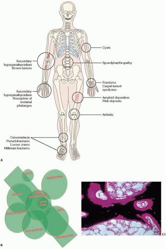
FIGURE 4.1. Renal Osteodystrophy at a glance. (A,B) Clinical and roentgenographic abnormalities in renal-related bone disease. (C) Osteomalacia showing increased volume and surface of osteoid. (C1) (Continued)


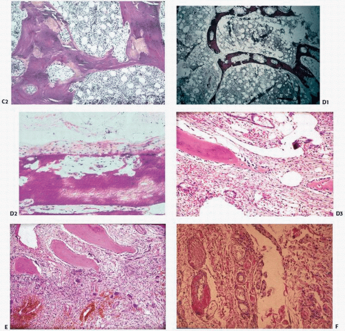
FIGURE 4.1. (Continued) Von Kossa stain. (C2) H&E stain. (D1) Hyperparathyroidism showing increased bone resorption as evidenced by lytic holes in the trabecular bone. (D2) Subperiosteal osteoclastic bone resorption, and (D3) high remodeling bone with fibrotic marrow. (E) Brown tumor showing giant cells clustering around areas of hemorrhage. (F) Amyloid deposition (pink) deposition in vessel walls.


TABLE 4.1 Histomorphometric and Histologic Classification of Renal Osteodystrophy


Type


Percent (%) of Patientsa/b


Histologic Findings


Pathogenesis


1. High turnover


38/9


Increased bone remodeling; (osteitis fibrosa cystica) paratrabecular fibrosis


Secondary hyperparathyroidism Aluminum (Al) may or may not be present


2. Low turnover “Adynamic renal disease”


36/60


Little bone remodeling or unidentifiable


Low levels of parathormone Some cases with Al or iron deposits


3. Mild disease


13/21


Mild bone remodeling No significant paratrabecular fibrosis


Early or treated hyperparathyroidism


4. Mixed disease


11/4


Features of both secondary hyperparathyroidism and osteomalacia


Secondary hyperparathyroidism


5. Osteomalacia


2/6


Increased osteoid defective mineralization


Aluminum and/or iron deposits Vitamin D problems Unknown causes


Other findings


Osteosclerosis


Osteoporosis


Calcifications


β2-Microglobulin amyloidosis


Cutaneous calciphylaxis


a Percent of patients on hemodialysis.


b Percent of patients on peritoneal dialysis.


Osteonecrosis is an additional musculoskeletal complication after renal transplantation, and by magnetic resonance imaging (MRI) has been detected in up to 25 percent of cases (9). Both allograft organ rejection and its associated hypersensitivity reactions and the use of corticosteroids have been implicated in its pathogenesis.

Prominent periosteal new bone formation in metatarsals, femora, and pelvic bones has been reported in up to 25 percent of ROD cases. Often referred to as periosteal neostosis, it is usually symmetrical, and biopsy findings of marked increased bone remodeling are consistent with a parathyroid effect (10).

When the myriad endocrine functions of the kidney are considered, it is not surprising that the potential for metabolic disease is present (Fig. 4.2). With chronic renal failure comes the retention of phosphate and often metabolic acidosis, the latter a condition that in its chronic state can lead to bone dissolution and osteopenia, if not osteomalacia. With phosphate retention comes a rise in the phosphate concentration in plasma, which can lead to increased levels of calcium phosphate compounds in the circulation, a so-called metastatic calcification depositing in both organs and soft tissue. The ensuing hypocalcemia may trigger secondary hyperparathyroidism and its many associated changes, which include intense bone resorption at the periosteal, cortical, and endosteal envelopes. Excessive resorption may result in brown tumors: hypervascular, fibroblastic, and giant-cell reactions that locally present as cystic lytic lesions. Histopathologic sections from patients with severe secondary hyperparathyroidism reveal intensive osteoclastic resorption with a tunneling osteoclastic resorption at all bone envelopes and a peculiar paratrabecular fibrosis of the marrow space, a condition that tends to be irreversible with correction of the underlying deformities. It should be noted that paratrabecular fibrosis, which is nonspecific, may be seen in myeloproliferative disorders as well as other metabolic bone conditions, such as Paget’s disease.

With a decrease in renal function comes a decrease in renal-based hydroxylase activity, leading to relative or absolute decreases in levels of 1,25-dihydroxyvitamin D. This latter change may lead to a skeletal resistance to PTH and a further decrease in serum calcium, which in turn leads to further activation of the hyperparathyroid bone disease pathway. Alternatively, decreases in calcitriol, a vitamin D compound that normally inhibits parathyroid gland function by suppression of secretion of PTH, leads to increases in circulatory PTH. Other decreases in hydroxylase activity may lead to other vitamin D disturbances, which may decrease the intestinal absorption of calcium.

In summary, with failing renal function, there are


  • phosphate retention,


  • ↓ calcitriol levels,


  • ↓ serum ionized calcium,


  • ↓ numbers of vitamin D receptors and ↓ numbers of calcium sensors in the parathyroid gland,


  • skeletal resistance to the calcemic action of PTH due to ↓ density of PTH receptors on osteoblasts; ↑ serum osteoprotegerin levels.


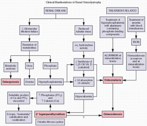
FIGURE 4.2. Biochemical (purple boxes) and osseous histologic (pink boxes) abnormalities in renal osteodystrophy.

Both the lack of availability of calcium and aluminum intoxication, which can be caused by either the water in dialysis fluid or the excessive use of aluminum-containing phosphate-binding gels in the treatment of hyperphosphatemia associated with renal disease, lead to further disturbances of mineralization, with aluminum deposition at the mineralization front (Fig. 4.3). Thus, osteomalacia may result from disturbances of vitamin D pathways as well as aluminum intoxication in patients with end-stage renal disease who are on hemodialysis. It is also well recognized that the accumulation of iron, resulting from the treatment of anemia associated with end-stage renal disease, may lead to significant hemosiderosis of the marrow as well as deposition of iron at the osteoblast-osteoid interface, within osteoblasts, and even at the osteoid-bone interface, resulting in osteomalacia (Fig. 4.4). Iron-induced bone disease has in part been alleviated by the administration of erythropoietin as a treatment for anemia in dialysis patients, precluding the need for significant numbers of transfusions.

In general, the analysis of undecalcified bone biopsy specimens and the use of special techniques, including histomorphometric
study of the uptake and activity of tetracycline at the bone surface, reveal at least five recognizable types of bone remodeling disease in ROD: (a) secondary hyperparathyroidism, (b) osteomalacia, (c) mixed uremic dystrophy (secondary hyperparathyroidism and osteomalacia), (d) adynamic or low-turnover disease, and (e) osteosclerosis (11). Stains for aluminum, iron, and amyloid should be performed, as all these may contribute to pathology.


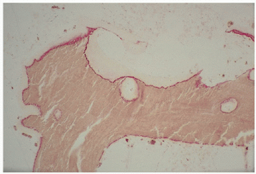
FIGURE 4.3. Aluminum (red) osteomalacia in renal-related bone disease develops at the osteoid-bone interface (“mineralization front”) and is directly associated with osteoid accumulation.


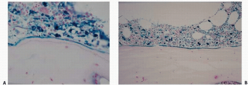
FIGURE 4.4. Iron (blue)-related bone disease in renal osteodystrophy includes marrow hemosiderosis with its potential impact on bone-lining cells as well as accumulation either along marrow-osteoid sites (A) or at the mineralization front (B), seen also in aluminum-induced osteomalacia.


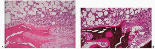
FIGURE 4.5. Osteitis fibrosa is the paratrabecular and tunneling fibrosis of the marrow seen in association with renal disease in patients who have biochemical evidence of secondary hyperparathyroidism. Fibrosis occurs at the surface and is associated with hematopoietic hypercellularity adjacent to it. (A) Hematoxylin-eosin (H&E) stain. (B) von Kossa stain, undecalcified sections.

Hyperparathyroid-related bone disease may be mild or severe (Fig. 4.5).

Mild hyperparathyroid disease is characterized by increased bone remodeling. Both osteoblasts (and the resultant osteoid) and osteoclasts are increased in number and amount. Because only 20 percent of bone surfaces normally would show these cells, any increase over 20 percent suggests a high remodeling state. In severe secondary hyperparathyroidism, there is intense osteoclastic activity including tunneling cones of bone resorption and paratrabecular fibrosis.

Osteitis fibrosa is characterized by increased bone turnover including an increased number and activity of osteoclasts, increased osteoblastic activity, and an increased quantity of osteoid (unmineralized bone matrix). Although other disorders involve accelerated bone remodeling, such as Paget’s disease, hyperthyroidism, and types of osteoporosis, the osteoclasts in hyperparathyroid bone disease penetrate into Haversian and Volkmann canals (forming characteristic “cortical cutting cones”), and tunnel into bone trabeculae (producing what is known as dissecting osteitis) (11). This unique pattern of bone resorption has numerous radiographic manifestations, the most characteristic of which is subperiosteal erosions of the phalanges and ends of other bones such as the clavicle and symphysis pubis.

Although the presence of deeply burrowing osteoclasts is suggestive of osteitis fibrosa, the diagnostic hallmark of the disorder is paratrabecular marrow fibrosis. In early osteitis fibrosa, fibrosis is often limited to paratrabecular regions, but as the disease progresses, fibrous tissue may occupy increasingly larger portions of the marrow space (4). In severe disease, microfractures and hemorrhage lead to macrophage accumulation, fibrous tissue growth, and, often, cystic degeneration called “brown tumors.” When
multiple, “brown tumors” represent the disorder von Recklinghausen described as osteitis fibrosa cystica, a rare observation in modern times due to early diagnosis and treatment of hyperparathyroidism.


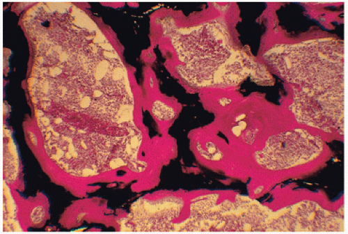
FIGURE 4.6. Osteomalacic bone in renal disease demonstrates marked accumulation of osteoid (red), which in normal states constitutes only 2 percent of the volume of bone and covers only 20 percent of its surface. (Undecalcified bone, von Kossa stain.)

The hallmark of secondary hyperparathyroidism using tetracycline labeling is an increase in the bone formation rate.

Osteoid accumulates, but the primary defect is not mineralization failure but rather hyperactivity of the skeleton. The collagen is mineralized in an irregular fashion, resulting in woven bone. The number of mineralizing sites—the extent of tetracycline uptake—as well as the mineral apposition rates or the distance between two labels administered separately in time is increased. Marrow fibrosis, although well established to be related to hyperparathyroidism, is poorly understood. One clinically significant correlation is its interference with the efficacy of erythropoietin in treating the anemia associated with uremic bone disease (12).

A second type of renal-related bone disease is characterized by frank osteomalacia, the hallmark of which is the marked accumulation of osteoid (Fig. 4.6). In these patients, uptake of the tetracycline label is very poor, and even with double labeling, is often seen as a smudged broad band of fluorescence. This condition is sometimes referred to as low-turnover osteomalacia and may be due to interference with vitamin D hydroxylation by dysfunctional renal tissue. Marked deposition of aluminum may also be found. The treatment of the aluminum accumulation with deferoxamine may correct the mineralization defect, and mineralization of the osteoid may ensue. Osteomalacia is best documented by marked increases in osteoid thickness, volume and bone surface coverage, and best established by a decreased apposition rate when the bone is tetracycline labeled. The osteomalacia type of renal-related bone disease has decreased in prevalence in recent years.

A third form is associated with both hyperparathyroid-related bone resorption, as previously described, and impaired mineralization, with detection of aberrant tetracycline labeling and mineralization (Fig. 4.7).

In a fourth type of renal disease, bone shows no discernible increased osteoclastic activity and no significant accumulation of osteoid, but it lacks identifiable cellular activity. Some authors refer to this type of bone as adynamic or low-turnover bone, although, as mentioned, the osteomalacic variant often carries this terminology. Low-turnover bone disease, the prevalence of which has increased in recent years, tends to be seen more frequently in patients undergoing continuous ambulatory peritoneal dialysis than in those on hemodialysis.


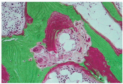
FIGURE 4.7. Osteoid accumulation and osteoclastic tunneling resorption in mixed uremic bone disease. Increased tunneling resorption substantiates secondary hyperparathyroidism. Osteoid accumulation when shown by tetracycline labeling to be indistinct and smudged with decreased mineralization rates is consistent with osteomalacia.

A fifth type of renal-related bone disease is osteosclerosis (Fig. 4.8). Approximately 20 percent of patients with ROD manifest osteosclerotic changes (Fig. 4.8). These changes include the “rugger jersey” spine, as well as sclerosis at the metaphyses of long bones, base of the skull, and pelvic and rib bones, as reviewed by Eastwood (13) and others (14,15). Cases of epiphyseal or bone-end sclerosis in which osteonecrosis has been ruled out have also been documented (16,17,18).

Ellis and Peart (19) studied 60 patients with chronic renal failure by analyzing undecalcified transcortical iliac bone biopsy specimens. They reported histologic findings on sections with osteosclerosis, as well as osteitis fibrosa cystica and osteomalacia. Their study, which demonstrated a 30-percent finding of osteosclerosis, noted that it was more evident in specimens in which osteomalacia was predominant. They noted that a combination of increased mineralized woven bone and osteoid matrix resulted in osteosclerosis and showed evidence of increased amounts of mineralized bone in cases in which there was excessive resorption. Indeed, PTH may have an anabolic effect, a feature exploited in the treatment of osteoporosis.

Studies have yet to reveal an accepted physiologic mechanism. Our evaluation of undecalcified bone biopsy specimens from ROD patients reveals an increase of osteoclasts per unit area of bone surface, but not a proportional amount of osteoclastic resorptive spaces. Therefore, it seems that there may be a dysfunction of the osteoclasts or the signaling process that activates them, or a barrier between the mineralized bone and the osteoclasts.

Treatment of ROD needs to address the specific pathologies present. Drugs such as cinacalcet, an oral calcimimetic agent, can activate the calcium-sensing receptor on parathyroid tissue and thus lower levels of parathyroid hormone. Hyperphosphatemia, a nearly universal complication of kidney failure and contributor to the development of secondary hyperparathyroidism, is usually treated with oral phosphate binders. Aluminum and iron can be chelated.


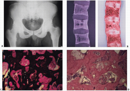
FIGURE 4.8. Osteosclerotic variant of renal osteodystrophy is characterized by diffuse or localized radiodense bone (A). Gross examination of the spine (B) and x-ray films of gross specimens may reveal diffuse obliteration of marrow space by thickened cortical and trabecular bone. Undecalcified biopsy specimens reveal abundant mineralized bone encroaching on the marrow space (C) (von Kossa stain). Foci of osteoclastic resorption document the ability of these patients to resorb bone, but suggest a dysfunctioning remodeling event (D) (H&E stain). (Continued)


Extraskeletal Calcifications (“Metastatic Calcification”)

Extraskeletal calcification is a well-known feature of renal bone osteodystrophy that may affect periarticular tissue, joints, soft tissue, internal organs, and arteries. Vascular calcifications have received great attention in recent years as cardiovascular mortality is very high in dialysis patients, accounting for nearly half of all deaths. Arterial calcification occurs at two distinct sites: in the tunica media, where it causes loss of vascular elasticity and compliance, and in the intima, where it is associated with atherosclerosis. Atherogenic factors in end-stage renal disease include oxidative damage to the endothelium, low-grade inflammation, abnormalities of lipoproteins, and/or hyperchromocystinemia. A recent pathologic study has confirmed that the coronary arteries of patients with end-stage renal disease contain more extensive calcium deposits than controls of similar age (11). Several studies using electron beam tomography have demonstrated the high frequency and rapid progression of coronary artery calcification in dialysis patients (12,13). Vascular calcification frequency in dialysis patients increases with age as in the general population and has been found much higher than in nonrenal controls of the same age. Most strikingly, a recent study has demonstrated both high frequency and rapid progression rate of coronary artery calcification in a relatively short series of young dialysis patients at an age at which calcification was nearly never observed in nonrenal patients. In this study, longer duration of dialysis treatment, high phosphatemia and serum calcium phosphorus product, low alkaline phosphatase blood level, and high intake in calcium-containing phosphate binders were associated with calcification, whereas serum calcium and intact PTH levels were not. As extent of coronary calcification measured by electron beam tomography is strongly correlated with obstructive coronary disease, this issue is receiving great attention. Another recent observational study found a positive correlation between carotid artery compliance and calcification, and reported calcium ingestion in renal patients. A large study previously demonstrated a significant increase in mortality risk associated with elevated serum phosphate concentration, especially for values exceeding 6.5 µg/dL, and calcium phosphate product (15). A number of data therefore point out that strict phosphate product in renal patients, without a large

increase in systemic calcium load, will limit soft tissue calcification and potentially reduce cardiovascular risk.


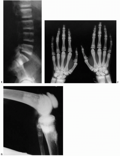
FIGURE 4.8. (Continued) Other classic roentgenographic features include alternating bands of lucency and sclerosis (“rugger jersey” spine), as seen in a lateral radiograph (E). In a frontal view of both hands and wrists, sclerosis is seen in the metaphyses and epiphyses of most of the short tubular bones, carpal bones, and distal ends of the radii and ulnae. There is expansion of the metaphyses of the metacarpals and mild widening of the growth plates, most apparent in the proximal phalanges of both thumbs (F). In a lateral view of the knee, areas of sclerosis are seen in the metaphyses and epiphyses of the femur and knee as well as in the patella (G).

Cutaneous calciphylaxis (calcific uremic arteriolopathy) refers to a particularly morbid skin and vascular calcification that is characterized histologically initially by barely perceptible calcifying vascular and septal panniculitis but in later-staged lesions with obvious epidermal ulceration, dermal necrosis, and mural vessel calcification (20) (Fig. 2.9). The classic triad in this disorder is arteriolar medial calcification, thrombotic subcutaneous ischemia, and subsequent skin necrosis.


Fractures

Patients with kidney disease have a higher risk of fractures than those without renal failure, the risk of fractures similar to that of a patient without kidney disease who is 10 to 20 years older (21). Hip fractures are the most common long-bone fracture and are frequently complicated by problems such as nonunion, implant failure, infection, hematoma, and prolonged wound drainage.

Multivariate analysis reveals greater risk with older age, female gender, diabetes, more years receiving dialysis, and cardiovascular disease; and lower risk with African American race, increasing body mass index, parathyroid hormone values in the fourth quintile (227.1 to 538.0 pg/mL), and renal transplantation during follow-up. Postfracture mortality rates are twice as high (22).


Aluminum Intoxication

Aluminum has been identified as a culprit in the development of osteomalacia and as a contributor to other abnormalities in renal-related bone disease. Aluminum intoxication may be caused by the natural presence of aluminum in water in certain geographic areas and by its presence in total parenteral nutrition solutions and even dialysis solutions, the latter situation having been largely corrected. Aluminum-binding phosphate gels, used to treat the hyperphosphatemia of renal failure in patients with end-stage renal disease, are a contributory factor. Other probabilities include slow accumulation of aluminum via oral absorption, as mentioned, through medication. The successful treatment of aluminum-related disease has been accomplished medically by the use of chelating agents, such as desferoxamine.

Aluminum is considered a trace substance in the normal human biologic state; the kidney is primarily the organ for excretion of aluminum. The incidence of aluminum toxicity in dialysis patients with skeletal symptoms has been reported to be as high as 30 percent; the manifestations include myopathies and also fractures. Although aluminum toxicity may be present in uremic patients, it is most commonly seen in patients on long-term hemodialysis. Although multiple fractures are the most commonly reported radiographic feature of aluminum toxicity, the diagnosis should be entertained in the face of any nontraumatic fracture of the large bones of the appendicular skeleton; nontraumatic fractures are not usually a feature of secondary hyperparathyroidism in these bones (23). Other symptoms of aluminum intoxication include encephalopathy and muscle weakness. The specific pathogenic mechanisms of aluminum-associated bone disease may include the prevention of synthesis and growth of hydroxyapatite crystals or the inhibition of osteoblast activity. The tissue localization at the mineralization front suggests competition with calcium for mineralization.

Aluminum is documented in tissue by the utilization of special stains in undecalcified bone sections (Fig. 4.3). In general, the extent of tissue demonstration is directly proportional to the amount of aluminum causing bone disease (24).

Identification of the type of underlying bone disease in cases of aluminum toxicity is important in that the identification of primarily secondary hyperparathyroid disease and correction of the hyperparathyroid state by parathyroidectomy in patients with concomitant aluminum deposition may lead to aluminum-induced osteomalacia after parathyroidectomy. Thus, both conditions—hyperparathyroidism and aluminum deposition—should be considered in clinical decision making. The potential for toxicity with aluminum is irrefutable. Although monitoring aluminum levels is possible, it is resource intensive.

Treatment of aluminum-induced bone disease is directed at minimizing exposure to aluminum phosphate-binding gels during the treatment of hyperphosphatemia associated with renal disease (25). In cases of already established disease, chelation therapy is employed (26,27).


Iron Excess

Iron may be the principal cause of osteomalacia (27) (Fig. 4.4). Evidence has indirectly suggested that iron may exert this effect. Gratwick et al. (28) found iron at marrow-osteoid interfaces, cement lines, and mineralization fronts in mildly osteomalacic bone from heavily transfused patients with β-thalassemia, but they did not examine the possibility that iron had impaired hepatic synthesis of 25-hydroxyvitamin D (29). de Vernejoul et al. (30) detected hyperosteoidosis and a mineralization defect in specimens from two of four patients with β-thalassemia, and demonstrated iron at some calcification fronts in both cases. Because serum concentrations of 25-hydroxyvitamin D were not measured when bone biopsy specimens were obtained, the authors declined to attribute osteomalacia to skeletal effects of iron.

Pierides and Myli (31) have recently suggested that iron might be a cause of osteomalacia in patients undergoing hemodialysis. In a series of 37 such patients, these investigators found that 24 of 41 specimens exhibited almost pure osteomalacia and that all 24 stained positively for aluminum at mineralization fronts. In seven of these cases, the aurintricarboxylic acid reaction yielded a purple instead of a red color. Gomori stain demonstrated concomitant iron deposition in each of these 7 and in 1 of the other 17. Each of the patients with iron-positive biopsy specimens had received numerous transfusions, and each had a myopathy. The serum ferritin concentration of each exceeded 2,500 ng/mL.

Excess of iron undoubtedly results from transfusions of red blood cells. Each unit of red cells provides approximately 250 mg of iron. To deplete body stores of iron, Edwards et al. (32) removed 2 to 11 g from homozygous women with early manifestations of hereditary hemochromatosis; Milder et al. (33) removed 12.7 to 40 g from patients with more advanced disease.

Does ingestion of iron provide a significant fraction of the total burden? Although considerable evidence suggests that intestinal absorption is appropriate to iron stores in hemodialysis (34,35), a recent report indicates that orally administered 1,25-dihydroxyvitamin D3 stimulates iron absorption in rats (36). If the same effect occurs in humans, consumption of this metabolite could contribute over time to pathologic iron accumulation. Detection of HLA-A3, the principal histocompatibility marker for the idiopathic hemochromatosis allele (37), might suggest other reasons for intestinal hyperabsorption of iron. However, because HLAA3 is demonstrable in only 70 percent of patients with idiopathic hemochromatosis (37), this marker is not universally applicable.


Excessive quantities of iron may disrupt normal function at multiple sites (32,33). Proximal muscle weakness (31,38) and the decline of plasma PTH concentrations (39) are possible consequences of iron toxicity. In turn, suppression of parathyroid function may be instrumental in the evolution of osteomalacia; prior parathyroidectomy (40) and suppression of PTH synthesis (41) have been implicated in the pathogenesis of aluminum-related cases.

Because aluminum and iron are both trivalent, possess small ionic radii (42), and congregate together in other tissue (43), it would not be surprising if their adverse effects on bone cells were comparable. To date, however, the skeletal effects of aluminum have received considerably more attention than those of iron. In pigs administered iron for a period of 36 days, de Vernejoul et al. (44) found accumulations of the metal in osteoclasts and osteoblasts and at marrow-osteoid interfaces, mineralization fronts, and cement lines. Bone formation was reduced because of an impairment in osteoid synthesis. These effects resemble those observed by Goodman et al. (45) in cortical bone of aluminum-intoxicated rats, but not those recorded by investigators who produced osteomalacia with aluminum (46,47,48,49). Consequently, if one attempts to relate osteomalacia to the skeletal burden of iron, iron most likely inhibits mineral transport by osteoblasts situated on widened osteoid seams. It is also possible that iron at mineralization fronts, perhaps in conjunction with citrate, prevents normal deposition of calcium compounds; again, a similar effect has been attributed to aluminum (42). Thus, toxic effects within cells or at bone-osteoid interfaces could produce deficient histopathologic bone changes.


Amyloidosis

Amyloid refers to a broad range of fibrillary, collagenous-type proteins with a distinct biochemical structure (50) (Fig. 4.9). The term amyloid, meaning starch-like, is a misnomer. Original observations that amyloid deposits would stain blue with the iodine reaction suggested the presence of starch or cellulose. Despite morphologic uniformity in virtually all cases, amyloid is now known to encompass a spectrum of secondary protein structure diseases.

All amyloid deposits share the following physical properties:


  • appearance of amorphous, eosinophilic deposits under light microscopy after hematoxylin and eosin (H&E) staining,


  • bright green birefringence under polarized light after staining with the dye Congo red,


  • regular fibrillary structure observed with electron microscopy,


  • β-pleated sheet structure demonstrated with x-ray diffraction.

Amyloids are associated with a broad range of clinical syndromes, which range from almost coincidental findings to systemic illness and serious morbidity and mortality (Table 4.2) (51,52,53).

Amyloid conditions can be both hereditary and nonhereditary. Because of the ubiquitous nature of some of these conditions, their prevalence is unknown. Although the incidence of amyloidosis is probably less than one percent, in areas where familial factors are significant, such as in Portugal or northern Sweden, considerably more cases are reported. Although historically, amyloids have been characterized on the basis of sites of tissue deposition, such as cardiac or reticuloendothelial, more biochemical information is now available about the exact structure of the proteins responsible.

Perhaps the most common form of amyloidosis is that associated with chronic infections. Therefore, amyloid may be seen in a broad range of inflammatory conditions, including osteomyelitis and even rheumatoid arthritis. This amyloid, often called secondary or acquired amyloid (AA) protein, is also seen in familial Mediterranean fever. It is a 76-amino acid molecule, and its reputed serum precursor is a 104-amino acid polymorphic protein with multiple isoforms. It may circulate in association with high-density lipoproteins such as apolipoprotein and behaves like an acute-phase reactant. In inflammation, it may increase significantly. It is synthesized in the liver.

Another form of amyloidosis, often referred to as idiopathic or primary, is associated classically with multiple myeloma. Because it is usually associated with the production by plasma cells of light chains, either κ or λ (heavy-chain-associated amyloidosis is also described), it may be seen in a broad range of plasma cell proliferations, including myeloma, monoclonal gammopathies, light-chain disease, and perhaps even other plasma cell dyscrasias and reactive processes. Interestingly, amyloid light-chain deposits may take the form of casts (in myeloma or cast nephropathy), precipitates (in light-chain deposit disease), crystals (in acquired Fanconi syndrome), or fibrils (in light-chain-associated amyloidosis) (54).

Genetic types of amyloidosis have been described in Mediterranean countries and also in Japan and northern Europe, including Sweden. The prototype is an autosomal dominant neuropathy, usually associated with the deposition of a prealbumin protein. Another hereditary amyloid is an apolipoprotein found in patients of Scotch-Irish-English background. Although amyloid deposition may occur in virtually any organ, the heart and endocrine organs are especially susceptible, particularly when deposition is systemic. In systemic amyloidosis, death usually occurs when amyloid deposits in the heart precipitate congestive heart failure or mortal arrhythmias.

Of particular interest in orthopaedic pathology, and renal disease in particular, are specific syndromes associated with the deposition of amyloid (55). In patients undergoing long-term hemodialysis, a known association between carpal tunnel syndrome and amyloidosis has recently been further elucidated by the demonstration of a β2-microglobulin protein as the culprit. Discovered in 1985, the β2-microglobulin has been shown to be the amyloid responsible for numerous synovial and osseous lesions, including cystic defects that can precipitate fractures (55,56). This amyloid protein is characterized by a molecular weight of 11,815 Da and is normally found in plasma at a level of 1 to 2 mg/L, but it increases during long-term dialysis to levels more than 50 times as high. Although not filtered by the dialysis membrane, it is not produced in all cases of dialysis, and therefore other factors are probable. Amyloid may also be seen in rheumatoid arthritis and may result in shoulder-pad syndromes. It is frequently associated with osteoarthritis and may, in fact, cause a frank myopathy.

Zingraff et al. (57), evaluating random sternoclavicular joint biopsy specimens in hemodialysis patients, found amyloid to be present in 9 of 22 patients, which suggests that its presence, even in asymptomatic clinical situations, is quite common (57). The patients with amyloid in the sternoclavicular specimens tended to be older and had undergone dialysis for longer periods of time compared with amyloid-negative patients. It is of interest that the sternoclavicular joint has been identified as a site of hemodialysis-associated amyloid deposition because it has previously been cited as a site of age-related amyloid deposition (58). In general, amyloid deposition may involve the synovium, tendons, ligaments, joint capsule, or articular cartilage. Patients may have pain in the affected joint as well as stiffness and soft tissue swelling. Palpable nodules may actually form, although this would be unusual.


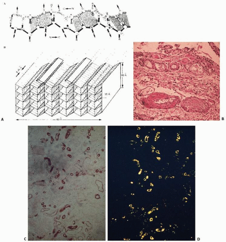
FIGURE 4.9. Amyloid composite. Fibrillary protein with a biochemical β-pleated structure (A) appears pink on routine tissue stain and is usually perivascular in location (B). With Congo red staining (synovial biopsy), amyloid tissue is pinkish red (C), and it is characterized by an apple green birefringence on polarized light microscopy (D). (Continued)


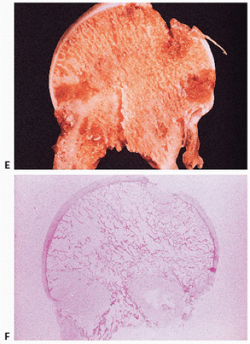
FIGURE 4.9. (Continued) Gross (E) and photomicroscopic (F) appearance of amyloid of the femoral head. (After Sack GH Jr, Dumars KW, Gummerson KS, et al. Three forms of dominant amyloid neuropathy. Johns Hopkins Med J. 1981;149:239-247.)

The deposition of β2-microglobulin has now been associated with a constellation of signs and symptoms, including, but not limited to, bone cysts (59), pathologic fractures (60), carpal tunnel syndrome (61), arthropathies (62,63), including spondyloarthropathies, and tumor masses (64) (Table 4.3).

Amyloidomas, tumoral masses composed of amyloid, occur less frequently in bone than in other tissues (65). Light-chain fibrils, AA fibrils, and β2-microglobulin components have been documented. Histologically, one may see corpora amylacea-like structures, in addition to the classic aggregates of amorphous-appearing eosinophilic material. Most, if not all, amyloidomas represent plasmacytomas, plasmacytoid lymphomas, or even multiple myeloma.

In hemodialysis, amyloid is predominantly deposited in perineural and periarticular structures, joints, bone, skin, and subcutaneous tissue. It is far less commonly seen in the liver, spleen, and even kidney. Kidney stones with β2-microglobulin deposits have been described. The incidence of carpal tunnel syndrome in hemodialysis patients has ranged from 2 to 31 percent and usually develops after an average of 8 to 9 years. Other known complications include infection and crystal-induced arthritis as well as periarticular calcifications. At least three distinct arthropathic syndromes have been described in association with β2-microglobulin amyloidosis: generalized arthritis, scapulohumeral periarthritis, and arthropathy with joint effusions (8).

Skeletal manifestations of amyloidosis usually are seen as a cystic change or pathologic fracture, particularly at the site of a tendinous insertion (Fig. 4.10). These cystic bone changes have been noted in the femoral heads, acetabulum, humerus, radial and carpal bones, and other bones, including the tarsal bones and pubic symphysis. Skeletal manifestations of β2-microglobulin amyloidosis are dependent on the duration of dialysis, age of the patient, and concomitant metabolic problems, such as osteomalacia and secondary hyperparathyroidism.

The diagnosis of amyloidosis should be suspected in any rheumatologic or orthopaedic condition associated with synovitis and pain, monoclonal gammopathies with renal disease, peripheral neuropathies, unexplained hereditary neuropathies, and conditions underlying chronic inflammation.

In addition, age-related localized deposition of amyloid has been demonstrated in degenerative articular and periarticular tissue and in traumatized tissue such as torn rotator cuffs (66).

The diagnosis of amyloid is usually established by a tissue biopsy in which the Congo red stain and polarized light microscopy are used. Although biochemical and immunohistochemical studies can be performed to classify further the type of amyloid, this is usually not necessary (50). The diagnosis is established by tissue, although the site of biopsy yielding the most diagnostic material is debatable. In systemic amyloidosis, tissue sites have ranged from gingiva to rectal mucosa and also the omental fat pad, small intestine, and skin. It should be noted that collagenous proteins are ubiquitous in connective tissue, and subtle amyloid-like changes giving the classic green birefringence on Congo red staining may be misleading.

Management includes treating the underlying condition if it is a secondary process. Ultimately, treatment should be directed at inhibiting the formation of amyloid fibrils or extracellular deposition of amyloid. Glucocorticoids have been of limited value, and other chemotherapeutic agents have been used, including colchicine and, most recently, melphalan and prednisone in combination. The use of more permeable artificial membranes in dialysis machines has been of some prophylactic value (62).


Cystinosis and the Fanconi Syndromes

Cystine crystal in tissue may be caused by a range of metabolic disorders (67,68) (Table 4.4). The classic condition is associated with an early onset and renal tubular dysfunction (the Fanconi syndrome) (69). Classic cystinosis is a recessively inherited metabolic disorder that is seen in approximately 1 person per 100,000 in the patient population. The condition results in a high intracellular content of nonprotein cystine crystals, presumably resulting from a defect in the carrier-mediated system that transports cystine out of light lysosomes. The result is the accumulation of cystine crystals within lysosomes and their histopathologically detectable accumulation in bone, cornea, conjunctivae, lymph nodes, leukocytes, and other internal organs (Fig. 4.11). It has been postulated that a component of the cystine molecule, perhaps the sulfhydryl group, can inhibit many sulfhydryl enzyme systems. For example, aberrant sulfhydryl enzyme systems responsible for the renal tubular reabsorption of amino acids in glucose may lead to glycosuria and aminoaciduria, one type of the so-called Fanconi syndrome.

Cystinosis is characterized by growth retardation, hypothyroidism, rickets, and photophobia resulting from cystine deposits in the cornea and conjunctivae. Its clinical course is characterized by unremitting destruction of kidney tissue. However, in the three types, infantile, late onset, and benign, considerable variations in the symptoms and
ocular and renal pathology are seen. Treatment is directed at reducing the amount of methionine (a dietary precursor of cystine) and cystine in the diet or using drugs that can reduce intracellular cystine levels. When possible, renal transplantation should be performed to bypass the genetically determined defect in the intracellular environment.


TABLE 4.2 Nomenclature and Classification of Human Amyloid and Amyloidosis


Amyloid Protein


Protein Precursor


Protein Type of Variant


Clinical Manifestations


AA


SAA


Reactive (secondary); FMF; familial amyloid nephropathy with urticaria and deafness (Muckle-Wells syndrome)


AL


κA, e.g., K111


Ak, Ag, e.g., AK111


Idiopathic (primary), myeloma- or macroglobulinemia-associated


AH


IgG1 (y1)


Ayl


ATTR


Transthyretin


e.g., Met 30


e.g., Meg 111


TTR or lie 122


Familial amyloid polyneuropathy (Portugal)


Familial amyloid cardiomyopathy (Denmark)


Systemic senile amyloidosis


AapoA1


apoA1


Arg 26


Familial amyloid polyneuropathy (Iowa)


AGel


Geisolin


Agn 187


Familial amyloidosis (Finland)


ACya


Cystatin C


Gin 68


Hereditary cerebral hemorrhage with amyloidosis (Iceland)


β-Protein precursor, e.g., βPP695


Alzheimer disease, Down syndrome


Gin 818


Hereditary cerebral hemorrhage with amyloidosis (The Netherlands)


2M


β2-Microglobulin


Associated with chronic dialysis


AScr


Scrapie protein precursor 33-35, cellular form


Scrapie protein 27.30


Creutzfeldt-Jakob disease, etc.


e.g., Lau 102


Gerstmann-Straussler-Scheinker syndrome


ACal


(Pro) calcitonin


(Pro) calcitonin


In medullary carcinoma of the thyroid


AANP


Atrial natriuretic factor


Isolated atrial amyloid


AIAPP


Islet amyloid polypeptide


In islets of Langerhans, diabetes type II; insulinoma


FMF, familial Mediterranean fever.


After Husby G. Amyloidosis. Semin Arthritis Rheum. 1992;22:67-82.


TABLE 4.3 Syndromes and Clinical Manifestations of β2-Microglobulin Amyloid Deposition


Carpal tunnel syndrome


Skeletal manifestations


Osteoporosis


Bone cysts


Destructive spondyloarthropathy


Pathologic fracture


Tumorlike masses in bone and soft tissue


Arthropathy


Generalized arthritis


Effusive arthritis


Scapulohumeral periarthritis


Camptodactyly


Renal calculi


Modified after Kleinman KS, Coburn JW. Amyloid syndromes associated with hemodialysis. Kidney Int. 1989;33:568-575.


The identification of cystine crystals in tissue is diagnostic (Fig. 4.11). However, crystals may be lost during routine laboratory processing because of the solubility of cystine in the acid solutions and formalin routinely used. They are best observed in their natural state, frozen or alcohol-fixed. In fact, when crystals are dissolved, cystinosis may be suspected if abundant cells with foamy cytoplasm are noted; these foamy areas represent spaces previously occupied by crystals. The crystals themselves may have numerous shapes, but most frequently clusters of brick- or block-shaped birefringent crystals are seen. Rectangular or needle-shaped accumulations may also occur. The crystals are usually intracellular, and the crystals within the clusters range from 0.1 to 1.0 µm in length or diameter. Electron microscopic studies have localized the cystine crystals
within membrane-limited organelles, having the appearance and enzyme markers of lysosomes, that is, containing acid phosphatase. The specific localization of crystals within lysosomes has suggested a classification of cystinosis as a lysosomal storage disease.


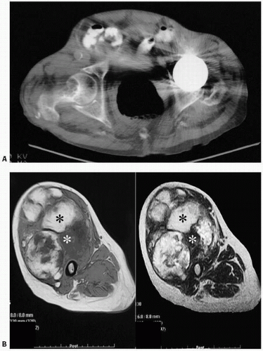
FIGURE 4.10. Amyloid. (A) Computed tomographic scan of the hip: well-defined lucency in the right femoral neck of a patient undergoing long-term renal dialysis. The opposite femoral head has been replaced because of osteonecrosis. The radiographic finding in amyloidosis is usually of one or more well-defined lucencies surrounded by minimal sclerosis. The lesions tend to occur in the ends of long bones. Metaphyseal and diaphyseal lesions can be found at times. The most common sites include the proximal femora, long bones at the knee, long bones of the upper extremity, and spine. (B) Axial T1-weighted and T2-weighted spin echo MR images of the right thigh show the mass involving the entire anterior compartment as well as the tensor fasciae latae. The mass is again noted to demonstrate both hypointense and hyperintense areas.


Cystinuria

Cystinuria is a recessively inherited metabolic condition seen in approximately 1 patient in 7,000. It is caused by the impaired reabsorption of dibasic amino acids—cystine, lysine, ornithine, and arginine. Because the solubility of cystine in urine is low, only cystine crystals are seen in the urine. In the heterozygous state, although cystine is increased in the urine, it is generally not increased enough to exceed solubility and be routinely detected.

Cystinuria usually occurs in patients between the ages of 20 and 40 years, with men affected more severely, although the sex predilection is equal. The kidneys bear the brunt of the damage, often with obstruction and subsequent infection and renal failure. Renal calculi composed of cystine crystals may have to be removed surgically. Clinically, affected patients are usually shorter than normal. Treatment is directed at hydration and alkalinization of the urine.


Oxalosis

The deposition of oxalate crystals in tissue may be due to either a primary metabolic defect or a secondary disturbance. Oxalate, a dicarboxylic acid, is highly insoluble in humans. A metabolic end product, it is excreted almost exclusively in the kidney.

In the primary inherited forms of oxalosis (“primary hyperoxaluria”), the main defect is primarily due to overproduction in the liver (70). This is a group of autosomal recessive disorders. Type I, the most common form, has a prevalence of one to three cases per million, and is caused by a deficiency of the liver-specific enzyme alanine glyoxylate aminotransferase (AGT), which catalyzes glyoxylate to glycine. Type II lacks glyoxylate reductase-hydroxypyruvate (GRHPR), which reduces glyoxylate and hydroxypyruvate to glycolate and d-glycerate, respectively. The defect in type III is in the liver enzyme 4-hydroxy-2-oxoglutarate aldolase (HOGA) (Fig 4.12).

The median age of onset is 5.5 years, with many patients having advanced renal disease at the time of diagnosis. Thus, many patients go unrecognized for many years. Major sites of crystal deposition are the kidneys, vascular walls, and bones leading to fractures.


TABLE 4.4 Some Distinguishing Characteristics of the Three Major Types of Cystinosis


Nephropathic


Intermediate


Benign


General symptoms


Onset


6-10 mo


18 mo – 17 y


No symptoms


Growth


Impaired


Variable


Normal


Skin pigmentation


Usually fair


Variable


Normal


Rickets


Present


Variable


Absent


Bone marrow cystine crystals


Present


Present


Presenta


Ocular symptoms


Retinopathy


Present


Variable


Absent


Crystalline deposits in cornea and conjunctiva


Present


Present


Present


Photophobia


Present


Variable


Absentb


Renal symptoms


Tubular dysfunction (Fanconi syndrome)


Present


Often incomplete


Absent


Glomerular failure


Present


Present at later age than in nephropathic type


Absent


Inheritance


Autosomal recessive


Autosomal recessive


Autosomal recessive


a Cystine crystals could not be demonstrated in bone marrow of two reported cases of benign cystinosis.


b Photophobia occurs in benign cystinosis if it is a part of the ophthalmologic condition that causes the patient to consult an ophthalmologist.


Modified from Goldman H, Scriver CR, Aaron K, et al. Adolescent cystinosis: comparisons with infantile and adult forms. Pediatrics. 1971;47:979-988.


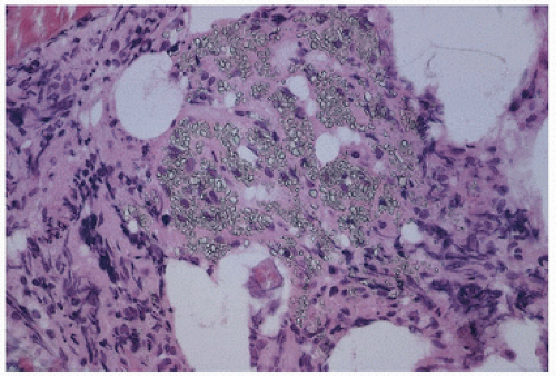
FIGURE 4.11. Cystine crystals in tissue.

Secondary oxalosis occurs as a result of excessive dietary intake or poisoning with oxalate precursors.

The roentgenographic appearance is highly dependent on the presence and degree of renal failure. The earliest specific radiographic features are fine radiodense transverse lines located symmetrically in areas of rapid growth. Radiolucent defects in the metaphyseal regions of long bones have been noted, which tend to be most prominent in areas of hematopoietic rather than fatty marrow, which tend to accrue more bloodborne oxalates. The lesions may be seen in the growth plate and in the intervertebral discs. Both primary and secondary oxalosis are characterized by crystalline deposition, which may be seen microscopically in marrow, osteoid, bone, and articular cartilage (Fig. 4.13). The identification of crystals depends on polarized light microscopy, which reveals highly refractile particles. Although these crystals have a characteristic needle shape and often form starlike clusters, they do not stain with von Kossa stain and may require special procedures for identification. Some investigators have used oxidation of oxalate to carbonate by hydrogen peroxide in the presence of silver nitrate (AgNO3), which gives a characteristic brown color. When oxalate stones or large crystalline deposits are present, chemical analysis may be used, but, in general, the crystalline deposits are microscopic and require finer techniques, such as x-ray diffraction or electron diffraction, the latter offering an exact method of identification of extremely small quantities of calcium oxalate in bone biopsy specimens. Oxalate stones are whitish or pale yellow. Microscopically, there may be no cellular response, a mononuclear cell reaction, or a giant-cell reaction, similar to that seen in other crystalline deposition disorders. Adjacent osteoclastic resorption of bone (secondary hyperparathyroidism) has been noted, but this change, as well as osteomalacia, is expected in the usual setting of chronic renal failure.

The natural course of this disease in the predialysis era was early death from uremia. The current definitive treatment of choice is thought to be combined liver-kidney transplantation.


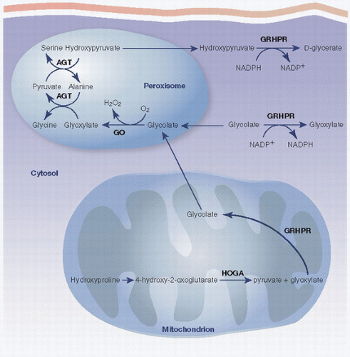
FIGURE 4.12. Oxalate metabolism.


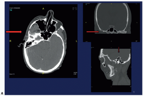
FIGURE 4.13. Oxalosis. Lytic lesion in the right infratemporal fossa (A). (Continued)


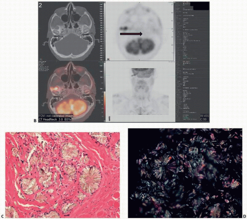
FIGURE 4.13. (Continued) Pet scan shows bilobed focus of hypermetabolic activity (standardized uptake values initially 4.4 and 3.0, and 8.1 and 5.8 on delayed imaging) (B). Histology of the crystals with flower and star shapes (C). polarized light microscopy (D).


Remodeling Metabolic Bone Disease


Paget’s Disease

Paget’s disease is a skeletal disorder associated with characteristic clinical, roentgenographic, and pathologic changes, the hallmark of which is intense activation of osteoblasts and osteoclasts and resultant abnormal remodeling of bone (71,72).

It has been estimated that 1 million people in the United States have Paget’s disease, and 10 to 40 percent of these have one or more family members affected. Paget’s disease preferentially targets the axial skeleton:

Pelvis 70 percent

Femur 55 percent

Lumbar spine 53 percent

Skull 42 percent

Tibia 32 percent.

The diagnosis of Paget’s disease is usually made on a radiographic image (focal osteolysis, coarsening trabecular and cortical bone, and bone enlargement). Bone scans best demonstrate, if present, lesions at other sites, Paget’s disease being very hot on technetium scanning, reflecting both increased bone formation and increased vascularization.

The abnormal results of laboratory tests seen in Paget’s disease are manifestations of high tissue turnover in skeletal bone; elevation of alkaline phosphatase reflects an increase in osteoblast activity, and excretion of collagen in the urine, detected as urinary hydroxyproline or pyridoline cross-links, is a manifestation of the high rate of breakdown of bone.


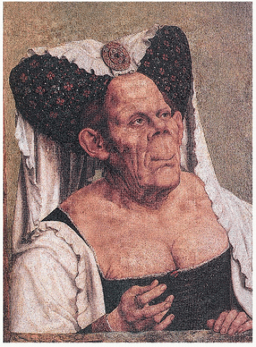
FIGURE 4.14. Grotesque facial features are due to enlarged frontal skull bone and maxilla. (After Quinten Metsys [1465/66-1530], The National Gallery, London.)

Because serum markers of bone turnover show less biologic variability than urinary markers, serum bone alkaline phosphatase and procollagen 1 N-terminal peptide (PINP) seem to best correlate with activity in Paget’s disease. Urinary N-telopeptide (NTX) has been used as a marker of resorptive activity in the disease and has been correlated with serum pyridinium cross-links.

Paget’s disease has been well described since the late 19th century, although paleontologic studies and paintings such as the one by Metsys, dating from the 15th century, suggest that it existed long before Paget’s original description (73) (Fig. 4.14). Paget’s disease, a common disorder, is rarely seen before the age of 40 years. However, a rare condition with the roentgenographic and histopathologic features of Paget’s disease, termed hyperphosphatasia, probably unassociated, is seen in childhood. Paget’s disease primarily affects older people and is found in as many as 10 percent of persons in certain geographic distributions by the eighth decade. It is more common in men and particularly in whites. Estimated to involve 2 to 3 percent of the U.S. population older than the age of 50 years, it is common in England and regions settled by European migration, such as the United States, Australia, New Zealand, Argentina, and South Africa (74).

Paget’s disease is rarely encountered in China, Japan, Iran, India, Scandinavia, Africa, or the Middle East (75). The identification of viral inclusions resembling an RNA-type virus resembling paramyxoviruses such as measles has raised interesting theories, as yet unproven, of a viral association. BCL-2, c-fos, and interleukin 6 are highly elevated in Paget’s disease. Some have suggested an association with pet or livestock ownership (birds, dogs, cats, and cattle) (75). In one large Italian study, a statistically significant association with rural (vs. urban) residence and dog ownership was shown (76).

Inherited as an autosomal dominant trait with high penetrance, a linkage with chromosome 18q near the polymorphic locus D18S42 has been shown. Recurrent mutations of the gene encoding sequestosome 1 (also known as SQSTM1 or p62) have been documented (77).


TABLE 4.5 Symptoms of Paget’s Disease


Bone pain


Active bone disease (periosteal stretching)


Involvement of joints with exacerbation of osteoarthritis


Fissure fractures


Complete fractures


Gout


Pseudogout


Osteosarcoma


Neurologic dysfunction


Deafness


Cranial nerve palsies


Spinal cord dysfunction


Brainstem and cerebellar dysfunction


Cardiovascular dysfunction


Increased cutaneous and skeletal blood flow


High-output heart failure


Other


Nephrolithiasis


Hypercalcemia


Retinal angioid streaks and blindness


Modified after Freeman DA. Paget’s disease of bone. Am J Med Sci. 1988;295:144-158.


In fact, genomic scans of pagetic families have identified 10 different mutations in the gene encoding sequestosome 1. Because p62 is a protein that mediates a diverse array of cellular signaling pathways involving molecules such as RIP, TrKA, TRAF6, and aPKCs and their response to various stimuli such as tumor necrosis factor α(TNF-α), interleukin 1, and the receptor activator of nuclear factor κB (RANKL) (78), the frenetic bone signaling that these mutated signals imply makes credible sense to the frenetic bone remodeling observed histologically.

Like many orthopaedic conditions, it is far more common as an asymptomatic condition. Symptoms may be diverse (Table 4.5).

In general, Paget’s disease may be characterized as a focal disease with sharp demarcation between normal and affected sites. There is slow spread through the marrow and bone with rare extension across joint spaces. Once established in a patient, new foci are generally not seen (79). However, Paget’s disease can recur in an old site after therapy and can be transferred from one part of the skeleton to another as a result of autogenous bone grafting (80).

The distribution of Paget’s disease has been studied by x-ray surveys and bone scans. Bone scans are more sensitive than x-ray studies because technetium localizes in areas of increased mineralization, the hallmark of bone remodeling seen in Paget’s disease (81) (Table 4.6). In addition to asymptomatic patients, approximately 10 percent of patients have monostotic involvement; this group usually responds to treatment. Polyostotic Paget’s disease frequently involves vertebrae, and these patients are particularly at risk for tumors and complications, such as high-output cardiac failure and neurologic changes.

The clinical feature of Paget’s disease in up to 40 percent of patients is bone pain, with areas involved often being warm and tender (Fig. 4.15). If the end of a bone around a joint is affected, arthritis may be the presenting symptom. Because of mechanical weakness resulting from the abnormal deposition of woven bone, cortical remodeling, and intense osteoclastic resorption, pagetic bone is prone
to fracture, and, in fact, fracture may be the presenting symptom. Other associated changes include calcific periarthritis and uremia, neurologic symptoms including deafness, vascular steal syndromes, platybasia (flattening of the base of the skull), and basilar invagination. Immobilization can lead to hypercalciuria, hypercalcemia, and nephrolithiasis. Roentgenographically, the changes are variable, depending on the stage and site of involvement (Fig. 4.16). Purely lytic lesions in Paget’s disease have been described, particularly in the skull, so-called osteitis circumscripta. In the initial stages of Paget’s disease in long bones, a lytic wedge advances along the cortex of the bone. Biopsies at this stage may show predominantly osteoclastic bone resorption mimicking hyperparathyroidism. Eventually, the resorptive phase progresses to one of intense bone remodeling, including osteoblastic bone formation. The resultant deterioration of the normal architecture of the bone leads to roentgenographically detectable coarsening of cortical bone, lack of demarcation between cortical and trabecular bone, and other such findings. The bone scan in this phase is intensely hot (Fig. 4.17). In many patients, Paget’s disease after a prolonged period of time enters a dormant phase, with little detectable osteoblast or osteoclast activity. In this stage, the disorder can be diagnosed by profound thickening and irregular cement lines throughout the involved bones. Fracture after biopsy of osteolytic Paget’s disease (82,83) and acute osteolysis after surgery potentially predisposing the patient to pathologic fracture (84) have been described.


TABLE 4.6 Pagetic Involvement of Different Bonesa


Skeletal Location


From Bone Scans (%)


From Roentgenograms (%)


Pelvis


72.3


68.2


Lumbar spine


58.2


48.8


Femur


55.3


54.7


Thoracic spine


44.7


37.0


Sacrum


43.5


40.6


Skull


41.7


44.1


Tibia


34.7


30.6


Humerus


31.2


28.8


Scapula


23.5


19.4


Cervical spine


14.1


12.9


Clavicle


11.1


9.4


Facial bones


11.1


8.2


Calcaneum


10.0


6.4


Patella


7.0


3.5


Hand


6.4


5.2


Foot


5.1


3.5


aExpressed as percentage of patients.


Modified after Meunier PJ, Salson C, Mathieu L, et al. Skeletal distribution and biochemical parameters of Paget’s disease. Clin Orthop. 1987;217:37-44.


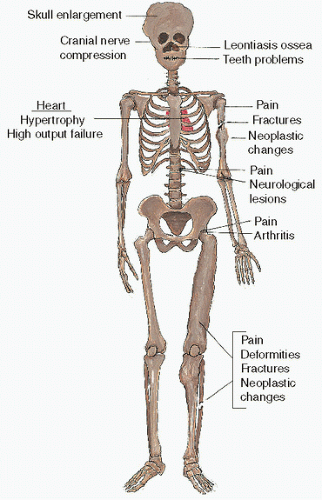
FIGURE 4.15. Clinical symptoms of Paget’s disease.

Classic radiographic manifestations of Paget’s disease involving the skeleton are bone deformity, thickening of cortical bone, and coarsening of the bone trabeculae. The disease most often involves the inner portion of the pelvis (iliopectineal line) and the calvaria of the skull. Any site of the skeleton may be involved, and at times just a small portion of a bone (e.g., the spinous process of a vertebra) may be involved by Paget’s disease.

Excluding tumor and fracture, Paget’s disease is the only condition in which the skeletal imaging is very abnormal but the fatty marrow is intact on a T1 MRI signal.

In the long bones, Paget’s disease always extends from the epiphyses to the diaphysis. Paget’s disease can extend from the trochanters of the femur to the shaft of the bone without involvement of the femoral head. The same applies to the humerus and its tuberosities and other apophyses in the skeleton.

The involved bone is usually enlarged, deformed in shape, and sclerotic. In a small group of patients, the involved area of the bone is seen mainly as a lucent lesion. The diseased area, as it progresses toward the shaft, has a pointed edge (which has been compared with an
advancing flame or the tip of an arrow). The deformity in the contour of the bone is the consequence of fractures (complete or incomplete). Fractures in bones with Paget’s disease heal in an irregular fashion, resulting in deformities (osteitis deformans). The deformity in the contour of the end of the bone can at times result in nonunion, narrowing of the adjacent joint space, and osteophyte formation. This form of degenerative joint disease has been labeled Paget’s arthritis, and it can occur when only one bone is involved, even though it is more common when both bones surrounding a joint are affected.


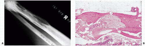
FIGURE 4.16. Roentgenographic (A) and histologic (B) changes of Paget’s disease. Areas of sclerosis and lucency extend from the distal end of the bone into its shaft. The proximal end of the area of Paget’s has a pointed edge.


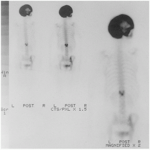
FIGURE 4.17. Hot skull (bone scan).

Paget’s disease in the spine usually causes increased density in the periphery of the vertebral body and accentuation of the trabeculae in the central portion of the body. This has been referred to as a “window frame” appearance. The vertebral body is usually larger in the anteroposterior diameter or taller than those in the adjacent, uninvolved vertebrae. A vertebra involved by Paget’s disease should be differentiated from an “ivory vertebra,” seen in cases of lymphoma as a homogeneously dense vertebral body without deformity in its contour, and also from vertebral hemangiomas, which are characterized by thickened vertical trabeculae without the peripheral sclerosis seen in Paget’s disease. Thickening of the vertebral body and neural arch in Paget’s disease can cause spinal stenosis.

The calvaria is the portion of the skull most commonly involved by Paget’s disease. Usually, the diploic cavity between the inner and outer tables is widened. One or more areas of sclerosis may be present. These areas of sclerosis at times are poorly outlined, leading to a comparison with “cotton balls.” Often, instead of areas of sclerosis, the involvement of the calvaria results in one or more areas of lucency (osteoporosis circumscripta). Lytic Paget’s disease in the peripheral skeleton is rare; however, osteoporosis circumscripta is not an unusual finding.

The base of the skull may also be involved. Sclerosis and bone thickening in the mastoids can result in deafness and loss of balance. Blindness can be caused by entrapment of the optic nerves by bones affected by Paget’s disease.

Paget’s disease is characterized grossly by hyperemia and architectural distortion of the normal contour and internal structure of bone. Hyperemia may lead to high-output cardiac failure and the feeling of warmth in clinically involved sites within bone. An increase in subchondral vessels has been noted in patients with Paget’s disease in comparison with other arthritic-type patients. Rongstad et al. (85) estimated vascularity to be increased sixfold in a resected femur.

The bones in Paget’s disease are grossly abnormal on visualization and lead to clinically discernible conditions: joint incongruity and degenerative joint disease, osseous deformities of the skull, protrusio acetabuli, coxa vara, and femoral bowing (Fig. 4.18).

Microscopically, Pagetic bone foci are sharply demarcated from normal bone.

The histopathologic features in Paget’s disease are dependent on the stage. In early Paget’s disease, identified by acute lysis of bone as seen roentgenographically in the advancing lytic wedge, osteoclastic resorption may predominate and create an appearance remarkably similar to the microscopic changes seen in hyperparathyroidism (81). In active, ongoing Paget’s disease, the constellation of findings is characteristic (Fig. 4.19). There is intense remodeling of bone as manifested by increased osteoblast and osteoclast activity. Normally, less than 20 percent of bone surface shows cellular remodeling. This is greatly increased in Paget’s disease (86) (Table 4.7), revealing bone


formation and bone resorption. Bone trabeculae become both thick and thin. Cortical bone remodeling is also irregular, with loss of the distinction between cortical and trabecular bone. Osteoclasts are increased in number, large, and markedly multinucleated.


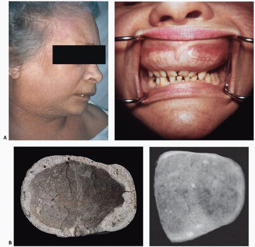
FIGURE 4.18. Composite of gross bone changes. (A) Paget’s disease involving the maxilla appears as a bulging upper lip (left), and oral examination reveals an enlarged distorted maxilla with loose teeth (right). (Courtesy of Ernest Baden, M.D., D.D.S.) (B) Calvarium showing irregular thickening of the skull, loss of diploic architecture, and a granular pumicelike appearance (left). Specimen x-ray film (right) shows irregular density with alternating densities and lucencies. (C) Proximal end of femur (left) and specimen x-ray slice of hip and acetabulum (right) reveal loss of normal cortical and cancellous architecture with replacement by coarsened, thickened bundles of bone. Grossly, bone may appear irregular, coarse, and pinkish, in contrast to the normal smooth, ivory appearance of cortical bone. (D) Vertebrae involved by Paget’s disease reveal sclerosis, typically more marked at the vertebral end plate, giving rise to a “picture frame” contour (left). External contour and internal architecture of bone are greatly distorted. Gross specimen and specimen x-ray film (right). (E) Undecalcified transcortical iliac bone biopsy specimen stained with von Kossa stain showing loss of demarcation between cortical and trabecular bone distorting the internal architecture of the ilium (left). Higher power (right) reveals irregular surfaces, increased surface osteoid, marrow fibrosis, and hypervascularity.


FIGURE 4.18. Continued


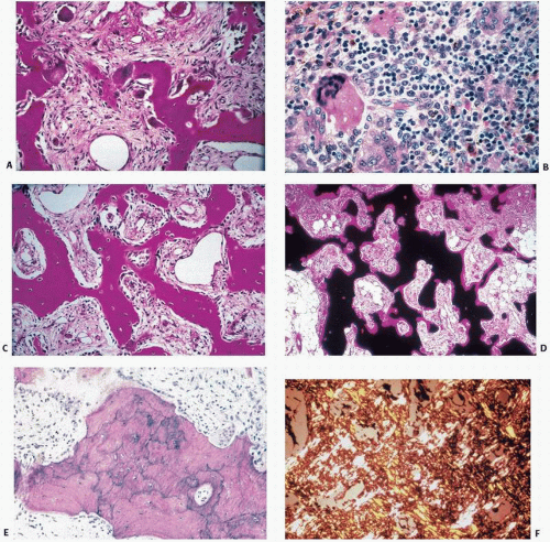
FIGURE 4.19. Paget’s disease at a glance.

• Abnormal osteoclasts

Large and multinucleated (A)

Numerous osteoclasts (B)

• Increased osteoblast activity

Increased numbers of osteoblasts (C)

• Increased bone remodeling.

Increased activation frequency (D)

• Woven bone

Irregular cement lines (E)

Irregular collagen orientation (F) (Continued)

Cement lines, the perimeter markers of previously resorbed bone units (Figs. 1.12 and 1.13), reflect the abnormal remodeling and are abnormally wavy, irregular, and curliform. Pagetic cement lines are an indication of the woven bone produced in this disease (Fig. 4.20). Woven bone is characterized by irregularly oriented collagen, seen under polarized light in states of increased bone remodeling. Although characteristic of Paget’s disease, it is also seen in embryonic bone and in both primary and secondary bone tumors.

Osteoclast remodeling, fibrosis, and cement lines may be quite dramatic in Paget’s disease and are the hallmark in active disease (Fig. 4.21). In inactive disease, the diagnosis may be obscure. On biopsy, the only hallmark is the presence of curliform cement
lines (Fig. 4.22) in now “burned-out” dense and (relatively) inactive tissue.


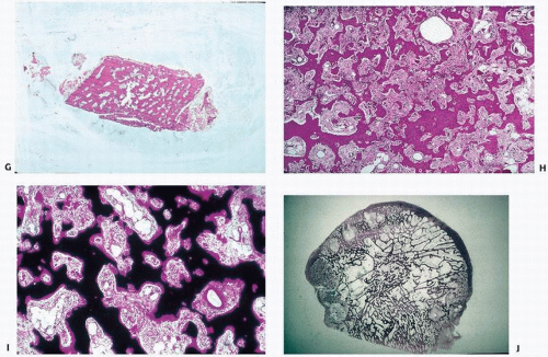
FIGURE 4.19. (Continued)

• Poor cortical/cancellous bone demarcation (G)

• Stromal fibrosis (H)

• Increased vascularity (I)

• Focal sharp demarcation from normal bone (J)


TABLE 4.7 Iliac Trabecular Bone Morphology and Histomorphometry Data in Normal Bone and Case Examples of Idiopathic Osteoporosis, Osteomalacia, Secondary Hyperparathyroidism, and Paget’s Disease


Disorder (No. Cases)


Bone Volume


Percentage of Osteoid Volume


Osteoid Surface


Osteoclasts per Millimeter of Bone Perimeter


Normal


22


<2


20


0.02


Osteoporosis (56)


15.9 ± 6.4


1.4 ± 13.7


17.9 ± 13.7


0.05 ± 0.09


Osteomalacia (5)


17.7 ± 4.9


43.1 ± 14.2


95.1 ± 3.9


0.27 ± 0.36


Secondary hyperparathyroidism (5)


21.6 ± 2.8


26.7 ± 17.5


83.2 ± 7.8


0.51 ± 0.37


Paget’s disease, active (3)


58.3 ± 22.2


6.6 ± 1.7


64.0 ± 29.2


1.15 ± 0.35


Modified after Vigorita VJ. The tissue pathologic features of metabolic bone disease. Orthop Clin North Am. 1984;15:613-629.


Paget’s disease may involve one or more bones. Interestingly, within a given bone, the involvement may be patchy (Fig. 4.23), which may lead to false-negative biopsy results in cases that are roentgenographically obvious.

Biopsy is usually indicated to confirm the diagnosis in symptomatic individuals when the diagnosis is uncertain. Patients may present with anemia, and pathologists evaluating biopsy specimens of the ilium, sternum, or pelvis for hematologic assessment should not overlook the fact that bone may reveal subtle abnormal remodeling changes indicative of Paget’s disease. The large multinucleated osteoclasts of Paget’s disease are reminiscent of virally induced changes seen in other illnesses, such as paramyxovirus infection (measles) and cytomegalic virus infection, giving indirect support to the current etiologic theory for Paget’s disease, which is a measles-type viral infection. In fact, viral

inclusions have been shown in osteoclasts in Paget’s disease by Rebel et al. (87) and Reddy et al. (88). However, osteoclast inclusions are by no means specific and have been shown in giant cells in giant-cell tumors (89), pyknodysostosis (90), osteopetrosis (91), familial expansile osteolysis (92), and in macrophage-like cells in primary oxalosis (93).


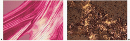
FIGURE 4.20. Polarized light histopathology of normal bone (A) and pagetic bone (B). Normal bone reveals the predictable lamellar, sheet-like orientation of collagen. In Paget’s disease, this is distorted, resulting in less refractile, interrupted wisps of collagen, “woven bone.”


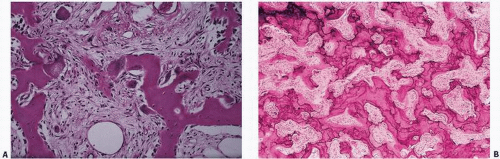
FIGURE 4.21. Active Paget’s disease with marked fibrosis and osteoclast activity (A) and pronounced irregular cement lines (B).


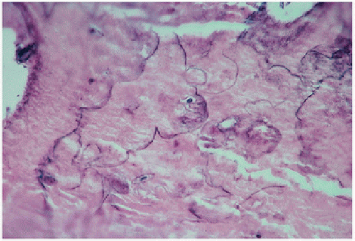
FIGURE 4.22. Inactive, “end-stage” or burned-out Paget’s disease. Only the curliform cement lines remain as a marker of the disease.


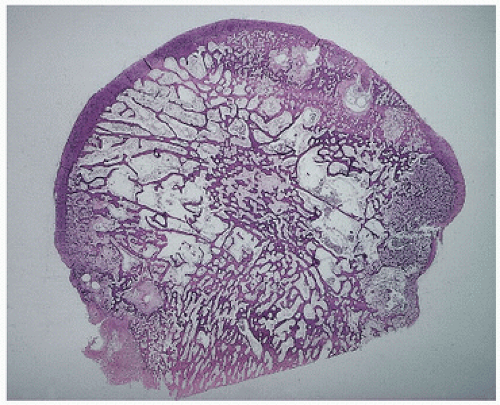
FIGURE 4.23. Coronal section through a femoral head with degenerative joint disease and Paget’s disease. The circumscribed dense areas represent pagetic foci. Intervening bone was nondiagnostic.

Viral candidates for initiating Paget’s disease have included measles virus, canine distemper virus, respiratory syncytial virus, simian virus 5, and parainfluenza virus type 3. To date, studies have failed to culture a virus (94).

Hoyland et al. (95) have proposed a paramyxovirus-linked modulation of osteoclast activity by interleukin 6 in genetically susceptible hosts.

Most recently, Reddy et al. (88) have shown by reverse transcriptase and polymerase chain reaction techniques that measles virus nucleocapsid transcripts are detected in pagetic bone marrow mononuclear cells. They further identified measles virus transcripts in very early osteoclast precursors (the colony-forming unit granulocyte-macrophage, or CFU-GM, progenitor cells). This supports the argument of a virally induced osteoclast dysfunction. But why is Paget’s disease a local disease? Here, microenvironmental factors, such as cytokine expression, may be critical.

The numerous ultrastructural studies performed on osteoclasts in Paget’s disease have created the following profile (87):


  • Osteoclasts are irregularly shaped with multiple extensions and much infolding of cytoplasmic membrane. The infolding presumptively indicates abnormally increased surface activity and motility of these cells.


  • The presence of calcified fragments within the cytoplasm under the ruffled border shows that the cells may actually phagocytose whole pieces of bone, which is a highly abnormal form of bone resorption.


  • The presence of vesicular mitochondria suggests a high turnover rate for osteoclasts.


  • Nuclei are highly polymorphic and frequently contain many nucleoli. Nuclear inclusions are present in several nuclei per osteoclast and are filament-like structures. Some are microcylindric in shape. They are usually grouped together in parallel bundles and sometimes packed in paracrystalline arrays.


  • Cytoplasmic inclusions can be filament-like structures, some of which are organized into bundles, like those found in the nucleus. No paracrystalline arrays, however, are usually found in the cytoplasm. The cytoplasm may also contain envacuolated glycogen.


  • In the fibrous tissue surrounding the osteoclasts in Paget’s disease, cells similar to mononuclear osteoprogenitor cells are often found. These are often massed together and contain no nuclear inclusions.

The treatment rationale for Paget’s disease is based on the ability of drugs to blunt or slow down the bone remodeling activity (72). Because osteoclasts are known to have receptors for calcitonin, the drug had been widely used. However, bisphosphonates are currently the first treatment of choice and are indicated in patients with pain localized to an affected site in which the presumption is that the pain is related to increased metabolic activity. Although evidence that long-term suppression of bone turnover remains controversial, bisphosphonates can, in the short term, normalize bone turnover.

The therapies of choice for treating Paget’s disease are the three most potent bisphosphonates: risedronat (Actonel), alendronate (Fosamax), pamidronate (Aredia), and zoledronic acid (Reclast). Reclast and Aredia are intravenously administered. Bisphosphonates (such as alendronate and etidronate) most likely produce their effect by binding bone mineral, thereby inhibiting both the formation and dissolution of calcium phosphate crystals (96). Bisphosphonates have a high binding affinity for hydroxyapatite crystals and are therefore selectively concentrated in bone, a feature that makes them excellent bone-scanning agents and clinically useful in the treatment of heterotopic ossification.

Bisphosphonates also have profound effects on osteoclasts. They accelerate osteoclast apoptosis and cause the ruffled borders of the cell to become rounded and dislodged from the bone surface (see Chapter 3).

Bisphosphonates, it has been suggested, are not without risk (97). Oversuppression of bone resorption may be deleterious, and recent cases of osteonecrosis of the jaw associated with the use of bisphosphonates in treating osteoporosis need further investigation.

Calcitonin also inhibits bone turnover, but is less commonly used due to nausea and flushing in some patients and the potential to develop neutralizing antibodies.

Analgesics, nonsteroidal anti-inflammatory drugs, and antineuropathic drugs may control pain that is unresponsive to bisphosphonates.

It should be noted that pain in a Paget’s disease patient may be the result of increased metabolic activity, joint involvement leading to osteoarthritis, or an unrelated musculoskeletal problem.

Long-term disodium etidronate in the treatment of Paget’s disease has been associated with the development of osteomalacia (98).

More directed therapy entails addressing the underlying mutation. In this regard, some success has been seen with recombinant osteoprotegerin (the osteoclast “decoy” factor) in treating juvenile Paget’s disease (98).

Reasonable indications for the application of antiresorptive treatment for Paget’s disease would include symptoms such as constant bone pain; headache with skull involvement; back pain; or other neurologic symptoms related to pagetic spine involvement, pagetic arthritis, and fissure fractures. Prophylactic therapy to avoid progression to fracture, arthritis, or nerve compression in known active lesions would be prudent as would treatment of pagetic hypercalcemia due to immobilization and before elective surgery.

Skeletal remains have suggested that Paget’s disease originated in Britain around 1000 A.D., and its prevalence has been studied extensively. However, there is recent evidence that the disease is in decline at least in those patients with severe or polyostotic disease (99,100).


Paget’s Sarcoma

Both sarcomas and benign tumors such as giant-cell lesions are known to arise in bone affected by Paget’s disease (Table 4.8). Of historical interest, at least one of Sir James Paget’s originally described patients died of sarcoma. Its incidence has been estimated at approximately 0.7 percent of patients with Paget’s disease. Clinically, patients experience progressively increasing pain with or without a mass or pathologic fracture. Swelling is often present. The usual pagetic elevated alkaline phosphatase shows a gradual increase, which should be a cause for concern. Patients typically have long-standing Paget’s disease, and the condition is therefore seen in later adult years, usually in the seventh decade, which explains the significant percentage of osteosarcoma cases occurring in the adult years. It is more common in polyostotic than in monostotic Paget’s disease, and, with the exception of the spine (a common site for Paget’s disease but not for Paget’s sarcoma), generally follows the distribution of Paget’s disease.


The bones most commonly affected in decreasing order of frequency are the pelvis, femur, humerus, and tibia (Fig. 4.24) (101).

Radiographically, change is observed in the usually coarsened bone affected by Paget’s disease (Fig. 4.25). Although most tumors are lytic, lysis, sclerosis, and mixed patterns may be seen. Soft tissue extension is common with extensive bone destruction.

Microscopically, the tumor is a highly pleomorphic, often spindlecell sarcoma. Osteogenic sarcoma is the usual salient feature. However, many different tumors, including fibrosarcoma, malignant fibrous histiocytoma, chondrosarcoma, and lymphoma, have been described. Hadjipavilou (102) recently summarized the incidence of the various histologic types (Table 4.8). Some prefer the term Paget’s sarcoma rather than a histologic classification to emphasize the unique link to an underlying metabolic disorder, the extremely poor prognosis, and interest in specific classification for therapeutic trials.


TABLE 4.8 Paget’s Tumors


Osteosarcoma


22%-90%


MFH


26%


Fibrosarcoma


3%-25%


Chondrosarcoma


1%-15%


Giant-cell sarcoma


3%-10%


Unspecified


9%


Lymphoma


3%


Hemangiosarcoma


<1%


MFH, malignant fibrous histiocytoma.


Modified after Hadjipavlou A, Lander P, Srolovitz H, et al. Malignant transformation in Paget’s disease of bone. Cancer. 1992;70:2802-2808.


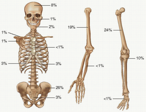
FIGURE 4.24. Skeletal distribution of Paget’s sarcoma.

The extremely poor prognosis in Paget’s sarcoma has been attributed to several factors by Mankin and Hornicek (101):


  • late stage at presentation compared with conventional osteosarcoma


  • higher percentage of metastatic disease at presentation


  • more rapid metastatic spread


  • higher risks in the use of chemotherapy due to higher age and poorer health of patients


  • risks associated with Paget’s disease-related high-out left ventricular heart failure


  • more axial location of Paget’s sarcoma compared with conventional osteosarcoma


  • harder detection of the tumors due to patient age-related mental frailty

Treatment (often futile), directed at surgical eradication, consists of limb salvage if possible and amputation if necessary. Chemotherapy and even radiation have been used with limited success. Little progress can be cited in the treatment of Paget’s sarcoma over the years with a dismal rate of survival of 14 percent at 2.5 years (101).

Benign tumors have also been described arising in Paget’s disease (103,104). These include giant-cell lesions (104) and

desmoplastic fibroma (105). The most extensively evaluated benign lesion is the so-called Avellino tumor (Fig. 4.26).


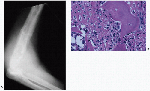
FIGURE 4.25. Paget’s sarcoma. Roentgenogram of Paget’s sarcoma of the humerus with superimposed destructive lytic lesion (A), proven to be an anaplastic sarcoma on biopsy (B).


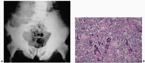
FIGURE 4.26. Giant-cell lesion in Paget’s disease. An 86-year-old white male immigrant from Avellino, Italy, with worsening pain in both extremities and weakness and progressive inability to ambulate. Physical examination revealed a large, left-sided, soft, immobile, painless mass predominantly involving the left buttock. Pelvic x-rays reveal diffuse pagetic changes of the pelvis, lumbar spine, sacrum, both pubic rami, and both proximal femora, and thickening of the iliopectineal line, all of which are classic for Paget’s disease of bone (A). A large lytic lesion was observed eroding the left iliac wing, L4, L5, and the sacrum. Computed tomogram was remarkable for a left-sided soft tissue mass invading the iliac crest, lumbar spine, and spinal canal. Pathology revealed multinucleated giant cells of various sizes and shapes, often with peculiar clear inclusions (B).

Only gold members can continue reading. Log In or Register to continue

Stay updated, free articles. Join our Telegram channel

Jul 24, 2016 | Posted by in PATHOLOGY & LABORATORY MEDICINE | Comments Off on Metabolic Bone Disease: Part II

Full access? Get Clinical Tree

Get Clinical Tree app for offline access