http://evolve.elsevier.com/Edmunds/NP/
DRUG OVERVIEW FOR TREATMENT—SELECTED CONDITIONS
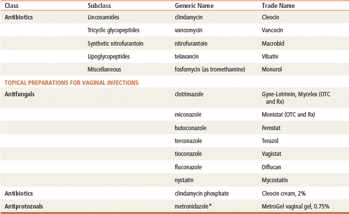
∗Metronidazole is classed as both an antibiotic and an antiprotozoal.
Therapeutic Overview
The student and the beginning clinician may be overwhelmed by the numerous antibiotics that are available. Table 58-1 serves as a general summary of common offending organisms for various infections. Both the first treatment choice and the alternative treatment suggestions listed are based on a compendium of various authorities.
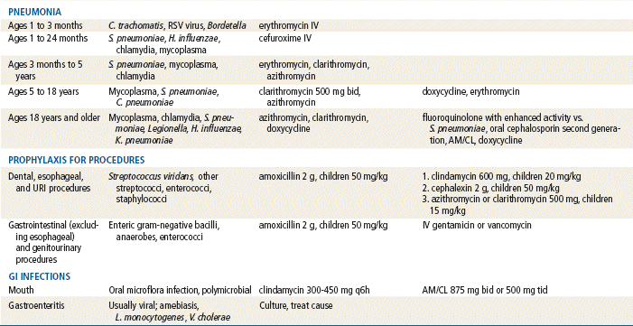
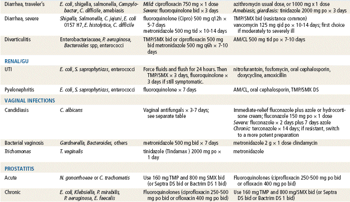
TABLE 58-1
Empirical Antimicrobial Treatment∗
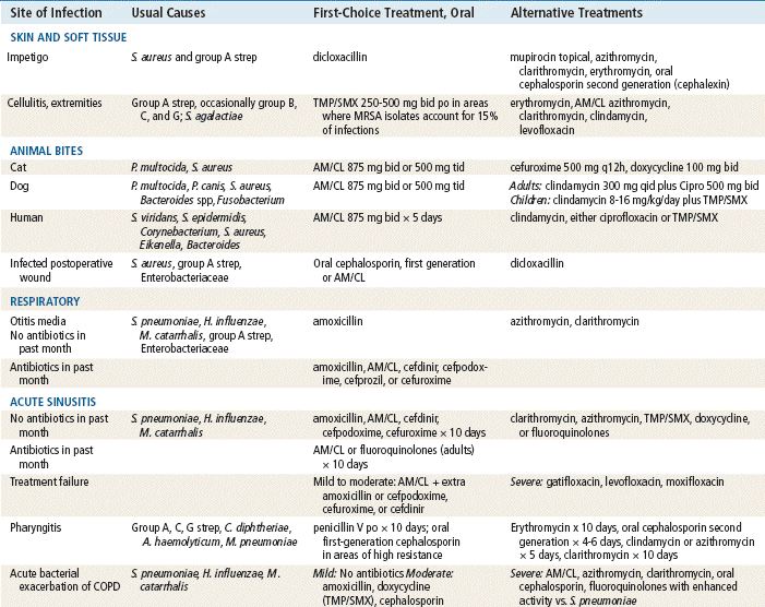
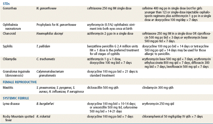
Fluoroquinolones with enhanced activity vs. S. pneumo: gatifloxacin, levofloxacin, and moxifloxacin.
∗Treatment recommendations, dosages, and therapy may vary, depending on age, pregnancy, or lactation. Consult the latest CDC recommendations at www.CDC.gov.
This chapter presents antimicrobial therapy according to disease- or site-specific recommendations. Treatments presented include recommendations for both children and adults when available. These are general guidelines for simple infections, with no complicating factors. See up-to-date specific drug information and check dosages before prescribing, especially for children and the elderly, because recommendations often vary with changing resistance patterns.
Skin and Soft Tissue Infections, Including Impetigo
Guidelines are provided by the Infectious Diseases Society of America (www.idsociety.org):
Impetigo is a contagious infection of the skin that is common in children. The lesions are macules, vesicles, bullae, pustules, and honey-colored crusts that usually appear on the face and other exposed skin. Systemic antibiotics usually are required. However, mupirocin (Bactroban) can be used for topical treatment of mild impetigo.
SSSI guidelines from the Infectious Diseases Society of America suggest vancomycin and telavancin for hospitalized patients with methicillin-resistant Staphylococcus aureus (MRSA).
Cellulitis
Although most cellulitis seen in primary care is easily treated, the provider must rule out the following complicated patients for whom they should seek referral:
Animal Bites
The location and type of the wound are important factors, as is the type of animal, in selection of appropriate treatment for the bite wound. Unprovoked animal bites should raise the suspicion of rabies. Cat bites are more likely to become infected than are human or dog bites. Human bites by children are not likely to become infected; infection is more likely with bites by adults. Dog bites are very unlikely to become infected.
Give the patient a tetanus/diphtheria booster if not vaccinated within the previous 5 years. Do not suture/glue the wound closed if it has been longer than 12 hours since the bite occurred, or if the injury is a hand wound or a cat bite. Prophylaxis is recommended for bites on the hand or in the genital region, for human or cat bites, for crush and puncture wounds, and for the treatment of patients with impaired immune systems.
Respiratory Tract Infections
By far, the most common causative agent is a virus, which causes an acute self-limiting disease that should not be treated with an antibiotic. Influenza is caused by a virus and can be treated with the drugs listed in Chapter 15. Evidence suggests that antibiotics, especially broad-spectrum antibiotics, continue to be overused for adult URIs.
Otitis Media
Signs and symptoms that indicate a need for antibiotic treatment include otalgia, fever, otorrhea, or a bulging yellow or red tympanic membrane. Simple effusion (i.e., presence of fluid in the middle ear with no signs or symptoms of acute infection) does not have to be treated with antibiotics. Much controversy has arisen about treatment with antibiotics in the past. However, doctors and patients are becoming comfortable with the new approach of treating with an antibiotic only if clearly indicated. Patients with tympanic membrane perforation, chronic or recurrent infection, craniofacial abnormalities, or immune compromise should be referred to an ENT specialist for treatment. How recently the patient has been treated with antibiotics will affect the antibiotic choice.
Sinusitis
This infection has a very similar pattern to that of otitis media, although perhaps more unusual bacteria are seen. The Infectious Diseases Society of America released new guidelines advising providers against routine antibiotic treatment of sinusitis, because most cases are caused by viruses. When antibiotics are used, the guidelines suggest amoxicillin-clavulanate instead of amoxicillin alone. In some cases, the new guidelines call for shorter treatment times. The common cold frequently involves the sinuses. Evidence of bacterial infection includes prolonged symptoms without improvement for 10 to 14 days or longer, fever higher than 102°F (39°C), unilateral pain, sinus tenderness, or tooth pain. Nasal discharge is not a reliable indicator of bacterial sinusitis. With viral URIs, the nasal discharge may change from clear to yellow during the first few days of the infection. Suspect a bacterial infection if the discharge changes to green. If the patient starts to improve and then worsens, this may indicate that a secondary bacterial infection is present.
An analysis of six studies involving almost 2500 patients found that corticosteroid nasal sprays offer small benefit in relieving acute sinusitis symptoms after 3 weeks at fairly high doses. Researchers found that the corticosteroid sprays work as well as antibiotics, which were given to 90% of patients but benefited only 1 in 15.
Unfortunately, younger children may not present with the classic signs or symptoms of sinusitis. Congestion and cough that last for longer than 10 days, high fever, and purulent nasal discharge are the most likely symptoms.
Because the causative organisms are similar to those of otitis media, treatment is very similar. Decongestants and mucolytics may improve drainage. Nonpharmacologic remedies such as drinking hot fluids, applying moist heat, inhaling steam, and using saltwater nasal sprays or rinses may decrease congestion. Research is mixed on the effectiveness of normal saline rinses.
Pharyngitis
Guidelines have been provided by the Infectious Diseases Society (www.idsociety.org):
Once again, virus (including mononucleosis) causes most cases of pharyngitis. The classic infection to be treated with antibiotics is group A β-hemolytic streptococci. It is extremely difficult to accurately diagnose a bacterial pharyngitis by history and physical alone. A throat culture is necessary. Symptoms such as pharyngeal pain, dysphagia, fever, tonsillar exudate, and lymphadenopathy with absence of cough increase the likelihood of a diagnosis of strep. Approximately one half of patients with these symptoms will have strep throat on culture. In areas with increased resistance to penicillin, an oral cephalosporin is more likely than penicillin to be effective.
Because of diagnostic vagueness, the provider has the option of waiting 2 days for culture results or treating empirically. Immediate empirical treatment is tempting because of symptom relief and decreased transmission but will result in substantial overtreatment. To avoid this dilemma, a rapid antigen detection test can be used. If used carefully, these tests can be very accurate. If the rapid test is positive, initiate antibiotic treatment. If it is negative, await culture results before initiating antibiotic treatment.
Acute Bronchitis
Bronchitis is a poorly defined illness, involving a cough. A virus causes almost all cases of bronchitis. Rarely, a child older than 5 years will have bronchitis caused by M. pneumoniae or C. pneumoniae. If the child does not get better in 10 to 14 days, consider treatment with a macrolide.
Acute Bacterial Exacerbation of COPD
Many acute exacerbations of COPD are viral. Patients may have any one of a wide variety of bacteria, depending on previous hospitalizations and so forth. These patients are likely to be frail, to have a history of smoking, and to have been previously treated with many antibiotics. A sputum culture may be difficult to obtain but will be very useful. Obtaining a sputum sample after a nebulization treatment may be necessary. Try to get the patient to stop smoking.
Community-Acquired Pneumonia (CAP)
The Infectious Diseases Society of America has recently released guidelines for CAP (www.idsociety.org). It is helpful for the clinician to differentiate disease origins by differentiating between bacterial and atypical organisms.
Treatment lasts until the patient is afebrile for at least 5 days for pneumococcal pneumonia, often for 10 to 14 days. Pneumonia caused by Enterobacteriaceae, Pseudomonas, or Staphylococcus is treated for 21 to 28 days.
Because so many different bacteria can cause pneumonia, determining empirical treatment can be problematic. Mycoplasma or chlamydial infections are common, and a macrolide is recommended. However, penicillin-resistant pneumococci occur regularly and may be resistant to a macrolide or doxycycline. Frail elderly patients may require treatment with one of the fluoroquinolones.
Prophylactic Regimens for Dental, Oral, Respiratory Tract, or Esophageal Procedures
These regimens are performed for patients with risk for endocarditis or with prosthetic devices, including knee, hip, and shoulder replacements. Research is mixed concerning use of prophylactic antibiotics in these situations.
Many clinicians give antimicrobial prophylaxis before patients receive vascular grafts or orthopedic prostheses. Others recommend prophylaxis before dental or genitourinary procedures are performed. Prophylaxis is not recommended for dialysis catheters, ventriculoperitoneal shunts, cardiac pacemakers, and defibrillators.
GI Infections
Mouth infections can be caused by a large variety of bacteria but often respond to penicillin. Because uncontaminated cultures are difficult to obtain, treatment with a broad-spectrum agent occasionally may be necessary.
Traveler’s Diarrhea
If the patient is traveling to an area with endemic infections and may be unable to adequately observe safety precautions (e.g., bottled water), a course of antibiotics may be provided so the patient is able to initiate treatment immediately on onset of symptoms. Treatment also may include loperamide and/or Pepto-Bismol. A transcutaneous immunization with a heat-labile toxin in a patch in two doses taken 2 to 3 weeks apart prior to travel is being tested to protect individuals against E. coli traveler’s diarrhea.
Severe Diarrhea
Symptoms of severe diarrhea include more than six liquid stools per day, temperature greater than 101°F, tenesmus, blood, and fecal leukocytes. Patients always require a stool for fecal leukocytes and culture. Treatment is based on culture results.
C. difficile is often a consequence of treatment with all antibiotics. The four antibiotics most likely to cause C. difficile infection are ampicillin, amoxicillin, cephalosporin, and clindamycin. The incidence of infection is high with these drugs because of their widespread use in children. Fluoroquinolones also are implicated in C. difficile–associated diarrhea, again because of widespread use. Tetracycline use is least likely to cause this problem. The presentation of C. difficile varies from mild illness to rapid clinical deterioration. It often is associated with copious liquid diarrhea, although it can be less acute in onset and moderate in amount of liquid stool produced. Suspect C. difficile in any patient who develops diarrhea after taking an antibiotic. Onset of symptoms can be delayed for weeks after treatment with an antibiotic. Because of the potential for severe symptoms, treat promptly.
Clostridium difficile is treated with metronidazole 500 mg po three times daily for 10 days for mild to moderate infection. More severe infection is treated with vancomycin 125 mg po four times daily for 10 days. Treatment of Clostridium difficile is covered in Chapter 27 on histamine blockers. Vancomycin and metronidazole are discussed in detail at the end of this chapter.
Stay updated, free articles. Join our Telegram channel

Full access? Get Clinical Tree


