Thyroiditis


Thyroiditis


Paul W. Biddinger


Thyroiditis encompasses a heterogeneous group of inflammatory diseases of the thyroid. The study of these inflammatory conditions can be challenging because of the different approaches to classification and various synonyms for a particular type of thyroiditis. Classification can be based on whether the clinical course is acute, subacute, chronic, or subclinical (Table 4.1). Other approaches to classification include the type of inflammatory response, the etiologic or pathogenic mechanism (e.g., autoimmune, infectious), or the effect on thyroid function.

This chapter is structured on the basis of predominant inflammatory cell type and utilizes the nomenclature for thyroiditis as shown in Table 4.1. The most common clinically significant form of thyroiditis is chronic lymphocytic thyroiditis, which describes histopathologically the autoimmune condition commonly called Hashimoto thyroiditis. In this chapter, chronic lymphocytic thyroiditis and Hashimoto thyroiditis are considered synonymous. Subacute forms of thyroiditis are divided according to the predominant inflammatory response (granulomatous or lymphocytic). The rare fibroinflammatory process known as Riedel thyroiditis is also included in this chapter. Diffuse toxic hyperplasia, or Graves disease, could qualify for consideration as an inflammatory disease of autoimmune etiology. However, it is covered in Chapter 5 as a hyperplastic disorder.


ACUTE THYROIDITIS


Definition and Etiology

Acute thyroiditis is characterized by an inflammatory infiltration with a predominance of neutrophils. An alternate term is suppurative thyroiditis. This rare form of thyroiditis is usually caused by bacterial infection. Common infectious organisms include Staphylococcus aureus, Streptococcus pyogenes, Streptococcus pneumoniae, other streptococcal species, Eikenella corrodens, or a mixture of oropharyngeal flora.1,2,3,4,5,6 Various other aerobic or anaerobic bacteria, mycobacteria, and parasites have also been reported as etiologic agents.1,7,8,9 Fungi can cause thyroiditis with acute, chronic, and/or granulomatous inflammation, and fungal infections are discussed below in the section on granulomatous thyroiditis. Usually, the thyroid is secondarily involved either by an inflammatory process arising elsewhere in the neck or by a generalized septic process. Individuals with increased risk include those with preexisting thyroid disease such as carcinoma, chronic lymphocytic thyroiditis, or multinodular goiter; those with a pyriform sinus fistula; and immunocompromised or debilitated individuals.10 Acute thyroiditis may be secondary to trauma or a rare complication of fine needle aspiration (FNA).11,12,13

In children, acute thyroiditis is usually associated with a pyriform sinus fistula.5,14,15 Instances of acute thyroiditis owing to pyriform sinus fistula have also been reported in adults.14,15,16,17,18 Pyriform sinus fistula is thought to be a developmental remnant of the thymopharyngeal duct (see Chapter 2). The thymopharyngeal duct is a structure that forms as the thymus descends from the third pharyngeal pouch during fetal development. A persistent duct can result in a fistula, or more accurately a sinus tract, that typically extends in an anteroinferior direction from the pyriform sinus, ending blindly in the thyroid or perithyroidal tissue. Most cases occur on the left side.5,15,19,20 Infections of the upper respiratory tract can be complicated by extension into a pyriform sinus fistula. These fistulae are usually demonstrated with a radiopaque fluid. A thyroglossal duct remnant is another potential means of a suppurative process reaching the thyroid.21 Infections of thyroglossal duct cysts are relatively common, but the infectious process rarely involves the thyroid gland itself.22,23


Clinical Features

Common signs and symptoms of acute thyroiditis include fever, chills, sore throat, hoarseness, dysphagia, and neck pain. Neck pain is often unilateral and radiates to the jaw or ear. Acute thyroiditis is usually associated with a euthyroid state, but thyrotoxicosis has also been reported in some cases.17,24,25,26,27


Pathology

The size of an affected gland ranges from normal to mildly enlarged. Enlargement may be focal or diffuse, reflecting the extent of inflammation. Microscopic examination reveals a predominately neutrophilic infiltration (Fig. 4.1). The concentration of neutrophils can vary according to the causative agent and the immune status of the patient. Necrosis with microabscess formation is common in cases caused by pyogenic bacteria. Immunocompromised individuals may show minimal inflammatory response. FNA and culture are typically used to confirm a clinical diagnosis of acute thyroiditis. Aspiration yields evidence of acute inflammation, either overtly as purulent exudate or more subtly with a combination of cellular debris, fibrin strands, and neutrophils.28


Differential Diagnosis

Acute thyroiditis can present as a neck mass. The differential diagnosis of a painful anterior neck mass includes infected thyroglossal duct or branchial cleft cyst, lymphadenitis, cellulitis, and infected cystic hygroma.4 Subacute granulomatous thyroiditis is typically associated with thyroid pain and fever, and although thyroid enlargement is usually diffuse, some cases present with localized, nodular enlargement (see Subacute Granulomatous Thyroiditis section below). Acute thyroiditis can also mimic a primary thyroid neoplasm, particularly anaplastic carcinoma if enlargement is rapid, or an extrathyroidal neoplasm with local extension.29,30,31,32 Older adult age, history of dysphonia, right thyroid lobe involvement, large lesional size, sterile cultures of thyroid aspirates, and lack of improvement after antibiotic therapy favor neoplasia over acute thyroiditis.


Table 4.1 Thyroiditis Classification by Inflammatory Response and Clinical Course


Predominant Inflammatory Cell


Name


Synonyms


Subcategories


Clinical Course


Neutrophil


Acute thyroiditis


  • Acute suppurative thyroiditis


  • Infectious thyroiditis


  • Bacterial thyroiditis


  • Mycobacterial thyroiditis


  • Fungal thyroiditis


  • Parasitic thyroiditis


Acute


Macrophage/histiocyte


Subacute granulomatous thyroiditis


  • de Quervain thyroiditis


  • Subacute thyroiditis


  • Painful subacute thyroiditis


  • Postviral thyroiditis


  • Giant cell thyroiditis


  • Subacute nonsuppurative thyroiditis


  • Pseudotuberculous thyroiditis


  • Struma granulomatosa


Subacute


Infectious granulomatous thyroiditis


  • Infectious thyroiditis


  • Tuberculosis


  • Fungal thyroiditis


Subacute to chronic


Sarcoidosis Granulomatous vasculitis


Subacute to chronic to subclinical


Other granulomatous reactions


  • Reaction to hemorrhage


  • Reaction to surgery


  • Foreign body reaction


Palpation thyroiditis


  • Multifocal granulomatous folliculitis


Subclinical


Lymphocyte


Chronic lymphocytic thyroiditis


  • Hashimoto thyroiditis


  • Autoimmune thyroiditis


  • Struma lymphomatosum


  • Classic


  • Fibrous variant


  • Atrophic or fibrous atrophy variant


  • Juvenile variant


  • Hashitoxicosis variant


Chronic


Silent thyroiditis


  • Sporadic thyroiditis


  • Painless thyroiditis


  • Painless sporadic thyroiditis


  • Painless thyroiditis with hyperthyroidism


  • Silent thyrotoxic thyroiditis


  • Subacute lymphocytic thyroiditis


  • Atypical subacute thyroiditis


  • Spontaneously resolving hyperthyroidism


  • Lymphocytic thyroiditis with spontaneously resolving hyperthyroidism


Subacute


Postpartum thyroiditis


  • Painless postpartum thyroiditis


Focal lymphocytic thyroiditis


  • Nonspecific thyroiditis


  • Focal autoimmune thyroiditis


Subclinical


Invasive fibrous thyroiditis


  • Riedel thyroiditis


  • Fibrosing thyroiditis


  • Sclerosing thyroiditis


Chronic


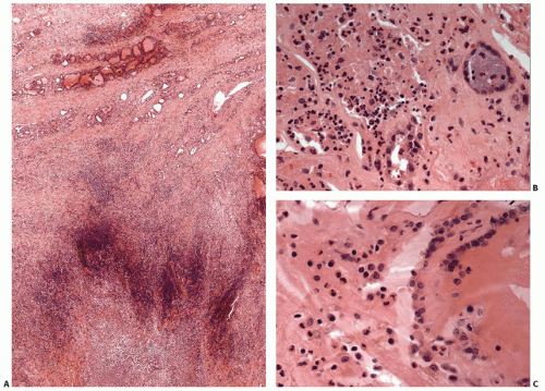
FIGURE 4.1. Acute thyroiditis secondary to a bacterial infection that initially affected extrathyroidal tissues. A: Low-power photomicrograph showing abscess (lower) with extension of inflammatory process into thyroid (upper). B and C: Medium- and high-power photomicrographs showing abundant neutrophils associated with damaged and necrotic follicles.


GRANULOMATOUS THYROIDITIS


Subacute Granulomatous Thyroiditis (Subacute or de Quervain Thyroiditis)


Etiology

Viral infection is the most suspect etiology given the common prodromal signs and symptoms, and several studies offer support.33,34,35,36 The incidence is highest in the summer when the incidence of enteroviral infections is also highest.37 One study of SGT demonstrated intrathyroidal activated T cytotoxic/suppressor cells and interferon γ (INF-γ) positive lymphocytes consistent with a viral infection.38 Several viruses have been implicated as causative agents including coxsackievirus, adenovirus, mumps virus, Epstein-Barr virus, rubeola (measles), Varicella zoster (chicken pox), cytomegalovirus, influenza, rubella, and human foamy virus.39 However, the etiologic association with viral infection is not conclusive because several studies have failed to find evidence of viral infection.39,40,41,42

Some cases of subacute granulomatous thyroiditis have been associated with antiviral therapy, particularly INF.43,44,45,46 There have been recent reports of subacute granulomatous thyroiditis among individuals receiving etanercept, an antagonist of tumor necrosis factor α.47,48 Certain individuals may have a genetic predisposition for this disease, including those with HLA-B35 haplotype.49,50,51,52,53,54


Clinical Features

Subacute granulomatous thyroiditis generally affects young to middle-aged adults.55,56,57,58,59 Children are rarely affected.60 Women are affected more commonly than men by a ratio ranging from 2 to 4:1.57,58,59 Patients usually experience a prodrome of low-grade fever, myalgias, and fatigue, and present with fever and neck pain. Subacute granulomatous thyroiditis is the most common cause of a painful thyroid gland.56,61 However, a significant number of patients do not experience pain or tenderness. C-reactive
protein and erythrocyte sedimentation rate are usually elevated.62 A minority of patients have significant elevation of antibodies to thyroglobulin (Tg) or thyroperoxidase (TPO), but these elevated titers are usually transient.58,59,63,64,65

Subacute granulomatous thyroiditis commonly presents with hyperthyroidism and, after a period of weeks to several months, passes through a euthyroid phase. Rarely, patients may present with thyroid storm.66,67 The euthyroid stage is typically followed by a hypothyroid stage that may last for weeks to months before resumption of normal thyroid function.10,64,68 Spontaneous return to euthyroidism occurs in most patients within 6 to 12 months, but between 2% and 15% of patients experience persistent hypothyroidism.57,64,69,70,71 Recurrence has been reported in about 2% of cases after a latency that may exceed 10 years.72,73 Conversion to chronic autoimmune thyroiditis or Graves disease has been reported.74,75,76,77,78


Pathology

The thyroid gland is enlarged, typically about twice its normal size. The enlargement is often asymmetric, even though the inflammatory process usually affects the entire gland. A small percentage of cases present as a solitary nodule.79,80 Areas with more pronounced changes appear as tan to yellow-white, ill-defined nodules that are firm to palpation (Fig. 4.2).

Microscopic findings exhibit temporal variation. The early stage is characterized by follicular damage with loss of epithelium and colloid. Acute and chronic inflammatory cells fill the residual follicles and extend into the surrounding interfollicular areas. During the early stage, neutrophils are the predominant inflammatory cell and microabscesses may be present.

Over time, the inflammation attains a granulomatous and chronically inflamed appearance with epithelioid and nonepithelioid macrophages, multinucleated giant cells, lymphocytes, plasma cells, and variable degrees of fibrosis (Fig. 4.3).81,82,83 Giant cells are typically associated with disrupted follicles and may encircle residual colloid. Distinct granulomata or lymphoid follicles are not characteristic of subacute granulomatous thyroiditis. Reparative changes become more apparent during the latter course of the disease as fibrous tissue replaces the areas of follicular destruction. With time, the fibrotic areas are partially or fully restored to follicular tissue. Zones of active inflammation can coexist with areas of fibrosis as the inflammatory process spreads to previously uninvolved thyroid parenchyma.


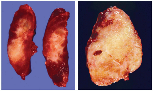
FIGURE 4.2. Cut surfaces of two different thyroids with subacute granulomatous thyroiditis. The inflammation and fibrosis in the thyroid pictured on the left resulted in a lesion clinically interpreted as a nodule.

Controversy exists regarding the nature of multinucleated giant cells seen in subacute granulomatous thyroiditis. Some have thought that they represent altered follicular cells or macrophages surrounding extravasated colloid that collectively simulate giant cells (pseudogiant cells).33,84 However, most consider them to be macrophages (histiocytes) that have fused into a single multinucleated cell comparable with those seen in other types of granulomatous inflammation. Immunohistochemical studies have found that most giant cells are positive for CD68, lysozyme, α1-antitrypsin, and vimentin although being typically negative for Tg, epithelial membrane antigen, and cytokeratin.81,85 An ultrastructural study revealed that most multinucleated giant cells had structures identical with those of infiltrating macrophages.85


Cytopathology

Cases of subacute thyroiditis are rarely seen as resection specimens because most are diagnosed on the basis of clinical features and laboratory tests and are treated medically. Increased use of sonography and FNA have also decreased surgical procedures.86 In the early stage of disease, fine needle aspirates contain neutrophils in addition to mononuclear inflammatory cells and
degenerated follicular cells.82,83,87 Oncocytic follicular cells are absent. In the latter stages, characteristic findings include multinucleated giant cells, strands of fibrous tissue, and a mixture of lymphocytes, macrophages, and possibly neutrophils.88,89,90 Follicular cells are usually scant to absent, and granulomata are rarely seen.


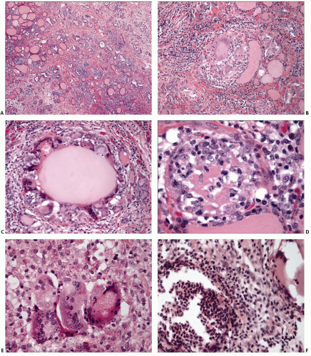
FIGURE 4.3. Subacute granulomatous thyroiditis. A: Low-power view showing decreased numbers of follicles. B and C: Medium-power views showing follicular damage with loss of epithelium and colloid and encirclement of follicle by multinucleated giant cells. D and E: High-power views showing granulomatous inflammation with predominance of macrophages and giant cells. F: High-power view showing mixture of acute and chronic inflammation with abundant neutrophils within an effaced follicle.


Differential Diagnosis

The differential diagnosis of subacute granulomatous thyroiditis includes sarcoidosis, palpation thyroiditis, and other forms of granulomatous thyroiditis that are discussed in the next section.


Other Types of Granulomatous Thyroiditis


Palpation Thyroiditis

Palpation thyroiditis is a very common finding in surgically resected thyroid glands. As the name implies, it appears to be caused by pressure-induced damage and rupture of follicles by squeezing during manual examination.91 Focally, palpation thyroiditis bears resemblance to subacute granulomatous thyroiditis, and an alternative name is multifocal granulomatous folliculitis. The damage associated with palpation thyroiditis is slight and not associated with clinical signs and symptoms, impairment of thyroid function, or significant release of Tg into the blood stream.92

Palpation thyroiditis is not evident grossly in most cases. Foci of hemorrhage are seen occasionally. Microscopic examination reveals focal damage limited to a single or small group of follicles. In the affected follicles, the epithelium is absent and a ring of multinucleated giant cells may encircle the residual colloid in its place (Fig. 4.4). The follicle may be totally effaced, with the colloid replaced by an infiltrate of macrophages, multinucleated giant cells, lymphocytes, and plasma cells. Palpation thyroiditis is distinguished from subacute granulomatous thyroiditis based on the very limited extent of changes. Typically, only single scattered follicles are affected, and there is absence of acute inflammatory cells and overt necrosis.


Tuberculosis

Tuberculosis of the thyroid is rare, and isolated tuberculosis is extremely rare because thyroid disease is usually associated with infection elsewhere in the body. Infection may occur through hematogenous dissemination or by direct extension from adjacent structures such as lymph nodes. The clinical presentation is variable. Normal thyroid function is usually maintained, and other thyroidal signs or symptoms may be absent, particularly if involvement represents miliary spread. Tuberculosis can also present as a nodule or abscess.93,94,95,96,97 Diagnosis of tuberculosis in nodular lesions is typically a surprise discovery during the evaluation for neoplasia.

The pathologic manifestations of thyroid tuberculosis are similar to those found in the lungs and other extrapulmonary organs. The typical histologic finding is multiple coalescing granulomata with and without necrosis (caseation).95,98 Abscess-like lesions can form when necrosis is abundant. Some lesions can exhibit marked fibrosis and therefore be particularly difficult to differentiate from subacute granulomatous thyroiditis. FNA plays an important role by allowing diagnosis without surgical resection. Tuberculosis should be included in the differential diagnosis when FNA reveals epithelioid granulomata, particularly when necrosis is also evident.99,100,101 Diagnosis can be confirmed by the demonstration of Mycobacterium tuberculosis with an acid-fast stain, culture, and/or DNA polymerase chain reaction.102


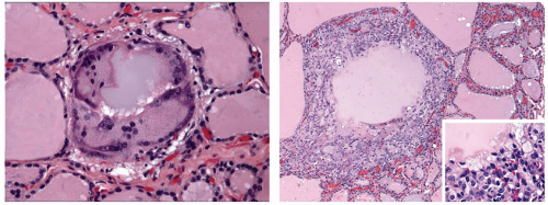
FIGURE 4.4. Palpation thyroiditis. Isolated follicle containing multinucleated giant cells (left). Disruption of a single follicle with a more intense surrounding inflammatory response (right). The high-power insert shows the predominance of macrophages.


Fungal Infection

Fungal infections of the thyroid can manifest with granulomatous inflammation, although a component of acute inflammation is often present and may predominate. Deep yeast infections such as histoplasmosis and coccidioidomycosis have been reported, but such cases are rare (Fig. 4.5).103,104 Aspergillus species are the most common cause of fungal thyroiditis and are usually part of a disseminated infection affecting immunocompromised individuals.8,105 The inflammatory response in these cases is not granulomatous but rather acute or absent (Fig. 4.6). The diagnosis of fungal thyroiditis rests on the demonstration of fungal yeast or hyphae, usually with the adjunct of specially stained sections.


Sarcoidosis

The thyroid may be involved in cases of sarcoidosis. Such instances seem to be rare, or at least rarely detected. Well-formed, nonnecrotizing granulomata, similar to those seen in other affected organs, are characteristic findings.106,107 The granulomata are typically located in the stroma and do not center on follicles, in contrast to subacute granulomatous thyroiditis and palpation thyroiditis. Inflammatory changes in sarcoidosis range from minimal to foci with effacement of thyroid architecture. Special stains for acid-fast bacilli and fungi are indicated to help rule out an infectious etiology.

Definitive diagnosis of sarcoidosis is problematic because of its uncertain etiology and the need to eliminate other granulomatous diseases. Discovery of nonnecrotizing granulomata in the thyroid may be just the initial step if similar granulomata have not been demonstrated in other tissues. Additional studies will be necessary to yield a diagnosis of sarcoidosis with reasonable confidence.

Sarcoidosis can present as a cold nodule that mimics a neoplasm.108,109 Coexistence of sarcoidosis and autoimmune thyroid
diseases has been observed.110,111,112,113,114 The most frequent coexisting disease is chronic lymphocytic (Hashimoto) thyroiditis. Although several cases of sarcoidosis with clinically overt chronic lymphocytic thyroiditis are relatively low, the incidence seems higher than by simple coincidence. The frequency of autoantibodies to Tg and/or TPO among individuals with sarcoidosis has been found to be as high114 as 27%. Rare cases of sarcoidosis associated with hyperthyroidism have also been reported.113,115,116,117


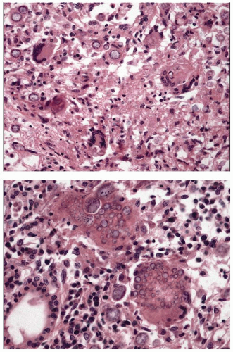
FIGURE 4.5. Coccidioidomycosis involving thyroid. An ill-defined granulomatous response is present focally (top) with scattered multinucleated giant cells. Overall, the inflammatory response in this case is predominately lymphoplasmacytic (bottom). Variably sized spherules are shown along with endospores within large spherules.


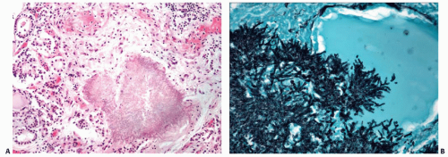
FIGURE 4.6. Fungal infection involving thyroid of an immunocompromised host. A: Medium-power photomicrograph showing aggregate of the fungus Scedosporium prolificans invading thyroid parenchyma with minimal inflammatory response (hematoxylin and eosin stain). B: High-power view showing fungal hyphae (Grocott methenamine silver stain).


CHRONIC THYROIDITIS


General Overview

Any discussion of chronic thyroiditis, particularly thyroiditis with predominately lymphocytic infiltrates, is fraught with difficulty related to the wide spectrum of histologic and clinical features and the attendant nomenclature. Table 4.1 illustrates the various names and synonyms that comprise these potentially confusing and sometimes overlapping classifications. A unifying approach is to view the various types of lymphocytic thyroiditis as manifestations of autoimmune disease, sharing the common feature of circulating antibodies to thyroid antigens. The prevalence of thyroid autoantibodies is relatively high in the general population, but the range of manifestations is broad.118 In many instances, autoimmunity is subclinical with no appreciable effect on thyroid function. The clinical course is variable because many cases remain subclinical for a prolonged time.119 Development of overt hypothyroidism is common after periods of time ranging up to a decade or more, but progression to overt disease does not seem to be an inevitable event.

The associated pathologic changes in turn range from focal lymphocytic infiltrates with minimal follicular damage to dense lymphoplasmacytic infiltrates with germinal centers and metaplastic epithelial changes and to atrophic glands with marked fibrosis. The dividing lines between categories in the spectrum of lymphocytic thyroiditis are not drawn sharply. Classification can be problematic, particularly in cases with early or limited inflammatory changes, and definitive evaluation will likely require integration of pathologic, clinical, and other ancillary studies. These confounding factors make it difficult to determine precisely the incidence of lymphocytic thyroiditis and its forms. However, it appears to be a common condition. Focal lymphocytic aggregates are seen in 10% to 45% of adult thyroid glands at the time of autopsy.120,121,122 Evidence of thyroiditis is more frequent among women, and in some groups of older women, >50% exhibit some degree of lymphocytic thyroiditis.


Chronic Lymphocytic Thyroiditis (Hashimoto or Autoimmune Thyroiditis)


Epidemiology

The disease has a peak incidence in the fifth decade of life, and affected individuals range from children to older adults.10,124,125 Although adults are more commonly affected than children, chronic lymphocytic thyroiditis is the most common form of thyroiditis among children and adolescents.124,126 Women are affected more commonly than men by a ratio ranging from 5 to 20:1.10,125,127

Only gold members can continue reading. Log In or Register to continue

Stay updated, free articles. Join our Telegram channel

Jul 9, 2016 | Posted by in PATHOLOGY & LABORATORY MEDICINE | Comments Off on Thyroiditis

Full access? Get Clinical Tree

Get Clinical Tree app for offline access