Chapter 5 The respiratory system
Development of the respiratory system
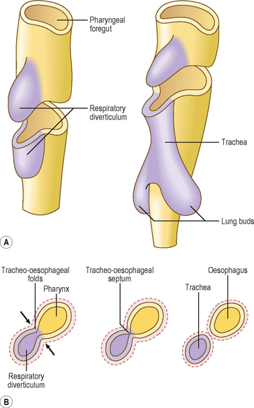
The epithelial component of the respiratory system (including that of the larynx, trachea, bronchi and lungs) develops from the ventral wall of the endodermal lining of the foregut as an outpouching, which grows into the surrounding splanchnopleuric mesoderm as primitive connective tissue surrounding the endodermal respiratory diverticulum (Fig. 5.1A). The same splanchnopleuric mesoderm also gives rise to the visceral pleura, cartilage, smooth muscle and blood vessels of the lower respiratory tract. The respiratory diverticulum develops at the junction between the cranial and caudal foregut. This diverticulum soon separates from the foregut by the development of bilateral longitudinal ridges, the tracheo-oesophageal folds, that fuse together to form the tracheo-oesophageal septum. This septum separates the ventral trachea from the dorsal oesophagus (Fig. 5.1B). Thus, the pharynx communicates with the larynx via the future laryngeal inlet.
The larynx is lined by epithelium derived from the endoderm, whereas the cartilages and muscles of the larynx arise from the fourth and sixth pharyngeal arches (see Chapter 11).
The lung buds are the paired structures that develop from a single midline outpouching, the respiratory diverticulum (Fig. 5.1A). Each lung bud divides many times to form the bronchial tree. The process by which the division of the airways is accomplished is known as branching morphogenesis and it is regulated by a variety of molecular signals. There are differences on the two sides: the right lung bud divides into three secondary bronchial buds, forerunners of the three lobes, whereas the left lung bud gives rise to two secondary bronchial buds, to become the two lobes on the left. Thus, a series of rapid further divisions of the airways takes place, penetrating the surrounding splanchnopleuric mesoderm and bulging into the coelomic cavity at the pericardioperitoneal canals (Chapter 3). These two canals are destined to become the right and left pleural cavities, and they lie on either side of the foregut (Fig. 5.2). The pericardial cavity (see Chapter 6) is separated from the primitive pleural cavities by the pleuropericardial folds on each side. The peritoneal cavity is separated from the pleural cavities by the pleuroperitoneal membranes (see Chapter 3). Whilst the splanchnopleuric mesoderm forms the visceral layer of the pleura, the somatopleuric mesoderm lines the thoracic walls and thus forms the somatic layers of the pleura.
Stay updated, free articles. Join our Telegram channel

Full access? Get Clinical Tree


