The Reproductive System


The Reproductive System


Perhaps one of the most important functions of humans is the ability to reproduce. While there is a plethora of literature related to the topic, many still find it a difficult and even embarrassing topic of conversation. Pathology of the reproductive system can interfere with an individual’s ability to contribute to the genetic pool.


● Physiologic Concepts (Male)


MALE REPRODUCTIVE ANATOMY

The reproductive role of the human male is to produce and deliver sperm to impregnate a female. To carry out these functions, a male has internal and external sexual organs. These structures include the testes, several tubules that carry sperm out of the testes, various glands, and the penis (Fig. 20-1).


The Testes

The testes are the male gonads. Testes develop during gestation in response to the production of androgenic hormones by the male embryo. The primary androgen is testosterone, the synthesis of which begins at approximately 8 weeks’ gestation.

During early gestation, the fetal testes are located in the abdominal cavity. At approximately 6 months’ gestation, the testes descend from the abdominal cavity through the inguinal canal into an external sac, called the scrotum. Associated blood vessels, nerves, and a supporting cord descend from the
abdominal cavity simultaneously. After descent, the abdominal opening of the canal closes. The scrotum sits dorsal to the penis, the male sexual organ and the male structure for urination. Because of their location outside the body, the testes remain at a lower-than-body temperature. This provides optimal conditions for spermatogenesis, or sperm formation.


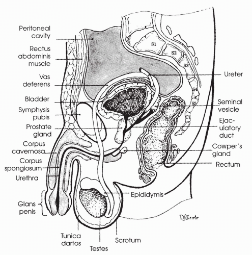
FIGURE 20-1 Side view of male genitourinary anatomy. (From Bullock, B. L. & Rosendahl, P. P. (1992). Pathophysiology: Adaptations and alterations in function (3rd ed.). Philadelphia, PA: Lippincott Williams & Wilkins.)


Seminiferous Tubules

Each testis is filled with hundreds of long, coiled tubules, called seminiferous tubules. Immature sperm arise from stem cells present in the tubular walls and then migrate into the tubule lumen. The seminiferous tubules include two types of cells: Sertoli cells, which line the inside of the tubules, and Leydig interstitial cells, which surround the outside of the tubules. The developing sperm receive essential support and nourishment from the Sertoli cells during their long maturation. The Leydig interstitial cells synthesize and secrete testosterone during gestation and after puberty. Testosterone is essential both for sperm maturation and Sertoli cell function.


The Epididymis, Vas Deferens, and Urethra

From the seminiferous tubules, sperm journey into another long tubule, the epididymis. The epididymis curves around the posterior side of the testes and ascends toward the peritoneal cavity. The epididymis leads into the vas deferens. The vas deferens enters the peritoneal cavity and widens to form a space called the ampulla, which has a glandular, convoluted structure on each side called the seminal vesicle.

At the ampulla, the vas deferens forms the ejaculatory duct. The ejaculatory duct passes through the prostate gland and joins with the internal urethra below the bladder. The internal urethra enters the penis and forms the urethra. Mucus-secreting glands line the urethra.


Sperm Maturation

Sperm at the entrance to the epididymis are immature and unable to fertilize an egg. By the time the sperm have traveled through the vas deferens (approximately 2 weeks), the sperm will have become fully mature. Mature sperm can be stored in the vas deferens and ampulla, where they remain viable for more than a month.


The Seminal Vesicles

With sexual excitement, the seminal vesicles secrete a mucuslike substance containing sugar, prostaglandins, and fibrinogen into the ejaculatory duct. The sperm use the sugar for energy, and the prostaglandins assist sperm in penetrating the female cervix. Prostaglandins may also cause contractions of the female genital tract, which propel the sperm in their journey to reach the egg.


The Prostate

The prostate is a walnut-shaped gland that sits beneath the bladder. During sexual excitement, the prostate secretes a thin, milky fluid containing various enzymes and ions into the ejaculatory duct. This mixture joins the sperm and seminal vesicle fluid. Prostate fluid is alkaline (basic). When it, along with the rest of the ejaculate, is deposited in the vagina of the female, it neutralizes the acidic vaginal secretions; this is important because sperm have poor motility in a low pH environment.


Neural Innervation of the Male Reproductive System

Afferent sensory neurons and efferent parasympathetic and sympathetic fibers are found throughout the male genitalia. Afferent sensory fibers are activated in response to tactile stimulation and send their information to the spinal cord. Parasympathetic nerves exit the spine at the level of the sacral area and innervate the arteries and arterioles of the penis. Parasympathetic fibers release the neurotransmitter acetylcholine, causing dilation of the blood vessels. Sympathetic
nerves exit the spine from the upper lumbar areas and innervate the smooth muscle of the vas deferens and ampulla. Sympathetic fibers release the neurotransmitter norepinephrine, which causes smooth muscle contraction. Sympathetic fibers also innervate and cause contraction of the prostate and the seminal vesicles. Descending neurons from higher centers of the brain, including the cerebral cortex, influence the firing of the parasympathetic and sympathetic fibers.


THE MALE SEXUAL ACT

Physical manipulation of the penis or sexual thoughts activate the parasympathetic and sympathetic nerves, causing sexual excitement. There are four stages of the male sexual act: erection, emission, ejaculation, and resolution. All stages can occur as simple spinal reflexes initiated by sensory stimulation. They do not require central nervous system involvement. Normally, however, both mental and physical stimuli contribute to sexual excitement. Inhibitory cerebral stimulation may interrupt the spinal reflexes at any point.


Erection

The penis hardens and becomes elongated during sexual excitement. An erection occurs following activation of the parasympathetic fibers to the penis that cause vasodilation and increased blood flow. As the arteries and arterioles of the penis fill up with blood, the veins draining the penis become compressed and occluded. Venous occlusion causes the spongy tissue in the shaft of the penis, the corpus cavernosum and corpus spongiosum, to become engorged. Engorgement of these tissues results in an erection. Parasympathetic stimulation also causes the glands lining the urethra to secrete mucus. Mucus lubricates the glans (head) of the penis, which facilitates and increases the pleasure associated with penetration of the female. During this stage, heart rate and respiratory rate increase.


Emission

When sexual excitement reaches a critical level, activation of the sympathetic nerves to the penis causes contraction of the vas deferens and ampulla. This contraction results in emission—the propulsion of the sperm out of the vas deferens and ampulla through the ejaculatory duct and into the internal urethra. During emission, sympathetic stimulation to the prostate and seminal vesicles causes release of prostate and seminal vesicle secretions into the ejaculatory duct. The combination of sperm, prostate secretions, and seminal vesicle secretions is called semen.


Ejaculation

With the addition of semen in the internal urethra, a feeling of fullness occurs. Sensory fibers traveling to the spinal cord transmit this feeling, resulting in
further activation of the sympathetic fibers and smooth muscle contraction of the ducts. Motor neurons to skeletal muscles at the base of the penis are also activated, leading to contraction of these muscles. The culminating responses are wavelike, rhythmic contractions associated with intense pleasure. During these contractions, semen is forcefully propelled through the urethra and out of the urethral opening. Emission and ejaculation comprise the male orgasm. Heart rate and respiratory rate reach a maximum at this time.


Resolution

After experiencing an orgasm, a male shows a reversal of sexual excitement, including disappearance of the erection and a return of heart rate and breathing patterns to normal.


SPERMATOGENESIS

Spermatogenesis (the formation of sperm) begins during puberty and continues throughout the lifetime of a male. Undifferentiated germ cells lining the seminiferous tubules undergo a programmed number of mitotic cell divisions, resulting in the production of the primary spermatocytes (immature sperm), which ultimately develop into the spermatozoa (mature sperm). Spermatogenesis requires approximately 2 months. From each primary spermatocyte, four viable sperm (each with 23 chromosomes) are produced. Spermatogenesis occurs in the seminiferous tubule under the control of two pituitary hormones—follicle-stimulating hormone (FSH) and luteinizing hormone (LH)—and the sex hormones, primarily testosterone.


Follicle-Stimulating Hormone

FSH is a protein hormone released from the anterior pituitary in response to a stimulating hormone from the hypothalamus: gonadotropin-releasing hormone (GnRH) (see Chapter 9). FSH binds to receptors present on the membranes of the Sertoli cells that line the seminiferous tubules, and activates a cyclic adenosine monophosphate (cAMP) second messenger system. The final effect of FSH is to cause Sertoli cells to proliferate and secrete various nutrients, ions, and proteins into the tubule that stimulate the continued proliferation and differentiation of the immature sperm.

Sertoli cells exert feedback on the hypothalamus and pituitary to control the further release of FSH by secreting the hormone inhibin. Inhibin levels rise with increased cell activity and inhibit the further release of FSH.


Luteinizing Hormone

LH is the second protein hormone released from the anterior pituitary in response to stimulation by GnRH. LH binds to the Leydig cells that surround
the tubule, and again through the activation of a cAMP second messenger system, stimulates the synthesis of the steroid hormone testosterone. Testosterone diffuses into the seminiferous tubules and binds to the Sertoli cells, stimulating them to continue to secrete the proteins, ions, and nutrients required to maintain proliferation and differentiation of the sperm. One protein that is manufactured by the Sertoli cells, androgen-binding protein, ensures that levels of testosterone remain high in the lumen of the seminiferous tubule. Mature Leydig cells usually develop at approximately 10 years of age in a boy.

Testosterone feeds back on the hypothalamus, and to a lesser extent on the anterior pituitary, to inhibit the further release of GnRH and LH. This feedback keeps the levels of circulating testosterone relatively constant. Besides being required for the successful formation of sperm, testosterone is also essential for the production of the male secondary sexual characteristics and the maintenance of the male libido (sex drive).


Stimuli Controlling GnRH Release

GnRH is released in small pulses throughout the day, resulting in relatively constant daily levels. Increases or decreases in GnRH release may occur seasonally and with different physical and psychological conditions such as anxiety or depression. Changes in the secretion of GnRH may affect sperm formation by affecting LH and FSH and may alter libido.


MALE SECONDARY SEXUAL CHARACTERISTICS

Male secondary sexual characteristics are under the control of the male androgens, especially testosterone. The effects of testosterone are described fully in Chapter 9. The male secondary sexual characteristics include the following:


  • Increased protein anabolism and muscle mass.


  • Increased bone growth and strength.


  • Male pattern of hair on the face, axillary, and pubic regions. Hair growth is thick on most areas of the body.


  • Increased metabolic rate, probably as a result of increased protein anabolism (buildup) and muscle mass formation. Increased metabolic rate raises the caloric needs of males, beginning at puberty, compared to females.


  • Proliferation and activation of sebaceous glands in the skin, which produce an oily substance called sebum. Increased amounts of sebum can cause acne, especially during teenage years.


  • A deepening voice, as a result of hypertrophy of the larynx.


  • Male pattern baldness, typically beginning with a bald spot on the top of the head. A genetic tendency influences male pattern baldness.


● Pathophysiologic Concepts (Male)


ERECTILE DYSFUNCTION

Erectile dysfunction (ED), previously called impotence, is the inability of a male to achieve or maintain an erection. ED can occur infrequently, frequently, or every time a man attempts sexual intercourse. When asked, approximately 35% of men 40 years of age and older, and more than 80% of men 70 years of age and older, report at least sporadic ED. Although in the past it was believed that ED was due mostly to psychological factors, it is now recognized that for most men physical causes are paramount (discussed later). With a greater awareness of the medical causes of ED, more open discussion of its prevalence, its effects on a man’s quality of life, and its treatment is becoming mainstream. Still, however, men frequently avoid seeking help for ED, thus making further education and services to promote men’s health and wellness necessary. Use of a tool such as The Sexual Health Inventory For Men, developed by Capparelli and Rosen, will encourage more dialogue. When ED is identified, extensive screening for coronary heart disease (CHD) should be initiated because of the high correlation between ED and CHD.


Physical Causes of Erectile Dysfunction

One of the main physical causes of ED is atherosclerosis of the penile arteries. With atherosclerosis, blood flow to the penis is reduced and there is an inability of the penile arteries to dilate during sexual excitement, thus limiting tumescence. Other physical causes of ED include systemic diseases such as hypertension, hyperlipidemia, renal failure, multiple sclerosis, Parkinson disease, stroke, hypothyroidism, acromegaly, and, most commonly, diabetes mellitus. Diabetes in particular is associated with atherosclerosis as well as neuropathy (nerve damage). At the cellular level, pathophysiologic changes contributing to ED include autonomic hypersensitivity, reduced production of nitric oxide by the prostate and the vascular smooth muscle of the penis, and endothelial cell dysfunction.

Besides physical factors, many drugs are known to interfere with a man’s ability to achieve an erection and/or an orgasm, including some antihypertensive and psychotropic medications. ED also may occur following surgery in the genital region, for example, after treatment for prostate cancer. Chronic or acute fatigue may also cause ED.


Psychological Causes of Erectile Dysfunction

Psychologically based ED occurs with activation of descending inhibitory impulses originating in the cerebral cortex. Psychological conditions associated with ED include stress, anger, worry, bereavement, fear of sexual failure, and depression.


Lifestyle Causes of Erectile Dysfunction

Lifestyle factors that may contribute to ED include smoking, alcohol abuse, recreational drug use, stress, and obesity. Smoking cessation and weight reduction have been shown to be effective in reducing the risk for CHD as well as ED.


Treatment of Erectile Dysfunction

There are many treatments available for ED, including mechanical aids and pumps and penile injections that cause local vasodilation. In addition, pharmaceutical advances have resulted in the production and marketing of several highly effective oral ED medications, the first of which was sildenafil citrate (Viagra). This type of drug acts to inhibit the enzyme phosphodiesterase that normally inactivates a second messenger required for relaxation of the penile arteries. By blocking phosphodiesterase, relaxation of the penile arteries is prolonged, allowing the penis to fill more fully with blood. An erection is achieved and augmented. ED medications are taken before attempting intercourse and enhance the normal sexual response; they do not stimulate an erection on their own. Side effects of oral ED medication may include headache, facial flushing, and visual abnormalities. Oral ED medications are contraindicated for men with certain types of heart disease or those taking other vasodilators such as nitroglycerin. Other medications may include adrenergic antagonists to potentiate parasympathetic neurotransmission and testosterone supplements.

For ED related to the side effects of prescribed medications, reevaluation of drug dosage or drug choice may reduce symptoms. ED associated with systemic disease or depression must be addressed directly. ED related to other psychological factors may be relieved by relaxation techniques, counseling, or sexual therapy. Studies suggest that in some men with ED, a strict weight loss and exercise program may help restore sexual function.


GYNECOMASTIA

Gynecomastia is the enlargement of breast tissue in males. It can result from excess production of estrogen in the male or the liver’s inability to break down normal male estrogen secretions. Gynecomastia is frequently seen during early puberty in some males and may be a normal development or may be related to excess body weight or a hormonal imbalance.


● Conditions of Disease or Injury (Male)

Table 20-1 includes a summary of male reproductive organ alterations. More detailed information about other alterations is included below.


TABLE 20-1 Summary of Male Reproductive Organ Alterations


Alteration


Overview


Cryptorchidism


  • Failure of one or both testicles to descent into the scrotum


  • Much greater risk for development of testicular cancer


  • May result in fertility issues


  • Causes: usually unknown; prematurity; testosterone deficiency; structural factors that impede gonadal descent; genetic predisposition


  • Clinical manifestations: testicle not palpable in scrotum


  • Treatment: hCG may stimulate descent; surgery; regular testicular self-exam


Hydrocele


  • Collection of plasma filtrate in the scrotum, outside the testes


  • Scrotal swelling causes reduced blood flow to testes


  • Causes: congenital, genital trauma, testicular tumor, idiopathic


  • Clinical manifestations: asymptomatic or scrotal swelling and discomfort


  • Treatment: identify cause and drain fluid


Varicocele


  • Abnormal dilation of vein in spermatic cord (usually left side)


  • Altered blood flow may cause infertility


  • Typically occurs between 15 and 25 years of age


  • Causes: valvular incompetence of spermatic vein or vena cava obstruction causing backflow and accumulation of blood; may be due to a renal tumor in older men


  • Clinical manifestations: asymptomatic or slight feeling of discomfort and testicular heaviness; torturous, dilated veins may be palpable


  • Treatment: testicular support garment; surgical ligation of the vein


Hypospadias/Epispadias


  • Hypospadias: Urethral opening located on ventral surface of the penis; in severe cases the opening may be in the scrotal or perineal area


  • Penis may be bowed downward (chordee), penile shaft may be rotated (torsion), foreskin may be altered


  • Complications may include infections, hematuria, calculi, painful intercourse


  • Causes: Congenital; genetic factors; hormonal variations; maternal ingestion of progestin; advanced maternal age


  • Treatment: surgical repair depending on severity; circumcism is not advised so the foreskin can be used for repair


  • Epispadias: congenital misplacement of urethral opening to the dorsal side of the penis; less common than hypospadias


Phimosis


  • Foreskin cannot be retracted back over the glans


  • Causes: poor hygiene; chronic infections


  • Clinical manifestations: edema, erythema, tenderness, purulent drainage


  • Treatment: circumcision


Paraphimosis


  • Foreskin is retracted; cannot be moved to cover the glans


  • Clinical manifestations: edema due to constriction


  • Treatment: surgery


Peyronie Disease


  • Lateral curvature of the penis during erection


  • Fibrous thickening of fascia in corpora cavernosa


  • May be associated with diabetes, keloids, beta-blockers


  • Clinical manifestations: painful erection, painful intercourse, erection problems distal to the fibrotic area


  • Treatment: surgery


Priapism


  • Prolonged penile erection


  • Idiopathic or associated with spinal cord trauma, sickle cell, leukemia, pelvic tumors, injections for impotence


  • Clinical manifestations: sustained, painful erection not associated with sexual arousal


  • Treatment: iced saline enemas, ketamine, phosphodiasterase 5 inhibitors, spinal anesthesia, aspiration of blood from corpus


Testicular Torsion


  • Rotation of the testicle on its vascular pedicle


  • Twisting of arteries and veins in the spermatic cord occludes circulation to the testicle


  • Most common in neonates and in adolescents


  • Clinical manifestations: pain and swelling


  • Treatment: surgical emergency if it cannot be reduced manually with scrotal elevation


BENIGN PROSTATIC HYPERPLASIA

Benign prostatic hyperplasia (BPH) is the noncancerous enlargement of the prostate gland. BPH is seen in more than 80% of men 80 years and older. BPH may cause compression of the urethra as it passes through the prostate, making urination difficult, reducing force of the flow of the urine stream, or causing dribbling of urine to occur. The cause of BPH is unclear but may be related to an imbalance between estrogen and testosterone in the prostate. The decrease in androgenic hormone production associated with aging produces an increase in dihydrotestosterone, the major prostatic intracellular androgen, and results in an androgen and estrogen imbalance. This imbalance causes changes in the periurethral glandular tissue of the prostate producing masses of fibrous glandular tissue that compresses the normal prostate tissue causing a nodular hyperplasia.


Clinical Manifestations


  • Increased frequency of urination, with delay in initiating urination and a reduction in the force of the urine stream.


  • Decreased diameter of the urinary stream.


  • As the condition progresses, the bladder may not empty completely, causing dribbling or urine overflow. The time required to void increases.


Diagnostic Tools


  • Diagnosis involves a good history and physical examination coupled with the use of imaging techniques. Biopsy of the prostate may be required to rule out neoplasia.


Complications


  • With advanced BPH, urinary tract obstruction may occur as urine is unable to pass through the prostate. Urinary obstruction can lead to urinary tract infections and, if unrelieved, renal failure.


Treatment


  • Mild prostate enlargement may not be treated, but followed in a “wait and see” manner.


  • Active surveillance involves annual monitoring for disease progression including a history, physical examination, and symptom score.


  • With more significant enlargement, medical therapy will be initiated. Alphablockers antagonize alpha-adrenergic receptors in the smooth muscle of the prostate causing relaxation of the muscle fibers thereby decreasing tension in
    the prostate. 5-alpha reductase inhibitors block the conversion of testosterone to dihydrotestosterone causing the prostate to shrink in size. A combination of the two types of medications significantly increases symptom improvement as opposed to monotherapy.


  • Surgical approaches that involve minimal invasiveness include transurethral incision of the prostate (TUIP). In this procedure, the gland is split in half surgically to reduce pressure on the urethra. Lasers may be used to split the prostate.


  • Other minimally invasive procedures to reduce the size of the prostate include transurethral needle ablation, transurethral vaporization, and transurethral microwave therapy.


  • If obstruction to urine flow is severe, transurethral prostatectomy (TURP) may be required to remove the enlarged prostate. Complications may include ED and incontinence.


  • A permanent catheter might be placed in patients unwilling to undergo or unable to tolerate surgery.


  • Annual digital rectal examinations and screening for prostate-specific antigen (PSA) are encouraged to identify a malignancy that may arise from hyperplastic cells.


INFLAMMATORY DISORDERS OF THE MALE REPRODUCTIVE TRACT

Inflammation of the male genital tract can occur anywhere between the testes and the urethral opening. Inflammation is usually due to a sexually transmitted disease or a urinary tract infection and is most commonly seen in sexually active men. Other causes of inflammation include systemic disease, such as mumps or trauma. Inflammation of the prostate may occur in older men with BPH. Common inflammatory conditions of the genitalia in men include the following.

Urethritis is an inflammation of the urethra. Urethritis is usually caused by a sexually transmitted microorganism, commonly Neisseria gonorrhoeae or Chlamydia trachomatis.

Epididymitis is an inflammation of the epididymis. Epididymitis is usually caused by a sexually transmitted microorganism, commonly N. gonorrhoeae or C. trachomatis. Epididymitis usually occurs from ascending urethral infection.

Orchitis is an acute inflammation of the testes. Orchitis usually develops following epididymitis or a systemic disease such as mumps.

Prostatitis is an inflammation of the prostate gland. Prostatitis is especially common in older men. It is often caused by an acute or chronic infection, usually ascending from the urethra. Prostatitis may be noninfectious and idiopathic in origin.


Clinical Manifestations


  • Urethritis may present with pain and burning on urination. A discharge from the penis may be present.


  • Epididymitis may present with acute scrotal or inguinal pain. Flank pain may be present. The scrotum may be inflamed and tender on the affected side.


  • Orchitis usually presents acutely with a very high fever (104°F) and swelling and redness of the testicle and scrotum. The individual appears very ill, and malaise is obvious.


  • Prostatitis from an ascending urinary tract infection usually presents with painful and frequent urination. Interrupted or slow urine stream and nocturia (urination at night) may be present. Fever and malaise are common. Low back or perineal pain is common, especially when standing. Digital examination reveals a very tender and enlarged prostate.


Diagnostic Tools


  • Diagnosis involves a thorough history and physical examination. Blood and urine cultures for the identification of an infectious organism may be required.


Complications


  • Epididymitis and orchitis may cause infertility, related to poor testicular blood flow, and infarct of the testicular cells.


Treatment


  • Antibiotic therapy is required for all bacterial or chlamydial infections.


  • Orchitis is treated with bed rest, analgesics for pain, and elevation of the testicles to increase venous drainage. Cold compresses may reduce initial inflammation. If a testicular abscess occurs, surgical removal of the testicle may be necessary.


CANCER OF THE MALE REPRODUCTIVE TRACT

Cancer of the male reproductive tract may develop in the penis, testes, or prostate.


Penile Cancer

Primary cancer of the penis is rare in the United States. It usually occurs in noncircumcised men, possibly related to accumulation of thick secretions (smegma) under the foreskin that increase the risk of sexually transmitted infections. It is slow growing and may occur anywhere on the penis. Primary penile
cancer usually develops in men between 40 and 80 years of age and is more common in African Americans than in Caucasians. Secondary penile cancer may occur from metastasis of bladder, rectal, or prostate cancer.


Testicular Cancer

Testicular cancer is rare, mostly occurring in young men between the ages of 15 and 35. It is usually a germ-cell (gamete) cancer but may develop from Leydig or Sertoli cells. The cause of testicular cancer is unknown, but there appears to be a genetic factor. Testicular cancer is more common in Caucasians and occurs more frequently in men with a history of cryptorchidism. Trauma and prenatal exposure to the synthetic estrogen diethylstilbestrol (DES) may increase risk.


Prostate Cancer

Prostate cancer is the number one cancer among American males and the second leading cause of death due to cancer in that population (the first is lung cancer). Prostate cancer is usually diagnosed in men older than 65 years of age; however, it is being diagnosed increasingly in younger men, perhaps as a result of more aggressive screening. Autopsy studies show that approximately 50% of men older than 50 years of age have some cancerous prostate cells; this finding has led to significant debate over recommended treatment, especially for elderly men with slow-growing, early-stage tumors.

The cause of prostate cancer is unknown, although both genetic and environmental factors are believed to play a role. The risk of prostate cancer is increased in men who have a first-degree relative with the disease, in African American men, and in men exposed to certain environmental or occupational toxins, such as cadmium. Prostate cancer appears to be related to lifelong levels of testosterone. Prostate cancers are testosterone dependent until late in the course of the disease.

Using clinical and biopsy results, prostate tumors are staged from A to D. Stage A tumors are well differentiated (A1) or moderately/poorly differentiated (A2) but restricted to the prostate gland. These tumors are asymptomatic, and their presence is reported in more than 80% of men older than 80 years of age. Stage A tumors cannot be felt on digital examination. Stage B tumors include a single nodule (B1) or a group of discrete nodules (B2) palpable on digital examination and confined to the prostate. Stage C tumors are large masses that fill the entire prostate gland (C1) and may extend beyond the edges of the gland (C2). Stage D tumors are metastatic, with cancerous cells found in the lymph nodes draining the pelvis (D1) and in other sites (D2), often the bone.


Clinical Manifestations


  • Penile cancer is characterized by a painless wartlike growth or an ulcerative lesion under the prepuce. It may also be a reddened lesion with plaque.


  • Testicular cancer is characterized by the development of a mass in the testis, which may become painful as it grows. Testicular heaviness or aching may occur. Gynecomastia may develop.


  • Prostate cancer may be asymptomatic or associated with increased frequency and urgency of urination, and a decrease in the force of the urine stream. Blood may be passed in the ejaculate, and in advanced disease, back pain may be present.


Diagnostic Tools


  • Biopsy of cells of the penis can diagnose and stage penile cancer.


  • Transillumination of the testes, ultrasound, and MRI may identify a testicular mass and support clinical findings of a testicular cancer.


  • Tumor markers commonly associated with testicular cancer include alphafetoprotein (AFP), beta human chorionic gonadotropin (hCG), and lactate dehydrogenase (LDH).


  • A digital rectal examination (DRE) may reveal a fixed, firm mass in the prostate, suggestive of a tumor. The mass is often painless with irregular borders and results in asymmetry of the prostate gland. Ultrasound may be used to pinpoint the location of a prostate tumor. A biopsy of prostate cells taken via a transurethral resection can confirm the diagnosis of prostate cancer. DRE should be conducted annually beginning at 50 years of age.


  • Prostate-specific antigen (PSA), a blood test that measures the level of a glycoprotein released by the prostate gland, can be used to identify the presence of even early-stage prostate cancer. Current recommendations are to perform a biopsy if the PSA level is greater than 4 nanograms per milliliter (ng/mL) of blood; levels greater than 10 ng/mL suggest cancer. However, PSA elevation may occur with noncancerous conditions such as prostatitis or benign prostatic hyperplasia (BPH). Likewise, in approximately 25% of men, PSA measurements may be in the normal range even when cancer is present. The need to treat stage A cancers detected by PSA assay is controversial, especially in elderly men.

A recently suggested strategy to address the issue of false-negative and false-positive PSA readings is to evaluate PSA levels on a sliding scale, depending on if the male is young (younger than 50 years) or older. For young men, a PSA of even 2.6 ng/mL may be considered significant, especially if a previous lower baseline measurement is available. In older men, a moderately elevated level may not call for aggressive follow-up. In addition, measuring free versus proteinbound PSA may allow for better discrimination of cancer versus benign source, with a lower ratio of free-to-bound PSA expected in men with cancer.


  • Measurement of PSA levels coupled with findings from a digital exam offer the most sensitive screening results. PSA levels should be drawn prior to the
    digital exam to minimize the risk of a false-positive reading due to manipulation of the prostate.


  • Transrectal ultrasound can be used to view the prostate using sound waves.


  • Diagnosis is confirmed with a biopsy.


Complications


  • Untreated, progressive penile cancer has an extremely high mortality rate (about 90%).


  • Testicular cancer may metastasize to the lungs, lymph nodes, liver, bone, or central nervous system.


  • Survival with prostate cancer depends on the stage at diagnosis. Most men diagnosed with stage D cancer die within 3 to 5 years.


  • ED and incontinence may develop as a result of any of the male reproductive cancers or may develop following treatment of the cancers.


Treatment


  • For penile cancer, treatment may range from excisional biopsy to a total penectomy. Radiation and chemotherapy may also be required.


  • For testicular cancer, surgery to remove the affected testis is performed. Radiation and chemotherapy are provided.


  • A chest radiograph and a lymph node biopsy are performed on men with testicular cancer to rule out metastasis.


  • Watchful waiting may be adequate for some elderly men with stage A prostate cancer.


  • Radical prostatectomy (surgical removal of the prostate) or radiation therapy is usually used to treat all stage B and C prostate tumors and all stage A tumors in young men. Treatment options can include proton-beam radiation, implanted radiation seeds, and cryotherapy. Stage D tumors are treated with hormonal therapy to slow the spread of the disease and palliative measures to reduce pain. Hormonal therapy includes antiandrogen drugs, estrogen therapy, and drugs that block the release of the hypothalamic GnRH (leuprolide). Orchiectomy (removal of the testes) may accompany hormonal therapy.


● Physiologic Concepts (Female)


FEMALE REPRODUCTIVE ANATOMY

The reproductive roles of the female include the monthly development and release of an ovum (egg), the provision of an appropriate internal environment if the ovum is fertilized by a sperm, and the carriage and nourishment
of an embryo and fetus until survival outside the womb is possible. Internal reproductive structures that allow a female to meet these roles include the ovaries, fallopian tubes, uterus, and vagina. After the birth of her infant, the female’s role continues as she nourishes the infant with milk produced in her breasts. The breasts are usually considered accessory reproductive organs. The clitoris is composed of erectile tissue and is located at the anterior portion of the female external genitalia. Although not essential for reproduction, the clitoris is important in providing a woman’s pleasure during the sexual act. The external genitalia consist of fatty tissue called the mons pubis and outer and inner folds of tissue, called the labia majora and labia minora, respectively. The opening of the urethra is located between the vagina and the clitoris. Internal female reproductive anatomy is shown in Figure 20-2.

Jun 17, 2016 | Posted by in PATHOLOGY & LABORATORY MEDICINE | Comments Off on The Reproductive System

Full access? Get Clinical Tree

Get Clinical Tree app for offline access