15 The pancreas and spleen
The pancreas
Surgical anatomy
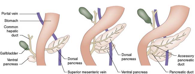
The pancreas develops from separate ventral and dorsal buds of endoderm that appear during the fourth week of fetal life. The ventral pancreas develops in association with the biliary tree, and its duct joins the common bile duct before emptying into the duodenum through the papilla of Vater (Fig. 15.1). During gestation, the duodenum rotates clockwise on its long axis, and the bile duct and ventral pancreas pass round behind it to fuse with the dorsal pancreas. Most of the duct that drains the dorsal pancreas joins the duct draining the ventral pancreas to form the main pancreatic duct (of Wirsung); the rest of the dorsal duct becomes the accessory pancreatic duct (of Santorini) and enters the duodenum 2.5 cm proximal to the main duct. In fetal life, the common bile duct and main pancreatic duct are dilated at their junction to form the ampulla of Vater. In extra-uterine life, only 10% of individuals retain this ampulla, although most retain a short common channel between the two duct systems.
The pancreas lies retroperitoneally, behind the lesser sac and stomach. The head of the gland lies within the C-loop of the duodenum, with which it shares a blood supply from the coeliac and superior mesenteric arteries (Fig. 15.2). The superior mesenteric vein runs upwards to the left of the uncinate process, and joins the splenic vein behind the neck of the pancreas to form the portal vein. The body and tail of the pancreas lie in front of the splenic vein as far as the splenic hilum, and receive arterial blood from the splenic artery as it runs along the upper border of the gland. The intimate relationship of the friable pancreas to these major blood vessels is the reason that bleeding is problematic after pancreatic trauma. The close association between the common bile duct and the head of pancreas explains why obstructive jaundice is so common in cancer of the head of the pancreas, and why gallstones frequently give rise to acute pancreatitis.
Surgical physiology
Exocrine function
The exocrine pancreas is essential for the digestion of fat, protein and carbohydrate. The pancreas secretes 1–2 litres of alkaline (pH 7.5–8.8) enzyme-rich juice each day. The enzymes are synthesized by the acinar cells and stored there as zymogen granules. Trypsin is the key proteolytic enzyme; it is released in an inactive form (trypsinogen) and is normally only activated within the duodenum by the brush border enzyme, enterokinase. Once trypsin has been activated, a cascade is established whereby the other proteolytic enzymes become activated in turn. Lipase and amylase are secreted as active enzymes. The alkaline medium required for the activity of pancreatic enzymes is provided by the bicarbonate secreted by the ductal epithelium.
Pancreatic secretion is stimulated by eating. Hormonal and neural (vagal) mechanisms are involved. Food entering the duodenum (notably fat and protein digestion products) releases cholecystokinin (CCK), which stimulates pancreatic enzyme secretion, while at the same time causing the gallbladder to contract and increase bile flow into the intestine. Acid in the duodenum releases the hormone secretin, which stimulates the pancreas to secrete watery alkaline juice.
Endocrine function
The islets of Langerhans are distributed throughout the pancreas. Although they account for only 2% of the weight of the gland, they receive 10% of its blood supply. Interaction between the endocrine and the exocrine pancreas is facilitated by the close proximity of islets and acini, and by a local ‘portal’ system in which blood draining from the islets enters a capillary network around neighbouring acinar cells before entering the tributaries of the portal vein. Four types of islet cell are recognized: A cells produce glucagon; B cells, insulin; D cells, somatostatin; and PP cells, pancreatic polypeptide. Glucagon and insulin have well-established physiological roles; the function of the other islet products is uncertain, but somatostatin and pancreatic polypeptidemay serve as local (paracrine) regulators, rather than as circulating (endocrine) messengers. Gastrin-producing (G) cells are not normally found in the pancreas, except in the rare Zollinger–Ellison syndrome.
Pancreatic pain
The parasympathetic nervous system has no role in the perception of pancreatic pain. Painful stimuli from the pancreas are transmitted by sympathetic fibres that travel along the arteries of supply to the coeliac ganglion, and from there to segments 5–10 of the thoracic spinal cord via the greater, lesser and least splanchnic nerves.
Congenital disorders of the pancreas
Annular pancreas is a rare cause of duodenal obstruction, resulting from failure of rotation of the ventral pancreas. In approximately 5% of individuals, the ducts draining the dorsal and ventral pancreas fail to fuse, giving rise to pancreas divisum. This means that the secretions of the larger dorsal pancreas have to drain to the duodenum through the smaller accessory duct. There is no evidence to suggest a strong association between pancreas divisum and acute or chronic pancreatitis. Rests of pancreatic tissue may be found at a variety of sites within the gut wall, but are most common in the duodenum, stomach and proximal small bowel. Such heterotopic tissue is usually asymptomatic, but can become a focus of inflammation in the rare condition of cystic duodenal dystrophy (‘groove’ pancreatitis) causing pain and duodenal obstruction.
Pancreatitis
Pancreatitis may be acute or chronic. After an attack of acute pancreatitis, the gland usually returns to anatomical and functional normality, whereas chronic pancreatitis is associated with a permanent derangement of structure and function. Some patients suffer from recurrent acute pancreatitis but enjoy relatively normal health between attacks.
Acute pancreatitis
Acute pancreatitis is a common cause of emergency admission to hospital. There are 100–400 new cases per million of the population each year in the UK, and the incidence continues to rise, possibly because of an increase in gallstone disease, alcohol misuse and obesity in the population. The disease is relatively rare in children, but all adult age groups may be affected.
Aetiology
Conditions associated with the development of acute pancreatitis are listed in Table 15.1 – gallstones and alcohol are by far the most important.
Table 15.1 Causes of acute pancreatitis
Class | Specific causes |
---|---|
Toxic | AlcoholMedicationTropical |
Genetic | Cystic fibrosisHereditary pancreatitisSPINK1 mutation |
Metabolic | HypercalcaemiaHyperlipidaemia |
Obstructive | |
Inflammatory | |
Physiological | Sphincter of Oddi dysfunction |
Gallstone pancreatitis
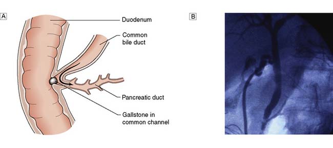
Gallstones are present in some 40% of patients in the UK who develop acute pancreatitis, but tiny gallstones or even microscopic crystals (so called microlithiasis) are thought to account for the majority of ‘idiopathic’ cases. Transient impaction of a gallstone within the common channel between the common bile duct and pancreatic duct causes obstruction of the pancreatic duct (Fig. 15.3) and a sequence of events within the pancreatic acinar cells resulting in intracellular activation of pancreatic enzymes, acinar cell damage and pancreatic inflammation.
Alcohol-associated pancreatitis
The proportion of cases of acute pancreatitis linked to alcohol varies in different parts of the world. In Scotland the figure is around 30%, whereas in some parts of France and North America it may be as high as 50–90%. The mechanism responsible is uncertain. Although there is no direct relationship between the quantity of alcohol consumed and the risk of pancreatitis (in contrast with alcoholic liver disease) alcohol consumption normally exceeds 50 g day (or 5 standard drinks).
Other causes
Acute pancreatitis occurs in approximately 5% of patients undergoing ERCP. Such cases are usually mild but can be life threatening. Trauma, particularly blunt abdominal trauma, may cause pancreatitis or pancreatic duct disruption. Hypercalcaemia is a rare cause of pancreatitis and is usually secondary to hyperparathyroidism. Hyperlipidaemia, due to familial hypertriglyceridaemia, is another rare cause of pancreatitis and in both of these metabolic conditions treatment is directed at the underlying cause. A small number of patients may have a family history of acute pancreatitis. Hereditary pancreatitis is a rare condition caused by mutations of the cationic trypsinogen gene and leads to the development of chronic pancreatitis at a young age.
Viral infections, including mumps, Coxsackie virus, rubella, measles and cytomegalovirus are the most common cause of pancreatitis in children. Bacterial infections are a very rare cause of pancreatitis. Ascaris worms are a relatively common cause of pancreatitis in areas of high prevalence, due to migration through the duodenal papilla from the common bile duct. Many drugs have been associated with acute pancreatitis although it is often difficult to prove a causal association and other causes always need to be considered. Azathioprine, mesalazine and simvastatin are responsible for the best documented cases.
Pancreatic neoplasms may cause obstruction of the pancreatic duct and lead to acute pancreatitis. Such neoplasms can be small and thus easily missed, even by CT. The presence of unexplained pancreatic duct dilatation or other suspicious clinical or radiological appearances should prompt further investigation, usually by endoscopic ultrasound.
Clinical features of acute pancreatitis
Constant, severe or agonizing pain in the epigastrium, with radiation through to the back, is usually prominent. Pain can also be experienced in either hypochondrium. Nausea, vomiting and retching are often marked. Clinical examination often reveals much less tenderness, guarding and rigidity than might have been expected from the patient’s history and the presence of generalized peritonism may warrant further investigation by CT to exclude other intra-abdominal pathology. Signs of systemic disturbance include tachycardia, hypotension or tachypnoea and are indicative of severe pancreatitis. Obstructive jaundice may be apparent in patients with pancreatitis due to an impacted gallstone, and if present should raise awareness of the possibility of co-existent cholangitis.
Diagnosis
The key to the diagnosis of acute pancreatitis is a high index of suspicion and measurement of the serum amylase concentration. Serum lipase is an alternative and has some advantages but is rarely available in UK hospitals. The usual diagnostic cut-off for serum amylase is three times the upper reference limit but the diagnosis should be considered in any patient with a relevant history, even where there is a non-diagnostic rise in serum amylase. A high serum amylase can be seen in patients with abdominal pain due to other pathology. The most common of these is mesenteric ischaemia due either to mesenteric vascular occlusion or small bowel strangulation, and when there is diagnostic doubt, an urgent CT is indicated to clarify the diagnosis. High serum amylase levels are also seen in some patients with perforated ulcer or ruptured aneurysm but rarely above the diagnostic threshold for pancreatitis. In all cases, if the history or clinical examination is atypical for acute pancreatitis, CT should be carried out to clarify the diagnosis.
Serum amylase levels fall rapidly in acute pancreatitis and have no relationship to the severity of the attack or the resolution of the disease. Patients who present some days after the initial onset of abdominal pain may have normal or near-normal serum amylase levels and although urinary amylase or serum lipase can be helpful in such cases, the diagnosis is again best clarified by CT.
Radiology
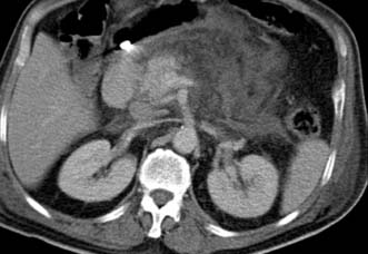
Initial diagnosis of acute pancreatitis is based on clinical features combined with serum amylase levels. The role of radiology in initial assessment is confined to cases where there remains diagnostic doubt, as when serum amylase is non-diagnostic or where the clinical history or examination findings are atypical (for example when there is evidence of generalized peritonitis). In these cases urgent contrast-enhanced CT is mandatory. In patients with acute pancreatitis, changes will be identified ranging from mild peripancreatic oedema through to extensive pancreatic necrosis (Fig. 15.4) but more importantly at this stage, other diagnoses which require urgent operative intervention, such as mesenteric ischaemia or perforated viscus, will be excluded.
Differentiation between gallstone- and alcohol-associated pancreatitis
It is important to clarify the aetiology of acute pancreatitis, primarily so that further attacks can be prevented where possible. In the UK, the majority of cases are due to gallstones and these may be present even where there is a clear history of alcohol excess. Therefore, all patients with acute pancreatitis should have an abdominal ultrasound carried out. Even when no stones are identified on ultrasound, other factors may suggest a gallstone diagnosis. In particular, elevated alanine transaminase (ALT) at admission of more than twice the upper reference limit is highly suggestive of a gallstone aetiology. In patients where no cause is found, further investigation by endoscopic ultrasound may detect microlithiasis or rare causes such as a small pancreatic neoplasm. This is best performed once the acute attack has resolved.
Assessment of severity
Approximately 80% of patients with acute pancreatitis will have a self-limiting illness which resolves within 48–72 hours. In such cases the main issue is identification and treatment of the underlying cause. The major challenge faced in the management of this condition is the 20% of patients who have a severe episode of pancreatitis in whom life-threatening complications can occur. Much effort has been directed at the early recognition of severe acute pancreatitis, the aim being to ensure these patients are adequately managed and placed in an appropriate high-dependency or intensive care environment. Many systems have been proposed of which the Glasgow Prognostic Score, APACHE II score and C-reactive protein have been most widely studied. None of these however have proved sufficiently accurate to influence management decisions, particularly in the crucial first 24 hours after hospital admission. Other attempts with serum or urine markers of severity have either proved disappointing or have not yet been tested in prospective studies. When faced with a patient with acute pancreatitis, the challenge therefore is to recognize the development of a severe attack which necessitates a clear understanding of the natural history of the illness.
The most important feature of severe acute pancreatitis is the development of multiple organ dysfunction syndrome (MODS). In most patients who develop severe acute pancreatitis this is identified by the presence of hypoxia, shock or more rarely, renal dysfunction around the time of admission to hospital. A minority of patients will develop such complications over the course of the next 48 hours and in these cases the presence of SIRS (systemic inflammatory response syndrome) characterized by a tachycardia, raised white cell count and/or tachypnoea is almost always present. It is very rare for a patient without SIRS at admission to develop MODS. Where SIRS persists for more than 48 hours, the risk of further deterioration is very high and such patients need close monitoring for the development of respiratory and renal complications. It is the group of patients with early persistent SIRS or MODS who are also at greatest risk of developing septic and other local complications of acute pancreatitis.
Contrast enhanced CT is used in some institutions to identify severe acute pancreatitis, as the presence of pancreatic necrosis or extensive fluid collections identifies a group at high risk of further complications. However it is increasingly recognized that the main determinant of outcome is early systemic organ dysfunction and the role of early CT is now mainly confined to cases of diagnostic doubt.
Management
Most attacks of acute pancreatitis will settle with conservative management involving intravenous fluids, analgesia and gradual re-introduction of diet when nausea and vomiting have settled. All patients with SIRS or early systemic organ dysfunction should be managed in a high-dependency or intensive care environment where adequate monitoring and specialist care is available.
Conservative treatment
• Pain relief. Severe pain requires the administration of opiates; there is no evidence to support the use of pethidine rather than morphine.
• Fluid resuscitation. Patients with severe acute pancreatitis often require large volumes of fluid to maintain adequate urine output and blood pressure. Adequate early resuscitation in such cases is the most important consideration in early management and where systemic organ dysfunction is present will necessitate invasive monitoring of venous and arterial pressure. Patients with MODS or SIRS will need oxygen therapy and continuous monitoring of oxygen saturation as well as urine output.
• Suppression of pancreatic function. There is no evidence that suppression of pancreatic function improves the outcome in acute pancreatitis. Nasogastric tubes are not routinely used. Fluids and diet are withheld until nausea and vomiting settle and in cases where systemic complications or other factors prevent recommencement of normal diet, nasoenteric feeding is commenced at an early stage. Suppression of pancreatic secretion by drugs such as octreotide or somatostatin is of no benefit in acute pancreatitis.
• Prevention of infection. Antibiotic prophylaxis has been advocated by some as a means of reducing the risk of infected pancreatic necrosis. Others have been concerned that the more widespread use of antibiotics will result in an increased incidence of severe fungal infection. The most recent consensus is that there is no evidence that prophylactic antibiotics reduce the incidence of infected pancreatic necrosis or mortality (EBM 15.1). It is important to recognize that patients with severe acute pancreatitis often have evidence of SIRS and the presence of a fever and raised white cell count are to be expected, even in the absence of infection. The only definite indication for early antibiotic therapy is when patients are suspected of having cholangitis, which may co-exist with gallstone pancreatitis. Jaundiced patients with SIRS are therefore managed with appropriate broad spectrum antibiotics while arrangements are made for urgent ERCP as described below.
• Inhibition of inflammatory response. Severe acute pancreatitis is one of many conditions where systemic organ dysfunction is driven by the systemic inflammatory response and there has been much experimental and clinical interest in the role of down-regulation of this response as a potential treatment for acute pancreatitis. The only agent that has been studied in large, prospective clinical trials was the platelet-activating factor (PAF) antagonist, lexipafant. Despite encouraging experimental data and promising results from initial clinical trials, a large international trial, recruiting 1500 patients, showed no difference in mortality.
• Nutritional support. Patients with severe acute pancreatitis who are unable to resume normal diet within 48–72 hours require nutritional support. This is best delivered by an enteral rather than parenteral route as meta-analyses of randomized trials (EBM 15.2) have demonstrated that TPN has a higher rate of complications and mortality. There are also sound experimental reasons to believe that enteral nutrition has additional benefits by potentially reducing the risk of bacterial translocation from the gut. There is no evidence that nasojejunal feeding is better or safer than nasogastric feeding but the nasojejunal route will be required where gastric stasis or outlet obstruction prevent effective nasogastric delivery.
• Other measures. A recent randomized trial assessed the role of probiotic therapy as an adjunct to early enteral feeding in the hope that this might reduce bacterial translocation from the gut more than enteral nutrition alone, thus potentially preventing later septic complications. Unfortunately, probiotic therapy actually increased mortality due to gut ischaemia and should no longer be used in these patients. Other proposed treatments which have been found to be of no benefit in prospective clinical trials include antiprotease therapy and peritoneal lavage.
15.1 Prophylactic antibiotics in severe acute pancreatitis
‘There is no evidence that prophylactic antibiotics (in acute pancreatitis) reduce the incidence of infected pancreatic necrosis or mortality.’
Bai Y, Gao J, Zou DW, Li ZS. Prophylactic antibiotics cannot reduce infected pancreatic necrosis and mortality in acute necrotizing pancreatitis: evidence from a meta-analysis of randomized controlled trials. Am J Gastroenterol 2008; 103(1):104–110.
15.2 Nutritional support in severe acute pancreatitis
‘In patients with severe acute pancreatitis who are unable to return to diet in 48-72 hours nutrition is best given by enteral rather than parenteral (TPN) route.’
Petrov MS, Whelan K. Comparison of complications attributable to enteral and parenteral nutrition in predicted severe acute pancreatitis: a systematic review and meta-analysis. Br J Nutr 2010; 103(9):1287–1295.
Endoscopic treatment
Gallstone pancreatitis is due to the transient impaction of a stone at the papilla causing pancreatic duct obstruction. There is good experimental evidence that the duration of this obstruction is an important determinant of the severity of an attack and so early removal of an impacted gallstone by ERC and sphincterotomy has long been proposed as a potential treatment option for patients. Unfortunately the results from randomized trials have been conflicting, and differences in study design have made direct comparisons difficult. Patients with evidence of obstructive jaundice and SIRS within the first 24 hours are suffering from or are at least at risk of developing cholangitis and there is broad consensus that these patients should have urgent ERC with sphincterotomy. However the majority of patients will pass the gallstone spontaneously and in this situation ERC carries additional risk and no potential benefit (EBM 15.3). The most recent meta-analysis of randomized trials shows no benefit from early ERC in patients with severe acute pancreatitis where cholangitis is not present. Early ERC with stone removal can however be life-saving in the patient with cholangitis and it is important to be alert to this possibility in patients diagnosed as having severe acute pancreatitis. Our own practice is to perform urgent ERC and sphincterotomy only in patients with acute pancreatitis associated with obstructive jaundice and SIRS but there is considerable variation in practice across the UK.
15.3 ERCP and sphincterotomy in severe acute pancreatitis
‘There is no evidence that early ERC and sphincterotomy gives benefit to those patients with acute severe pancreatitis unless cholangitis is present (evidence of obstructed biliary system and SIRS).’
Ayub K, Imada R, Slavin J. Endoscopic retrograde cholangiopancreatography in gallstone-associated acute pancreatitis. Cochrane Database Syst Rev 2004;(4):CD003630.
Surgical treatment
There is no role for surgical intervention in the first 1–2 weeks of an attack of acute pancreatitis, even in the face of deteriorating multiple organ failure. Complications that were managed surgically in the past are now being managed by radiological or endoscopic treatments. Acute pancreatitis is managed conservatively whenever possible, but surgery is indicated under the following circumstances:
1. Where an alternative diagnosis is suggested by CT following initial assessment.
2. Where gallstones are considered the likely cause, cholecystectomy is carried out after recovery from the acute attack. In patients with mild acute pancreatitis this is best carried out during the index hospital admission but following a severe attack, cholecystectomy is delayed until resolution of the inflammatory process. Even after severe acute pancreatitis it is usually possible to perform cholecystectomy by the laparoscopic route. In patients who have had no prior imaging of the common bile duct by MRCP or ERC, it is important to carry out operative cholangiography to exclude bile duct stones. In patients considered unfit for cholecystectomy, ERC with endoscopic sphincterotomy is carried out to protect against further attacks.
Complications
Infected pancreatic necrosis
Pancreatic necrosis itself is not an indication for surgical intervention, even when complicated by MODS. However, infected pancreatic necrosis is the most challenging complication of acute pancreatitis and management is complex, requiring the input of surgeons, interventional radiologists and endoscopists, as well as the critical care team. Infection occurs in up to 40% of patients with pancreatic necrosis and usually presents more than 2 weeks after symptom onset. The development of infection may be suspected where there is deterioration in systemic organ failure or where new organ failure develops in a patient more than 2 weeks after admission. However, infected pancreatic necrosis is not always complicated by organ failure and may present with worsening pain and fever associated with a rise in inflammatory parameters but with little evidence of systemic illness.
The development of infected pancreatic necrosis may be associated with evidence of gas within a pancreatic collection seen on CT, but this is not always the case and it can be difficult to make this diagnosis on radiological grounds alone. In some centres, if infected pancreatic necrosis is suspected, fine needle aspirates are taken from pancreatic collections under CT or ultrasound guidance to establish whether or not infection is present, with early surgical intervention where infection is proven. Others prefer to act on clinical grounds and where infection is suspected place a percutaneous drain under CT guidance prior to definitive surgical, endoscopic or radiological management.
There is much variation in practice in the management of this life-threatening complication of acute pancreatitis and the variety of approaches reflects variation in local expertise and experience and a lack of good evidence supporting any one approach over another. Where infected pancreatic necrosis is confirmed or strongly suspected, the common approaches to management are briefly described below.
Surgical debridement
Conventional management of infected pancreatic necrosis involves wide debridement of devitalized pancreatic and peripancreatic tissue and either placement of wide-bore drains for postoperative lavage or abdominal packing and planned re-exploration. This is clearly a major undertaking in the critically-ill patient and there has been an increasing trend towards measures to delay surgery until patients are stabilized and organ failure has resolved.
Percutaneous drainage/debridement
Following initial percutaneous drainage of infected pancreatic necrosis, patients will commonly show signs of initial improvement but subsequently deteriorate as drains block with necrotic debris. Rather than proceeding to conventional surgical debridement, several approaches have been described to facilitate removal of necrotic debris via the percutaneous drain track by radiological, laparoscopic or endoscopic means. A minimally-invasive surgical approach through a small flank incision has also been described. Retrospective studies tend to support the view that initial percutaneous drainage followed by conventional surgery or one of these minimally invasive techniques is associated with lower mortality and severe systemic complications than conventional surgery alone.
Endoscopic drainage
The retrogastric position of the pancreas allows endoscopic transgastric drainage of infected pancreatic necrosis, usually under control of endoscopic ultrasound. There is less experience with this approach than open surgery or percutaneous approaches but it is increasingly utilized in selected patients. More than one approach may be used at different times in an individual patient and the importance therefore of management within a specialist multidisciplinary environment cannot be over-emphasized.

Stay updated, free articles. Join our Telegram channel

Full access? Get Clinical Tree

