Chapter 10 The nervous system
The nervous system forms mainly from the ectoderm layer, at the beginning of the third week (see Chapter 1), the process known as neurulation. The neural plate forms as a thickening which is widest at its cranial end (Fig. 10.1A). Laterally, the plate edges thicken to form the neural folds (Fig. 10.1B). This is by a process of induction by the underlying notochord and somites. As these neural folds develop they turn towards each other forming the neural groove (Fig. 10.1C), and ultimately fusing as the neural tube. The neural tube structure begins in the cervical region and ends caudally. At the cranial end of the tube the brain develops, whereas the remainder of the tube gives rise to the spinal cord. At each end of the tube are the anterior and posterior neuropores which close in the middle and end of the fourth week respectively (Fig. 10.1D).
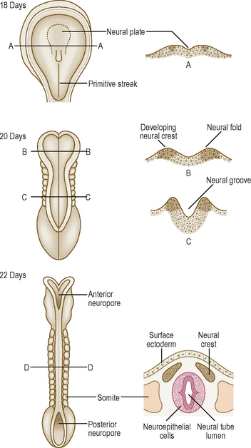
In addition to the neuroectoderm cells from which the neurons develop, neural crest cells develop on the edges along the length of the neural folds (Fig. 10.1). These cells detach themselves from the edges of the folds lying beneath the surface ectoderm, and migrate laterally to form a variety of structures. The principal derivatives of neural crest cells are:


Development of the spinal cord (Fig. 10.2A, B)
Most of the length of the neural tube gives rise to the spinal cord. As the wall of the tube thickens it comes to consist of three zones. The innermost is the neuroepithelial (ventricular) layer and, with the mantle layer, it forms the rest of the wall as well as the lining of the central canal of ependymal cells (Fig. 10.2A). The neuroepithelial cells in the mantle layer differentiate to become neuroblasts which will eventually be the neurons of the grey matter. The outermost layer of the developing spinal cord becomes the marginal zone and contains the axons entering and leaving the mantle zone (Fig. 10.2A). After myelination, this layer looks whitish and constitutes the white matter of the spinal cord. There is further development of the mantle zone through differentiation of the neuroblasts forming thickenings in the dorsal and ventral regions of the cord: the alar and basal plates (Fig. 10.2B). The alar plate becomes the sensory region (or dorsal horn) of the grey matter and the basal plate becomes the motor region (ventral horn). The lumen of the neural tube in the region of the spinal cord becomes diamond shaped. The pointed dorsal aspect of the tube is the roof plate, whilst the floor plate lies at the opposite pole. Dividing the alar and basal plates is the sulcus limitans
Stay updated, free articles. Join our Telegram channel

Full access? Get Clinical Tree


