14 The liver and biliary tract
The liver
Anatomy
The liver is the largest abdominal organ, weighing approximately 1500 g. It extends from the fifth intercostal space to the right costal margin. It is triangular in shape, its apex reaching the left midclavicular line in the fifth intercostal space. In the recumbent position, the liver is impalpable under cover of the ribs. The liver is attached to the undersurface of the diaphragm by suspensory ligaments that enclose a ‘bare area’, the only part of its surface without a peritoneal covering. Its inferior or visceral surface lies on the right kidney, duodenum, colon and stomach.
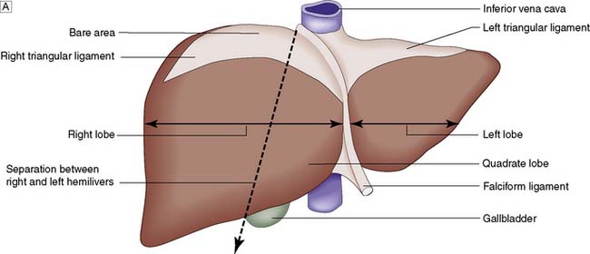
Topographically, the liver is divided by the attachment of the falciform ligament into right and left lobes; fissures on its visceral surface demarcate two further lobes, the quadrate and caudate (Fig. 14.1A). However, it is the liver segmental anatomy, as defined by the distribution of its blood supply, that is important to the surgeon.


Fig. 14.1 Anatomy of the liver.
A The broken line represents the separation between right and left hemilivers. B Segmental anatomy and venous drainage.
Segmental anatomy
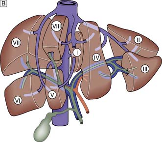
The portal vein and hepatic artery divide into right and left branches in the porta hepatis. Occluding either branch at surgery produces an easily visible line of demarcation that runs from the gallbladder bed behind and to the left of the inferior vena cava, thus separating the two hemilivers. Each hemiliver is further divided into four segments corresponding to the main branches of the hepatic artery and portal vein. In the left hemiliver, segment I corresponds to the caudate lobe, segments II and III to the left lobe (or left lateral section), and segment IV to the quadrate lobe. The remaining segments (V–VIII) comprise the right hemiliver (Fig. 14.1B).
Blood supply and function
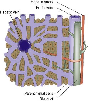
The liver normally receives 1500 ml of blood per minute and has a dual blood supply, 75% coming from the portal vein and 25% from the hepatic artery, which supplies 50% of the oxygen requirements. The principal venous drainage of the liver is by the right, middle and left hepatic veins, which enter the vena cava (Fig. 14.1B). In 25% of individuals, there is an inferior right hepatic vein, and numerous small veins drain direct into the vena cava from the caudate lobe (segment I). The functional unit of the liver is the hepatic acinus. Sheets of liver cells (hepatocytes) one cell thick are separated by interlacing sinusoids through which blood flows from the peripheral portal tract into the hepatic acinus to the central branch of the hepatic venous system. Bile is secreted by the liver cells and passes in the opposite direction along the small canaliculi into interlobular bile ducts located in the portal tracts (Fig. 14.2).
The liver has an important role in nutrient metabolism and is responsible for storing glucose in the form of glycogen, or converting it to lactate for release into the systemic circulation. Amino acids are utilized for hepatic and plasma protein synthesis or catabolized to urea. The liver has a central role in the metabolism of lipids, bilirubin and bile salts, drugs and alcohol. It is the principal organ for storage of a number of minerals and vitamins, and is responsible for the production of the coagulation factors I, V, XI, the vitamin K-dependent factors II, VII, IX and X as well as proteins C and S and antithrombin. The liver is also the largest reticuloendothelial organ in the body and its Kupffer cells play a role in the removal of damaged red blood cells, bacteria, viruses and endotoxin, much of which enter the body from the gut.
Summary Box 14.1 Surgical anatomy
• The liver is divisible into right and left hemilivers (each having four segments) using a line running from the gallbladder fossa to the inferior vena cava
• Each hemiliver receives a branch of the hepatic artery and portal vein; 75% of liver blood flow and 50% of its oxygen supply are provided by the portal vein
• The hepatocytes are arranged in lobules, each of which has a central branch of the hepatic vein and peripheral portal tracts (containing a branch of the hepatic artery, portal vein and bile duct)
• Liver anatomy allows the surgeon to perform right hepatectomy, left hepatectomy and extended right hepatectomy (i.e. resecting all of the liver to the right of the falciform ligament). Resection of individual segments is also possible.
Jaundice
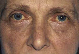
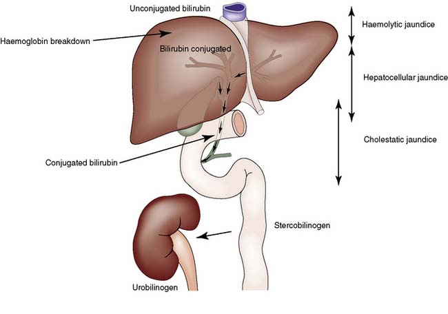
Jaundice is caused by an increase in the level of circulating bilirubin and becomes obvious in the skin and sclera when levels exceed 50 μmol/l (Fig. 14.3). It may result from excessive destruction of red cells (haemolytic jaundice), from failure to remove bilirubin from the blood stream (hepatocellular jaundice), or from obstruction to the flow of bile from the liver (cholestatic jaundice) (Fig. 14.4). Congenital non-haemolytic hyperbilirubinaemia (Gilbert’s syndrome) is a relatively rare cause of jaundice due to defective bilirubin transport; the jaundice is usually mild and transient, and the prognosis is excellent.
To the surgeon, the most important type of haemolytic jaundice is that caused by hereditary spherocytosis, in which splenectomy may be necessary (Ch. 15). Haemolytic jaundice may also occur after blood transfusion and after operative or accidental trauma, when haematoma formation produces a pigment load that exceeds hepatic excretory capacity.
Cholestatic jaundice due to intrahepatic obstruction of bile canaliculi may be a feature of acute and chronic liver disease and can be caused by drugs (e.g. chlorpromazine). This form of jaundice must be differentiated from that due to extra-hepatic obstruction, the cause of which has most surgical relevance. Extrahepatic obstruction most commonly results from gallstones or cancer of the head of the pancreas. Other causes include cancer of the periampullary region or major bile ducts, extrinsic compression of the bile ducts by metastatic tumour, iatrogenic biliary stricture and choledochal cyst.
Diagnosis
History and clinical examination
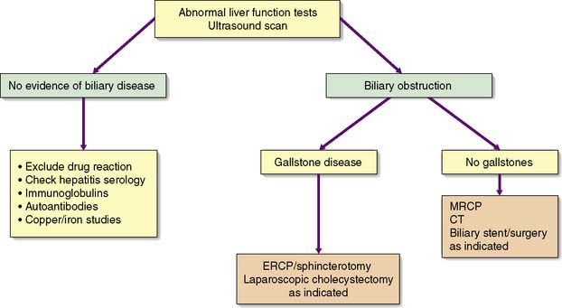
An accurate, rapid diagnosis of the cause of jaundice allows prompt institution of appropriate treatment (Fig. 14.5). The age, sex, occupation, social habits, drug and alcohol intake, history of injections or infusions, and general demeanour of the patient must be considered. A history of intermittent pain, fluctuant jaundice and dyspepsia suggests calculous obstruction of the common bile duct, whereas a history of weight loss and relentless progressive jaundice favours a diagnosis of neoplasia. Obstructive jaundice is likely if there is a history of passage of dark urine and pale stools, and if the patient complains of pruritus (owing to an inability to secrete bile salts into the obstructed biliary system). Hepatocellular jaundice is likely if there are stigmata of chronic liver disease, such as liver palms, spider naevi, testicular atrophy and gynaecomastia. The abdomen must be examined for evidence of hepatomegaly or gallbladder distension, and for signs of portal hypertension such as splenomegaly, ascites and large collateral veins (caput medusae) in the abdominal wall.
Biochemical and haematological investigations
Haemolytic jaundice is suggested if there are high circulating levels of unconjugated bilirubin but no bilirubin in the urine. Serum concentrations of liver enzymes are normal in these circumstances and the appropriate haematological investigations should be set in train.
In jaundice due to biliary obstruction, the circulating bilirubin is conjugated by the liver and rendered water-soluble; it can then be excreted in the urine and gives it a dark colour. As bile cannot pass into the gastrointestinal tract, the stool becomes pale and urobilinogen is absent from the urine. Obstruction increases the formation of alkaline phosphatase from the cells lining the biliary canaliculi, producing raised serum levels. This rise precedes that of bilirubin and its fall is more gradual once obstruction is relieved. Serum transaminase and lactic dehydrogenase levels may rise in obstruction. Conversely, swelling of the parenchyma in hepatocellular jaundice frequently produces an element of intrahepatic biliary obstruction and a modest rise in serum alkaline phosphatase concentration.
Full blood count and coagulation screen should be undertaken as a matter of routine and viral status should be determined. Anaemia may signify occult blood loss, and a low white cell or platelet count may indicate hypersplenism due to portal hypertension. Prolongation of the prothrombin time may be present in both hepatocellular and cholestatic jaundice, but should readily correct within 36 hours with the administration of parenteral vitamin K when jaundice is cholestatic.
Radiological investigations
If the clinical picture and biochemical investigations suggest that jaundice is obstructive, radiological techniques can be used to define the site and nature of the obstruction.
Ultrasonography
In skilled hands, this key investigation is safe, non-invasive and reliable. It is used to define whether the patient has bile duct dilatation or gallbladder distension due to obstruction, and to confirm the need for more invasive investigations. Ultrasonography will also detect gallstones and space-occupying lesions in the liver and pancreas, although overlying bowel gas may prevent a clear view of the pancreas.
Magnetic resonance imaging (MRI)
Magnetic resonance cholangiopancreatography (MRCP) has largely replaced other forms of invasive radiological imaging of the bile duct and pancreas. MRI has the advantage that it does not introduce infection into an obstructed biliary system or the pancreatic duct; it also enables assessment of the vascular anatomy and the parenchyma of the liver and pancreas. This is important in patients presenting with symptoms suggestive of malignant obstructive jaundice.
Endoscopic retrograde cholangiopancreatography (ERCP)
ERCP outlines the biliary and pancreatic systems by injecting contrast through a cannula inserted into the papilla of Vater by means of an endoscope passed into the duodenum. It gives more detailed information than ultrasonography and allows endoscopic treatment of gallstones, biopsy of periampullary tumours, and relief of obstructive jaundice by stent insertion. The investigation may be complicated by acute pancreatitis, and prophylactic antibiotics should be administered to reduce the risk of cholangitis for complex interventions. Haemorrhage and perforation are less frequent complications.
Percutaneous transhepatic cholangiography (PTC)
Used less often than formerly to assess obstruction of the upper biliary tree, PTC provides a clear outline of the biliary system by the injection of contrast through a slim flexible needle passed percutaneously into the liver. The technique may cause bleeding or bile leakage and can be complicated by bacteraemia and septicaemia; coagulation status must be checked and antibiotic cover should be given. It is almost exclusively undertaken before placement of a biliary drain or stent to relieve obstruction of the proximal biliary tree.
Computed tomography (CT)
Contrast enhanced CT can be used to identify hepatic, bile duct and pancreatic tumours in jaundiced patients. It often demonstrates the dilated biliary tree to the level of the obstruction, vascular abnormality or invasion and may show dissemination to adjacent lymph nodes. It is also used to assess viability of pancreatic tissue in severe pancreatitis.
Other radiological investigations
Positron emission tomography (PET-CT) has found an increasing role in staging hepatobiliary and pancreatic (HBP) malignancy. Isotopic liver scanning has been superseded by ultrasonography and CT. Selective angiography has been largely superseded by CT and MRI assessment of vascular anatomy but may be used for embolization of tumours or hemorrhagic complications of HBP disease.
Liver biopsy
Liver biopsy may be considered in patients with unexplained jaundice, in whom an obstructing lesion has been excluded radiologically. ‘Targeted’ liver biopsy can be conducted under ultrasound or CT guidance. Prothrombin time, platelet count and hepatitis B surface antigen (HBsAg) status must always be determined, and clotting abnormalities should be corrected before biopsy is undertaken.
Laparoscopy
Laparoscopy under general anaesthesia may be used in the evaluation of liver disease. In selected patients with malignancy of the liver, pancreas and biliary tree, it may have a role in the staging of the tumour to exclude peritoneal or hepatic dissemination.
Laparotomy
Laparotomy is no longer necessary to establish the cause of jaundice and is only undertaken to remove the causal lesion or relieve biliary obstruction. Intraoperative ultrasonography and operative cholangiography may give useful additional information in patients with neoplasia and biliary obstruction. Appropriate preoperative preparation is particularly important in jaundiced patients.
Congenital abnormalities
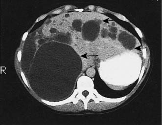
Up to 5% of the population has simple liver cysts. They are lined by biliary epithelium and contain serous fluid, but never communicate with the biliary tree. They rarely produce symptoms, are associated with normal liver function, and on ultrasound or CT have no discernible wall (Fig. 14.6). In the few patients who develop symptoms, cysts tend to recur following aspiration, and sclerosis by alcohol injection is of little value for large symptomatic cysts. Surgical management consists of deroofing and may be undertaken by laparoscopic means. Polycystic disease is a rare cause of liver enlargement and may be associated with polycystic kidneys as an autosomal dominant trait. In symptomatic patients, it may be necessary to combine a deroofing procedure with hepatic resection or to consider liver transplantation.

Fig. 14.6 CT demonstrating multiple biliary cysts appearing as hypodense areas within both lobes of the liver (arrowed).
• Jaundice is a yellowish discoloration of the tissues that becomes clinically apparent when serum bilirubin levels exceed 50 μmol/l (normal < 20 μmol/l)
• It may be due to excessive haemolysis, hepatic insufficiency or cholestasis; cholestatic (obstructive) jaundice is the type encountered in surgical practice
• The two most common causes of surgical obstructive jaundice are cancer of the head of the pancreas and stones in the common bile duct (choledocholithiasis)
• In cholestatic jaundice, the bilirubin has been conjugated by the hepatocytes and is therefore soluble in water and can be excreted in the urine; patients with obstructive jaundice typically have dark urine and pale stools and may have pruritus (thought to be due to the accumulation of bile salts)
• Obstructive jaundice is characterized by elevated serum alkaline phosphatase levels in addition to hyperbilirubinaemia, and may be accompanied by modest elevations in transaminase (aminotransferase) levels, reflecting liver damage.
Cavernous haemangiomas are one of the most common benign tumours of the liver (up to 5% of population) and may be congenital. Women are affected six times more frequently than men. Most haemangiomas are small solitary subcapsular growths found incidentally at laparotomy or autopsy, but they are sometimes detected on ultrasound examination as densely hyperechoic lesions that mimic hepatic tumours. These lesions rarely give rise to pain. Resection may be considered for symptomatic lesions exceeding 5 cm in diameter.
Liver trauma
After the spleen, the liver is the solid organ most commonly damaged in abdominal trauma, particularly following road traffic accidents. Stab injuries and gunshot wounds of the liver are also increasing in incidence. These are considered in Chapter 7.
Hepatic infections and infestations
Liver abscesses can be classified as bacterial, parasitic or fungal. Bacterial abscess is the most common type in Western medicine, but parasitic infestation is an important cause world-wide. Fungal abscesses are found in patients receiving long-term broad-spectrum antibiotic treatment or immunosuppressive therapy, and may complicate actinomycosis.
Pyogenic liver abscess
Infection from the biliary system is now more common due to the increasing use of radiological and endoscopic intervention. Infection may spread through the portal vein from abdominal sepsis (e.g. appendicitis, diverticulitis), via the hepatic artery from a septic focus anywhere in the body, or by direct spread from a contiguous organ (e.g. empyema of the gallbladder). Abscess formation may follow blunt or penetrating injury, and in one-third of patients the source of infection is indeterminate (cryptogenic). Common organisms are:
Clinical features
The onset of symptoms is often insidious and the patient may present with pyrexia of unknown origin. There is sometimes a history of sepsis elsewhere, particularly within the abdomen, and pain in the right hypochondrium. Other patients present with swinging pyrexia, rigors, marked toxicity and jaundice. The liver is often enlarged and tender.
Investigations
Plain radiographs may show elevation of the diaphragm, pleural effusion and basal lobe collapse. Leucocytosis is usually present and liver function tests are deranged. Ultrasonography or CT is used to define the abscess (which is often irregular and thick-walled) and to facilitate percutaneous aspiration for culture. ERCP may be useful if biliary obstruction is thought to be responsible.
Management
Untreated abscesses often prove fatal because of spread within the liver to multiple sites, and because of septicaemia and debility. The principles of treatment are percutaneous drainage of accessible abscesses under ultrasound or CT guidance, and antibiotic therapy selected on the basis of culture of blood or pus. It is exceptional to resort to surgical drainage. Percutaneously or surgically placed drainage tubes are left in place and the size of the cavity is monitored by CT or serial X-rays following the injection of contrast material. Multiple small abscesses may require prolonged treatment with antibiotics for up to 8 weeks. Investigation is required to detect the source (e.g. colonoscopy, ERCP for stone removal).
Amoebic liver abscess
Entamoeba histolytica is a protozoal parasite that infests the large intestine and is endemic in many tropical regions. Trophozoites released by the cyst in the intestine may penetrate the mucosa to gain access to the portal venous system and so spread to the liver. The abscess is large and thin-walled, is usually solitary and in the right lobe, and contains brown sterile pus resembling anchovy sauce.
Clinical features
Right upper quadrant pain may be accompanied by anorexia, nausea, weight loss and night sweats. Tender enlargement of the liver is invariable, although jaundice is uncommon. Other signs include basal pulmonary collapse, pleural effusion and leucocytosis.
Investigations
Ultrasonography and CT are used to demonstrate the site and size of the abscess, which often has poorly defined margins. The stools should be examined for amoebae or cysts. Direct and indirect serological tests to detect amoebic protein are available.
Management
Early diagnosis is important, and treatment may be commenced empirically in areas where the problem is endemic. Treatment consists of the administration of metronidazole (800 mg 8-hourly for 5 days) and usually results in rapid resolution. The abscess should be aspirated by needle puncture, if there is no clinical response within 72 hours. If untreated, an amoebic abscess may rupture into the peritoneal cavity or into a bronchus.
Hydatid disease
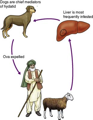
This less common infestation is caused in humans by one of two forms of tapeworm, Echinococcus granulosus and E. multilocularis. The adult tapeworm lives in the intestine of the dog, from which ova are passed in the stool; sheep or goats serve as the intermediate host by ingesting the ova whereas humans are accidental hosts (Fig. 14.7). The condition is most common in sheep- and goat-rearing areas. Ingested ova hatch in the duodenum and the embryos pass to the liver through the portal venous system. The wall of the resulting hydatid cyst is surrounded by an adventitial layer of fibrous tissue and consists of a laminated membrane lined by germinal epithelium, on which brood capsules containing scolices develop.
Clinical features
The disease may be symptomless, but chronic right upper quadrant pain with enlargement of the liver is the common presentation. The cyst may rupture into the biliary tree or peritoneal cavity, the latter sometimes causing an acute anaphylactic reaction due to absorption of foreign hydatid protein. Other complications include secondary infection and biliary obstruction with jaundice.
Investigations
Eosinophilia is common and serological tests, such as complement fixation, are available to detect the foreign protein. Hydatid cysts commonly calcify and may be seen on a plain film of the abdomen. Alternatively, they can be detected by ultrasound or CT of the liver and are recognizable by their thick wall, which may contain multiple daughter cysts.
Management
In asymptomatic patients, small calcified cysts may require no treatment. Patients can be treated successfully with albendazole or mebendazole but this may be prolonged. Large symptomatic cysts are best managed by complete excision, together with the parasites contained within. A laparoscopic approach is possible for simple accessible cysts.
Portal hypertension
Portal hypertension is caused by increased resistance to portal venous blood flow, the obstruction being prehepatic, hepatic or posthepatic (Table 14.1). Rarely, it results primarily from an increase in portal blood flow. The normal pressure of 5–15 cmH2O in the portal vein is consistently exceeded (above 25 cmH2O). Portal vein thrombosis is a rare cause and is most commonly due to neonatal umbilical sepsis. The most common cause of portal hypertension is cirrhosis resulting from chronic liver disease and is characterized by liver cell damage, fibrosis and nodular regeneration. The fibrosis obstructs portal venous return and portal hypertension develops. Arteriovenous shunts within the liver also contribute to the hypertension.
Table 14.1 Causes of portal hypertension
Obstruction to portal flow: |
Prehepatic |
Intrahepatic |
Posthepatic |
Increased blood flow (rare) |
Alcohol is the most common aetiological factor in developed countries, whereas in North Africa, the Middle East and China, schistosomiasis due to Schistosoma mansonii is a common cause. Chronic active hepatitis and primary and secondary biliary cirrhosis may result in portal hypertension, but in a large number of patients the cause remains obscure (cryptogenic cirrhosis).
Post-hepatic portal hypertension is rare. It is most frequently due to spontaneous thrombosis of the hepatic veins and this has been associated with neoplasia, oral contraceptive agents, polycythaemia and the presence of abnormal coagulants in the blood. The resulting Budd-Chiari syndrome is characterized by portal hypertension, caudate hypertrophy, liver failure and gross ascites.
Effects of portal hypertension
As a result of gradual chronic occlusion of the portal venous system, collateral pathways develop between the portal and systemic venous circulations. Portosystemic shunting occurs at three principal sites (Fig. 14.8). The most important is the development of varices in the submucosal plexus of veins in the lower oesophagus and gastric fundus. Oesophageal varices may rupture, to cause acute massive gastrointestinal bleeding in about 40% of patients with cirrhosis. The initial episode of variceal haemorrhage is fatal in about one-third of patients, and recurrent haemorrhage is common. Bleeding from retroperitoneal and periumbilical collaterals (‘caput medusae’) is troublesome during abdominal surgery, and collaterals may develop and cause bleeding at the site of stomas. Anorectal varices are not uncommonly found at proctoscopy but rarely cause bleeding.

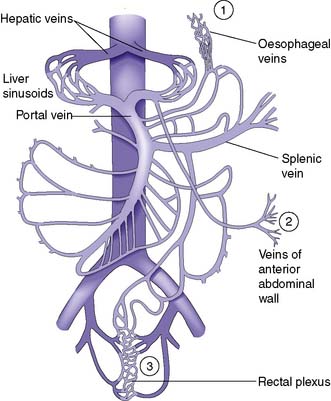
Fig. 14.8 The portal venous system.
Sites of portosystemic shunting are marked 1–3. Retroperitoneal communications also exist.
Progressive enlargement of the spleen occurs as a result of vascular engorgement and associated hypertrophy. Haematological consequences are anaemia, thrombocytopenia and leucopenia (with the resulting syndrome of hypersplenism). Ascites may develop and is due to increased formation of hepatic and splanchnic lymph, hypoalbuminaemia, and retention of salt and water. Increased aldosterone and antidiuretic hormone levels may contribute. Portosystemic encephalopathy is due to an increased level of toxins such as ammonia in the systemic circulation. This is particularly likely to develop where there are large spontaneous or surgically created portosystemic shunts. Gastrointestinal haemorrhage increases the absorption of nitrogenous products and may precipitate encephalopathy.
Clinical features
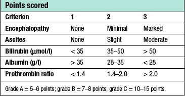
Patients with cirrhosis frequently develop anorexia, generalized malaise and weight loss. Clinical manifestations include hepatosplenomegaly, ascites, jaundice and spider naevi. Slurring of speech, a flapping tremor or dysarthria may point to encephalopathy, and this may be precipitated or intensified by the accumulation of blood in the gastrointestinal tract. The serum bilirubin may be elevated and the serum albumin depressed. Anaemia may be present and the leucocyte count raised (or depressed if there is hypersplenism). The prothrombin time and other indices of clotting may be abnormal. Clinical and biochemical parameters are used as the basis of the modified Child’s classification (Table 14.2). Patients allocated to grade A have a good prognosis, whereas those in grade C have the worst prognosis.
Table 14.2 Assessment of patients with portal hypertension using a modification of Child’s grading system

Patients with portal hypertension may be referred to a surgeon because of active uncontrolled bleeding from oesophageal varices, or for consideration of elective surgery for varices that have been resistant to non-surgical management.
Acute variceal bleeding
Patients presenting with acute upper gastrointestinal bleeding are examined for evidence of chronic liver disease (EBM 14.1). The key investigation during an episode of active bleeding is endoscopy. This allows the detection of varices and defines whether they are or have been the site of bleeding. It is important to remember that peptic ulcer and gastritis are common complaints that occur in 20% of patients with varices.
14.1 Variceal bleeding in cirrhosis: assessment and prophylaxis
‘Severity of cirrhosis is best described using the Child-Pugh score. If grade 3 varices are diagnosed, patients should have primary prophylaxis, irrespective of the severity of the liver disease. Pharmacological therapy with propranolol is the best available modality for primary prophylaxis, with variceal band ligation recommended in cases of intolerance or contraindication to propranolol.’
Jalan R, Hayes PC, British Society of Gastroenterology. Gut 2000; 46 suppl 3–4:III 1–III 15.
Management
The priorities in the management of bleeding oesophageal varices are summarized in Table 14.3.
Table 14.3 Priorities in the management of bleeding oesophageal varices
Active resuscitation |
Assessment of coagulation status |
Urgent endoscopy |
Control of bleeding |
Treatment of hepatocellular decompensation |
Treatment/prevention of portosystemic encephalopathy |
Prevention of further bleeding from varices |
Active resuscitation
The aim is to replace blood loss quickly with a view to urgent endoscopy. Many patients have coagulation defects from the outset, and thrombocytopenia is a common manifestation of hypersplenism. Fresh blood is preferred for transfusion purposes and the advice of the haematologist is sought regarding the use of fresh-frozen plasma (FFP) or platelet transfusion.
Endoscopy and control of bleeding
Endoscopy will reveal tortuous varices in three columns most prominent in the lower third of the oesophagus. Haemorrhage usually occurs from varices at the lowest few centimetres of the oesophagus. Rarely, bleeding occurs from varices in the gastric fundus. Although the synthetic form of somatostatin, octreotide, can be used to lower portal venous pressure and arrest bleeding, the injection of a sclerosant such as ethanolamine, or the application of bands is now used to arrest the bleeding at endoscopy (EBM 14.2). If haemorrhage is torrential and prevents direct injection, balloon tamponade may be used to stop the bleeding. The four-lumen Minnesota tube (Fig. 14.9) has largely replaced the three-lumen Sengstaken–Blakemore tube. The four lumina allow:
• aspiration of gastric contents
• compression of the oesophagogastric varices by the inflated gastric balloon
• compression of the oesophageal varices by the inflated oesophageal balloon
• aspiration of the oesophagus and pharynx to reduce pneumonic aspiration.
14.2 Control of variceal bleeding
‘Variceal band ligation is the method of choice for control of variceal haemorrhage, with endoscopic variceal sclerotherapy as second choice. If endoscopy is unavailable, vasoconstrictors such as octreotide or glypressin, or a modified Sengstaken tube may be used while more definitive therapy is arranged. In case of bleeding that is difficult to control, a modified Sengstaken tube should be inserted until further endoscopic treatment, transjugular intrahepatic portosystemic stent shunting (TIPSS) or surgical treatment, with the mode of treatment (surgical intervention such as oesophageal transection or TIPSS) determined by the specialist centre.’

Stay updated, free articles. Join our Telegram channel

Full access? Get Clinical Tree

