Chapter 4 The integumentary, skeletal and muscular systems
The integumentary system
The skin
By the end of the first month the single layer of ectodermal cells divide, forming a superficial layer of cells known as the periderm and a basal layer. The basal layer becomes the stratum germinativum which produces all the definitive layers of the epidermis (Fig. 4.1A, B). The cells of the periderm become keratinized, and are continuously lost in the amniotic fluid during fetal life. In a newborn infant, the sloughed cells of the periderm mix with the skin secretions to form the vernix caseosa that covers the entire skin. Later in fetal life, the peridermal cells are replaced by the stratum corneum and the melanocytes derived from the neural crest migrate into the epidermis (Fig. 4.1B, C).
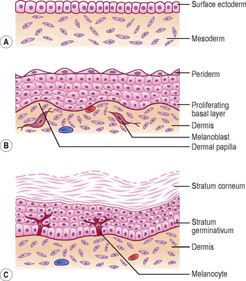
The dermis is derived from two sources: the somatic layer of the lateral plate of mesoderm and the dermatomes of the somites. The capillary network develops in the dermis to nourish the epidermis, and by 9 weeks, the sensory nerve endings grow into the dermis and epidermis. By 12 weeks, the outer layer of dermis projects into the overlying epidermis as dermal papillae (Fig. 4.1B).
The hairs and glands of the skin
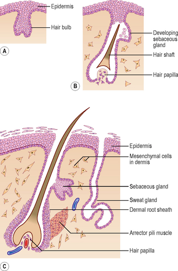
Hairs develop during the fetal period as proliferations of the stratum germinativum of the epidermis growing into the underlying dermis. The tip of the hair bud becomes a hair bulb and is soon invaginated by the mesenchymal hair papilla in which the vessels and nerve endings develop (Fig. 4.2A, B). The epidermal cells in the centre of the hair bud become keratinized to form the hair shaft, and the surrounding mesenchymal cells differentiate into the dermal root sheath (Fig. 4.2C). Small bundles of smooth muscle fibres called arrector pili muscles develop in the mesenchyme and are attached to the dermal sheath (Fig. 4.2C). Most sebaceous glands develop as buds from the side of the epithelial root sheath growing into the dermis; these glands produce an oily secretion that lubricates the hair and skin. The sweat glands develop as epidermal buds into the underlying dermis which become coiled to form the secretory part of the glands (Fig. 4.2C).
Mammary glands and teeth
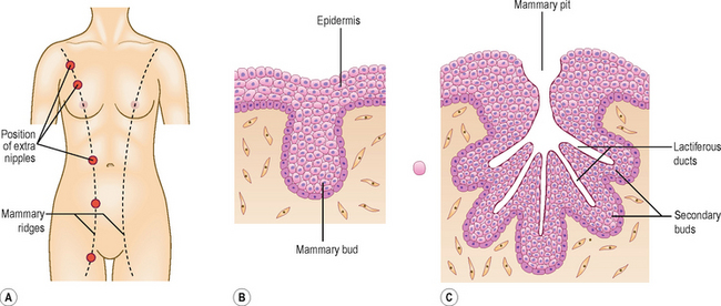
The mammary glands arise from a pair of epidermal thickenings called mammary ridges which appear in both sexes during the fourth week from the axilla to the inguinal region (Fig. 4.3A). Normally these ridges disappear except in the pectoral region where breasts develop, but part of the mammary ridges may persist to form an extra breast (polymastia) or supernumary breasts and nipples (Fig. 4.3A). The breast develops from a primary mammary bud which gives rise to several secondary buds that form the lactiferous ducts and their branches. The buds branch throughout the fetal period, become canalized and open into a small depression called the mammary pit of the nipple (Fig. 4.3B, C).
The teeth develop as tooth buds from the epithelial lining of the oral cavity along a U-shaped epidermal ridge called the dental lamina along the curves of the upper and lower jaws. The ectodermal tooth bud grows into the neural crest-derived mesenchyme, and passes through a ‘cap’ and a ‘bell’ stage. The enamel is produced by ameloblasts which are derived from the ectoderm; the underlying dentin and other tissues are derived from the mesenchyme and neural crest cells.
The musculoskeletal system
The mesenchyme gives rise to the musculoskeletal system. Most of the mesenchyme is derived from the mesodermal cells of the somites and the somatopleuric layer of lateral plate mesoderm (see Chapter 1). The mesenchyme in the head region comes from the neural crest cells. Regardless of their sources, a common feature of mesenchymal cells is their ability to migrate and differentiate into many different cell types, e.g. myocytes, fibroblasts, chondroblasts or osteoblasts. This differentiation often requires interaction with either epithelial cells or the components of the surrounding extracellular matrix.
The skeletal system
Development of the axial skeleton
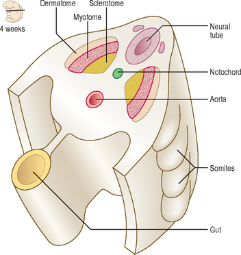
The axial skeleton is composed of the skull, vertebral column, sternum and ribs. This part of the skeleton is derived from the paraxial mesoderm, which is soon organized into the somites. The first somites appear on day 20 in the cranial region, and by 30 days approximately 37 pairs are formed. Somites appear as rounded elevations under the surface ectoderm on the dorsal aspect of the embryo from the base of the skull to the tail region. Each somite subdivides into two parts: the sclerotome and the dermomyotome (Fig. 4.4). The cells of the sclerotome give rise to the vertebrae and ribs, and those of the dermomyotome form muscle and the dermis of the skin.
The vertebral column
During the initial mesenchymal stage, the sclerotome cells migrate medially towards the notochord, and meet the sclerotome cells from the other side to form the centrum or vertebral body. Each sclerotome splits into a cranial and a caudal segment (Fig. 4.5A). The cranial half of the sclerotome consists of loosely arranged cells, whereas the caudal half contains densely packed cells. The caudal half of a sclerotome fuses with the cranial half of the sclerotome below to form the vertebral body (Fig. 4.5B
Stay updated, free articles. Join our Telegram channel

Full access? Get Clinical Tree


