Chapter 7 The digestive system
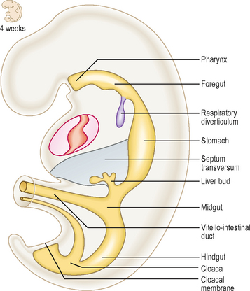
The longitudinal and lateral folding (see Chapter 1) of the embryo results in the incorporation of part of the yolk sac. Thus, the endoderm germ layer is incorporated into the embryo and forms the primitive gut tube (Fig. 7.1). In the anterior part of the embryo the incorporation of the endoderm into the head fold results in the formation of the foregut, whilst in the posterior part of the embryo the hindgut forms. The foregut is divided into a cranial and a caudal portion. The cranial portion develops within the head and neck, as the pharynx. This is particularly associated with the pharyngeal arches which are lined by endoderm. In the middle region the midgut forms and initially this is in direct continuity with the remaining yolk sac. The persisting connection becomes the vitello-intestinal (or vitelline) duct. Each region of the embryonic gut tube is supplied by its specific artery: the coeliac, superior mesenteric and inferior mesenteric arteries for the foregut, midgut and hindgut respectively. In addition to the primitive gut tube, the endoderm layer also gives rise to the parenchyma of the two large glandular organs associated with the gastrointestinal duct, the liver and the pancreas. The connective tissue, smooth muscle and serosal layer of the gut tube and the connective tissue of the pancreas arise from the splanchnopleuric lateral plate mesoderm (see Chapter 1).
Primitive gut tube
Mesenteries
As the gut tube is incorporated into the body of the embryo it comes to be suspended by a dorsal mesentery (Fig. 7.3). This is formed from the serosal membrane derived from the lateral plate splanchnopleuric mesoderm. As the gut tube is incorporated into the body of the embryo, ‘wings’ of amnion push in laterally (responsible for the lateral folding), and these squeeze the connection of the yolk sac and the gut tube. Eventually the endodermal gut tube is formed and it moves ventrally, leaving the overlying splanchnopleuric mesoderm still in contact with the posterior wall of the embryo. This suspensory structure is called the dorsal mesentery of the gut tube and extends from the lower part of the oesophagus to the cloacal part of the hindgut. It has two adjacent layers of serosal membrane. The two layers diverge on each side, forming the mesentery, which is reflected onto the dorsal wall of the embryo. Since the mesentery is related to the posterior wall of the embryo, close to where the dorsal aorta lies, there is the opportunity for ventral arterial branches of the abdominal aorta to lie between the two layers of the mesentery to gain access to the gut tube without piercing the future peritoneal membrane (Fig. 7.2). In the situation where the gut tube is invested by splanchnopleuric mesoderm all around, aside from the point at which the mesentery begins its investment, the gut tube is said to be intraperitoneal. If an organ is not suspended by a mesentery, thus outside of the peritoneum and in contact with the posterior abdominal wall, it is said to be retroperitoneal, e.g. kidney, parts of the duodenum, pancreas (Fig. 7.2).
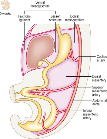
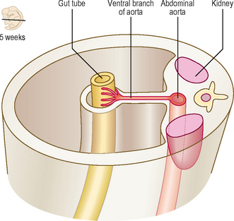
Fig. 7.2 A transverse section through the embryo showing the peritoneal and retroperitoneal structures.
With further development the dorsal mesentery is lost for some parts of the adult derivatives of the gut tube: duodenum and the ascending and descending colon. In this situation those parts of the tube are more firmly anchored to the underlying connective tissues. The dorsal mesentery related to the stomach is known as the dorsal mesogastrium, and elongates considerably, as the greater omentum.
For the caudal part of the foregut another mesentery develops, the ventral mesentery. The ventral mesogastrium is derived from the septum transversum, and there is, therefore, a ventral mesentery only for the portion of the gut tube adjacent to the septum transversum. Thus, the terminal part of the oesophagus, the stomach and the initial portion of the duodenum are invested by a ventral mesentery (Fig. 7.3). The liver develops within the ventral mesentery and divides the ventral mesogastrium into two parts. The mesentery lying between the stomach and the liver is the lesser omentum. The part of the mesentery lying between the liver and the ventral wall of the embryo is the falciform ligament. In all these cases the mesenteries are double layers, thus allowing for the possibility of other structures such as blood vessels and nerves to occupy those spaces between the two layers.
Foregut
The cranial (pharyngeal) portion of the foregut, and its subsequent development is described in Chapter 11. The remainder of the foregut is termed the caudal foregut.
Development of oesophagus
The initial component of the caudal foregut is the oesophagus. Initially, it too has a dorsal mesentery, which disappears, bringing the oesophagus into its adult position in the posterior mediastinum. As described in Chapter 5 the endodermal respiratory diverticulum arises as a ventral bud off the future oesophagus. Because of the growth of the thoracic organs the oesophagus is lengthened.
Development of stomach
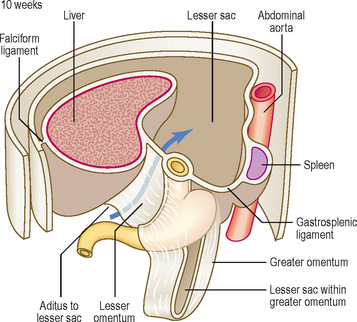
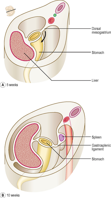
At first the stomach is merely a cylindrical tube, like the rest of the gut tube, passing craniocaudally, and it is invested by the dorsal and ventral mesenteries (Fig. 7.4A). Conventionally, the developing stomach is initially described as being fusiform or flask-shaped. The stomach undergoes a clockwise rotation through about 90° (Fig. 7.4B). The uneven growth is evident by the shorter lesser curvature and the longer greater curvature of the stomach; this is because the original posterior wall grows quicker than the anterior wall. This rotation of the stomach also results in the dorsal mesogastrium being pulled over to the left side (Fig. 7.5). Consequently, the small compartment lying between the stomach and the dorsal wall of the embryo develops as the lesser sac of the peritoneal cavity (Fig. 7.5). Developing within the dorsal mesogastrium is the spleen, and this is thus carried over to the left side so that it comes to occupy the left hypochondrium of the abdominal cavity in the adult. The portion of the dorsal mesogastrium that lies between the stomach and spleen becomes the gastrosplenic ligament, whilst the part lying between the spleen and the dorsal wall of the embryo becomes the lienorenal ligament (Fig. 7.5).
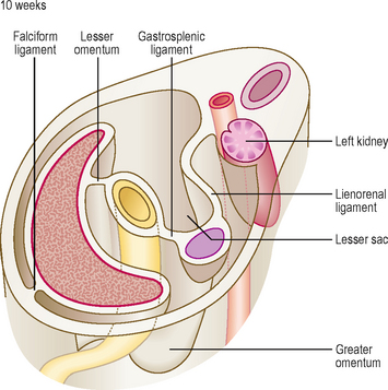
As the liver develops in the ventral mesogastrium and enlarges it is carried towards the right. This accentuates the separate compartmentalization of the lesser sac. The ventral mesogastrium of the foregut is carried rightwards with the liver. The border of the mesogastrium between the liver and the ventral wall of the embryo becomes the falciform ligament. The portion between the stomach and liver becomes the lesser omentum. There is a free border of the lesser omentum (that contains the hepatic artery, bile duct and hepatic portal vein) lying between the duodenum and the visceral surface of the liver (Fig. 7.6).