The Diabetic Foot
John M. Giurini
Introduction
It is currently believed that there are 23.6 million diabetic patients in the United States: 17.9 million diagnosed and another 5.7 million undiagnosed. There are another 57 million who are diagnosed with prediabetes with 1.6 million new cases occurring per year. With these staggering numbers, the cost of caring for patients with diabetes has also increased exponentially. In 1992, the total cost of care (direct and indirect) for the diabetic patient was $91.8 billion compared to $20.4 billion in 1987. In 2004, the cost has nearly doubled to $174 billion. Conservative estimates put the cost of diabetic foot ulcers at nearly $1 billion per year. The ability to improve outcomes, reduce amputations, and contain costs requires a thorough understanding of the risk factors leading to ulcerations and amputations.
The foot contains 26 bones that provide structure and support to the lower extremity and body. Foot function is controlled by the synergistic activities of 19 intrinsic muscles with origin and insertions contained entirely within the foot and 7 extrinsic muscles with their origins in the leg but with their insertions in the foot. These muscles are responsible for smooth transfer of weight-bearing forces throughout the gait cycle from heel strike to midfoot loading and finally to propulsion. There are also numerous ligaments that connect various bones to each other to provide structure and support. Alterations or injuries to these structures can result in changes in foot structure such as development of flatfoot deformities, bunions, hammertoes, or metatarsal deformities. They can also result in biomechanical deformities such as excessive pronation, which can lead to changes in weight bearing and the plantar distribution of forces.
Another important anatomical feature of the foot is that it can be divided into four layers. The first layer or superficial layer is comprised of the abductor hallucis, flexor digitorum brevis, and the abductor digiti minimi muscles. The second layer is formed by the flexor digitorum, the accessorius (also known as the quadratus plantae), and the lumbrical muscles. The third layer is comprised of the flexor hallucis brevis, the adductor hallucis, and the flexor digiti minimi muscle. The dorsal and plantar interossei muscles form the fourth and final layer. These muscle groupings are loosely separated by intramuscular septa and fascia creating separate tissue planes. In addition, the extrinsic muscles course through these layers. Infections on the plantar aspect of the foot are particularly problematic and dangerous. Aggressive infections can spread unimpeded between these layers or within tendon sheaths and spread up the leg very quickly. This can jeopardize the foot, leg, or the patient’s life.
Why are diabetic patients at such high risk for ulceration and amputation? One of the more common complications of diabetes is the development of peripheral neuropathy. The patient’s inability to perceive even the most minor of injuries to the feet can put the entire limb at risk of amputation. A small puncture wound or a superficial friction blister can quickly progress to a deep space infection and sepsis within a short time frame if undetected and left untreated. It is interesting to note that patients with rheumatoid arthritis (RA) develop similar foot deformities as patients with diabetes. Yet, the incidence of foot ulcerations in patients with RA is relatively low. This is due to the fact that patients with RA experience pain with walking or with the presence of a callus. Therefore, they will alter their gait or restrict their walking before developing ulcerations. The patient with diabetes, lacking this early warning signal, will continue to walk to the point of skin breakdown and potential infection.
While the actual mechanism of this nerve abnormality is not completely elucidated, it is believed to result from thickening of the basement membrane in the vasa nervorum, those small blood vessels providing nutrients to the nerves. This results in conduction deficits to the nerves, abnormal signaling, and eventual loss of transmission. Because the nerves of the feet are at the distal most edge of the circulation, these nerves are most vulnerable. Like all complications of diabetes, it is believed that patients exhibiting poorer degrees of glycemic control are most vulnerable to this complication. However, one of the most troubling facts is that clinically significant peripheral neuropathy can precede the diagnosis of diabetes by as much as 5 years.
Peripheral vascular disease is another common complication of diabetes mellitus. We will not spend much time in discussing this complication here as it is being covered
in greater detail elsewhere in this book. However, a few points deserve further discussion. The number of purely ischemic ulcerations in patients with diabetes is relatively small. It is estimated that ∼15% of diabetic foot ulcerations are purely ischemic in nature. The pattern of peripheral vascular disease is quite characteristic for patients with diabetes. Typically, stenoses occur in vessels between the knee and the ankle, that is, the posterior tibial, anterior tibial, and peroneal arteries. The vessels will then reconstitute at the level of the ankle to form patent segments of the dorsalis pedis and posterior tibial arteries. It is this fact that makes distal bypass surgery a feasible option in patients with nonhealing ulcerations of the foot.
in greater detail elsewhere in this book. However, a few points deserve further discussion. The number of purely ischemic ulcerations in patients with diabetes is relatively small. It is estimated that ∼15% of diabetic foot ulcerations are purely ischemic in nature. The pattern of peripheral vascular disease is quite characteristic for patients with diabetes. Typically, stenoses occur in vessels between the knee and the ankle, that is, the posterior tibial, anterior tibial, and peroneal arteries. The vessels will then reconstitute at the level of the ankle to form patent segments of the dorsalis pedis and posterior tibial arteries. It is this fact that makes distal bypass surgery a feasible option in patients with nonhealing ulcerations of the foot.
Another point to be kept in mind is the effect autonomic neuropathy has on the vascular system. Diabetic patients with nonhealing ulceration and with autonomic neuropathy will commonly present with warm, pink skin that appears to be well perfused. However, pulses may not be palpable. In this case, autonomic neuropathy has resulted in an autosympathectomy, resulting in maximum dilation of blood vessels. This leads to the clinical appearance of a well-perfused foot, even in the presence of critical limb ischemia. Many a diabetic foot has been ill-advisedly operated upon due to lack of recognition of this fact.
Early and aggressive surgical intervention in patients with diabetic foot disease has become more readily accepted and a standard form of treatment in patients with chronic foot ulcerations or severe deformities. This is a significant paradigm shift from only a few years ago when surgery was to be avoided at all costs in patients with diabetes.
The primary goal of surgery in patients with diabetes is to reduce the risk of lower extremity amputation (Table 1). This is achieved by eliminating the focus of osteomyelitis or correcting structural deformities, which may lead to ulcerations. Keeping this goal in mind will guide the surgeon in their selection of appropriate procedures. The end result of any surgical procedure in the diabetic patient is to eliminate the deformity, reduce the risk of amputation, and leave the patient with extremity that will support continued ambulation.
In addition, it is helpful to classify diabetic foot surgery into three distinct categories: elective, prophylactic, and urgent. Elective surgery implies the presence of a deformity that may pose a future risk to the patient and that can be corrected surgically. In many cases, these deformities can be managed nonsurgically and there are clinical situations where the conservative approach is in the best interest of the patient.
Prophylactic surgery by definition is preventative in nature. The patient has demonstrated a foot deformity and a recurrent or nonhealing ulceration that puts the limb at risk of amputation. In this case, local foot surgery should be considered to prevent major limb loss.
Table 1 Goals of Surgery in Neuropathic Foot | |||||
|---|---|---|---|---|---|
|
Urgent surgery is performed in those situations where limb loss is imminent. These patients will commonly present with foul-smelling ulcerations with drainage and cellulitis and other signs of sepsis, including fevers, chills, hyperglycemia, and hypotension. These patients require immediate surgical intervention to control sepsis, stabilize the patient, and to salvage as much of the foot and/or leg as possible. Once sepsis is controlled and the patient is stable, thoughts of reconstructive surgery may proceed.
Prior to any definitive elective or prophylactic surgery on the diabetic foot, it is assumed that it is free of acute infection. Any area of undrained sepsis must be adequately drained by promoting dependent drainage. The proper technique is to incise the wound such that the wound drains from distal to proximal as the patient lies recumbent in bed (Fig. 1). The use of multiple small stab incisions with penetrating Penrose drains should be avoided as they allow small pockets to collect and do not promote dependent drainage. In addition, prior to any elective or prophylactic surgery, all necrotic tissues need to be debrided to healthy granulation tissue. If present, any exposed or infected bone should be excised. The wound should be packed open and inspected daily for control of sepsis, resolution of cellulitis, and development of healthy granulation tissue.
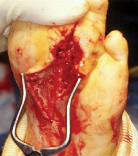 Fig. 1. Incision and drainage procedures should be performed so that dependent drainage is achieved from distal to proximal as the patient is recumbent in bed. |
The foot can be divided into three distinct anatomical regions for the purposes of surgical considerations: forefoot, midfoot, and hindfoot. Forefoot ulcerations involve the ball of the foot or the metatarsophalangeal joints (MTPJs). Because of the mechanics of the foot and the forces generated at this level, these comprise the vast majority
of ulcerations that occur in patients with diabetes.
of ulcerations that occur in patients with diabetes.
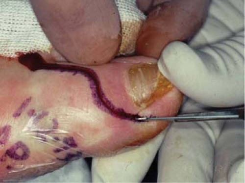 Fig. 2. The “lazy-S” incision provides good exposure while neutralizing tensile forces along the incision line. |
Midfoot problems most commonly occur in patients with advanced deformities resulting from Charcot joint disease. These can occur either in the medial arch, central part of the midfoot, or the lateral border of the midfoot and can be very challenging to manage.
Hindfoot problems are some of the most serious deformities that can occur in the diabetic patient. These can include the subtalar joint or the ankle joint or both. These deformities are manifestations of a severe and aggressive form of Charcot joint disease that can lead to severe instability, ulcerations, and difficulty in walking. Untreated, these almost always go on to major limb amputation. Even with early aggressive treatment, the incidence of major limb loss can approach 50%.
Forefoot Procedures
First Ray
The first ray comprises the great toe and first metatarsal. Because of the weight-bearing forces generated through these structures and potential abnormal mechanics, these are common sites of foot ulcerations. Excessive pronation can lead to transfer of weight-bearing forces through the medial longitudinal arch, the first metatarsal, and the hallux. In addition, structural deformities such as osteoarthritis, hallux limitus/rigidus, or a severe plantarflexion deformity of the first metatarsal make this structure particularly vulnerable to callus buildup, blister formation, and eventual ulceration. Common sites of ulceration can include: plantarmedial aspect of the hallux, distal tip of the hallux, directly plantar to the hallux interphalangeal joint (HIPJ), directly plantar to the first metatarsal head, or medial to the first metatarsal head.
Ulcerations of the hallux may be related to a deformity within the HIPJ or restriction of motion at the first MTPJ. This commonly occurs in osteoarthritis or hallux limitus where there is compensation at the HIPJ for the restriction of motion at the MTPJ. Both of these conditions can be addressed by performing an arthroplasty through the HIPJ. In this procedure, a lazy-S incision is made centered over the IPJ, exposing the joint (Fig. 2). The head of the proximal phalanx is freed by resecting the medial and lateral collateral ligaments. The head of the proximal phalanx is then resected transversely from dorsal to plantar just behind the condyles. This procedure alleviates pressure directly plantar to the joint as well as medial to the joint.
In cases where there is a significant degree of osteoarthritis at the first MTPJ with resultant restriction of motion, it is best to address the problem directly at that level. Resection of the base of the proximal phalanx (i.e., standard Keller procedure) results in restoration of this motion and resolution of the hallux ulceration. In this case, resection of the proximal one-third of the phalangeal base is performed rather than the head of the proximal phalanx.
Ulcerations plantar to the first metatarsal head result from prominent or enlarged sesamoid bones or plantarflexed position of the first metatarsal itself. They may also occur from a hallux hammertoe exerting a retrograde force on the first metatarsal. Successful surgery requires identifying the etiologic cause.
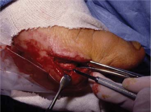 Fig. 3. The medial incision at the interface of the dorsal and plantar skin provides good exposure of the tibial sesamoid. Excision is facilitated with a #64 beaver blade. |
Prominent or enlarged sesamoids can be addressed by excising both the tibial and the fibular sesamoids. This is best performed through a longitudinal medial incision at the level of the first MTPJ. Once the joint capsule is incised longitudinally, the tibial sesamoid is easily visualized. It can be released from its soft-tissue attachments using a #64 beaver blade (Fig. 3). Once removed, the fibular sesamoid is visualized immediately lateral to the tibial sesamoid and can be removed in a similar fashion. The wound is closed in a layered fashion by first reapproximating the capsule, then subcutaneous tissue, and finally the skin with nonabsorbable suture material.
Stay updated, free articles. Join our Telegram channel

Full access? Get Clinical Tree


