Chapter Twenty-Six. The central nervous system
Introduction
The ability to respond appropriately to environmental change depends on rapid communication achieved by the nervous system and the endocrine system (see Ch. 28). The nervous system communicates by the rapid transmission of electrical signals. The endocrine glands secrete hormones into the bloodstream which modify the working of target organs. Normal functioning of these systems is critical for maintaining homeostasis (the ability of a regulated system to maintain itself close to a fixed point).
The nervous system controls function of every body system, every thought, action and emotion. It is only possible to give a brief outline of this complex system and to elaborate on important functions such as pain perception. This chapter concentrates on the central nervous system; Chapter 27 considers the peripheral and autonomic nervous systems and neural integration.
Organisation of the nervous system
The central nervous system (CNS) consists of the brain and spinal cord. It has an integration function, receiving messages from and sending messages to all parts of the body via the peripheral nervous system (PNS). The autonomic nervous system (ANS) is that part of the PNS that innervates the smooth muscle and glands of the viscera and the cardiac muscle. It can be subdivided into the sympathetic nervous system and parasympathetic nervous system. Sensory organs such as eyes and ears feed environmental information back to the brain (Fitzgerald et al 2006).
Neuroanatomy
Nervous tissue is made up of two cell types: the neurons (Fig. 26.1) and a group of cell types collectively known as neuroglia, the supportive connective tissue of the nervous system. The tissue is supplied by blood vessels.
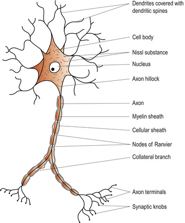 |
| Figure 26.1 Structure of a whole nerve fibre. (From Hinchliff S E, Watson R 1996, with permission.) |
Neurons
The structural units of the nervous system are the neurons. These specialised cells have the following characteristics (Marieb 2008):
• They have processes called axons and dendrites communicate with other cells.
• They conduct messages by nerve impulses from one body part to another.
• They are extremely long-lived but cannot undergo mitosis and divide.
• They have a very high metabolic rate needing continuous glucose and oxygen.
• They cannot survive more than a few minutes without oxygen.
Structure of neurons
The cell body
Each neuron has a cell body ( soma) with a large spherical nucleus surrounded by granular cytoplasm and contains all the usual organelles except centrioles. Neurotransmitters (NTs) are synthesised in the soma. Most neuronal cell bodies are located in the central nervous system (CNS), where they are clustered together in groups called nuclei. The few neuronal cell bodies in the parasympathetic nervous system (PNS) are called ganglia.
Dendrites
Dendrites are short, diffusely branching extensions which receive messages from other cells at synapses by conducting electrical signals called graded potentials towards and into the cell body. Dendrites are the input part of the neuron and there may be hundreds clustering close to the cell body. They provide an enormous surface area for reception of signals from other cells.
Axons
Each neuron has only one axon arising from the axon hillock on the cell body. The axon is the same diameter along its length and some are over 1 metre long such as those travelling from the spine to the foot. Axons may give off branches called axon collaterals and usually have terminal branches ending in synaptic knobs or boutons. Axons contain microtubules and microfilaments that transport substances to the cell body ( anterograde) and from the cell body ( retrograde). They conduct messages to other cells by electrical nerve impulses and by neurotransmitters which excite or inhibit other neurons by attaching to receptors on their plasma membrane.
Myelin sheaths
Larger nerve fibres are covered in a white, fatty, segmented sheath called the myelin sheath which protects and insulates fibres and increases the rate of impulse transmission. Messages can be transmitted up to a 100 times more rapidly than in unmyelinated fibres (Fig. 26.2). Myelin sheaths in the PNS are formed by Schwann cells which wrap themselves around the axon. Schwann cell protoplasm is squeezed out of the cell to leave the axon wrapped in a multilayered membrane. The external portion is called the neurolemma or Schwann sheath. Adjacent Schwann cells along the axon do not touch and the gaps between them, which occur at regular intervals, are called the nodes of Ranvier. Axon collaterals can only emerge at these nodes.
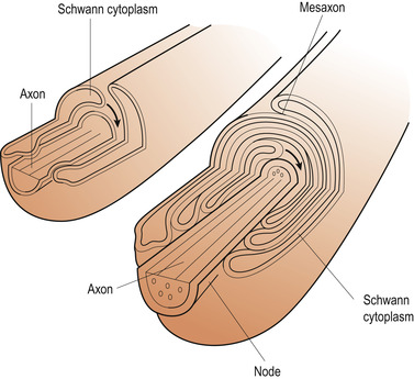 |
| Figure 26.2 Myelination in peripheral nervous system. Arrows indicate movement of flange of Schwann cytoplasm. (From Fitzgerald M T J 1996, with permission.) |
The myelin sheaths of the CNS are produced by cells called oligodendrocytes. Myelinated fibres form the white matter in the brain and spinal cord and the cell bodies form the grey matter. Cell bodies are outside the white matter in the brain and inside the white matter in the spinal cord.
Classification of neurons
Neurons may be classified structurally or functionally (Marieb 2008).
Structurally, neurons are grouped according to the number of processes extending from the cell body. There are three major groups:
1. Multipolar neurons are the most common type and have at least three processes, usually multiple dendrites and one axon. However, some do not have an axon.
2. Bipolar neurons have two processes—an axon and a dendrite. They are rare but are found in special sense organs such as the retina or in the olfactory mucosa.
3. Unipolar neurons have a single, very short process. They are found in the ganglia of the PNS where they act as sensory neurons.
Functional classification is into motor, sensory and association neurons.
• Motor neurons with cell bodies mainly in the CNS carry impulses to control effector organs such as the muscles or glands.
• Sensory neurons whose cell bodies are located in the sensory ganglia outside the CNS often have very long dendritic branches which gather impulses from the periphery such as the ends of the toes and fingers.
• Association neurons, often called interneurons, carry signals between motor and sensory neurons in complex networks.
Neuroglia
Neuroglial cells support and protect neurons, outnumbering them in a ratio of 5:1. As there are billions of neurons in the brain, the number of neuroglial cells is enormous. These cells are subdivided into four basic types (Fitzgerald et al 2006):
1. Astrocytes are star-shaped cells with long, fine processes arising from their bodies. They have one process against a neuron and other processes close to capillary walls.
2. Oligodendrocytes in the CNS and Schwann cells in the PNS are smaller than astrocytes and have fewer processes. They form myelin sheaths around nerve fibres.
3. Ependyma form a continuous layer of cells lining the ventricles of the brain and spinal cord central canal. They help to produce cerebrospinal fluid.
4. Microglia are small cells which are part of the immune system, acting as macrophages.
The structure of a nerve
The axons from single neurons are bound together to form nerves (Fig. 26.3). Nerves may contain afferent fibres (to the CNS), efferent fibres (from the CNS) or both when they are called mixed nerves. Each separate nerve fibre is embedded in a fibrous connective tissue sheath called the endoneurium. These are bound into groups by the perineurium, a connective tissue sheath and the complete nerve is surrounded by the epineurium. Each nerve has an arterial blood supply and venous drainage.
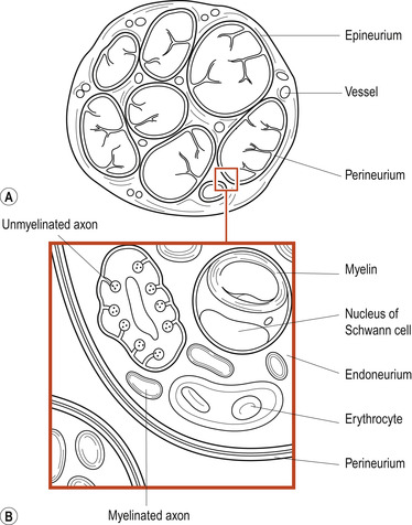 |
| Figure 26.3 Transverse section of a nerve trunk. (A) Light microscopy. (B) Electron microscopy. (From Fitzgerald M T J 1996, with permission.) |
Neurophysiology
The nerve impulse
Both nerve fibres and muscle fibres are excitable tissues which conduct electrical signals. When a neuron is stimulated, an electrical impulse is sent along the axon (Fig. 26.4). Bodies are electrically neutral (positive and negative charges are equal). Potential electrical energy is called voltage, which is measured in volts (V) or millivolts (mV). The flow of electricity from one point to another is called a current. Substances that hinder the flow are said to provide resistance. Ions, which are electrically charged particles, provide the currents and usually flow through an aqueous solution across a plasma membrane. Plasma membranes are studded with proteinaceous ion channels.
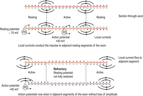 |
| Figure 26.4 Propagation of an action potential along a nerve fibre. PD, potential difference. (From Hinchliff S E, Montague S E 1990, with permission.) |
Polarisation
When a membrane is resting, its potential is polarised, i.e. the inside has a different electrical potential from the interstitial fluid. The resting potential of neurons averages −70 mV. This is maintained by the distribution and relative contribution of negative and positive ions. Inside the cell the positive ion is potassium and the negative ion is protein. In the interstitial fluid the positive ion is sodium and the negative ion is chloride.
The role of sodium ions
When the cell is stimulated, sodium channels open in the membrane and sodium ions rush into the cell, changing the membrane potential to +40 mV. This is called depolarisation. The action potential is the sum of all negative and positive charges stimulating the cell and proceeds down the axon in a wave. Behind the wave the cell pumps three sodium ions out in exchange for two potassium ions. The membrane potential falls to −90 mV (hyperpolarisation) and then recovers.
The period of hyperpolarisation is called the refractory period during which the cell cannot generate an action potential. Depolarisation increases the chance of a nerve impulse being generated but hyperpolarisation decreases it. Firing of a neuron is an all-or-nothing phenomenon, occurring only if the potential reaches a threshold. Strong stimuli result in more impulses, not stronger impulses.
Saltatory conduction of the impulse
Nerve fibres can be classified according to the speed of conduction of the action potential. The larger the nerve, the more rapidly it conducts impulses. Myelinated nerves conduct impulses more rapidly than unmyelinated nerves. The myelin sheath increases electrical resistance of a nerve but it is leakier at the nodes of Ranvier. The electrical current flows smoothly along each section of the sheath between nodes of Ranvier and it is only necessary to generate an action potential at the nodes. The impulse seems to jump from node to node. This is called saltatory conduction (Fig. 26.5). In a non-myelinated nerve, new action potentials have to be generated across each adjacent section of the nerve membrane.
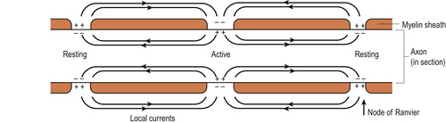 |
| Figure 26.5 Saltatory conduction in a myelinated nerve fibre. Local currents conduct the impulse from node to node. The action potential is regenerated at each node of Ranvier. (From Hinchliff S E, Montague S E 1990, with permission.) |
Classification of nerves by speed of conduction
Nerve fibres can be classified as follows:
• Group A fibres are myelinated and can conduct impulses at up to 120 metres per second (m/s). They are further subdivided into α, β, γ and δ (alpha, beta, gamma and delta) fibres.
• Group B fibres are myelinated. They are all preganglionic fibres of the autonomic nervous system.
• Group C fibres are non-myelinated and conduct impulses as slowly as 1 m/s.
The synapse
Synapses are junctions between the terminal bouton of the axon and its target tissue which may be a neuron cell body, a gland or a muscle (Fig. 26.6). Synapses enable transfer of information between one cell and another. They may be electrical or chemical (Fitzgerald et al 2006). The presynaptic neuron conducts impulses towards the synapse and the postsynaptic neuron transmits information away from the synapse.
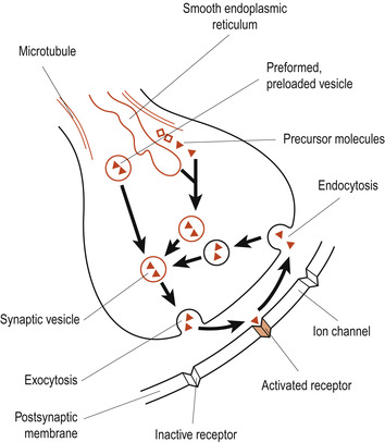 |
| Figure 26.6 Diagram to show origin and fate of synaptic vesicle and transmitter–receptor binding. (From Fitzgerald M T J 1996, with permission.) |
Electrical synapses
These synapses are bridged junctions corresponding to gap junctions in other cells. Protein channels connect the cytoplasm of adjacent neurons, providing electrical pathways through which ions can flow from one neuron to another. Such neurons are electrically coupled and communication between cells is extremely rapid. They synchronise interconnected neurons and are rare in the adult but much more common in the embryo, gradually being replaced by chemical synapses. They are responsible for stereotypical movements such as the jerky movements of the eyes. They remain abundant in some nervous tissues such as cardiac and smooth muscle.
Chemical synapses
Between the neuron and its target cell the synaptic cleft is a fluid-filled space into which NTs are released from the presynaptic membrane. These open and shut ion channels in the postsynaptic membrane. The electrical message of the action potential is conducted chemically across the gap. This is a reversible change and the NT is removed. Enzymes that degrade the NT are released into the synaptic cleft; the NT is taken up by the presynaptic membrane and then diffuses away from the synapse.
Neurotransmitters
Neurotransmitters (NTs) help neurons to communicate messages and regulate body activities and states (Marieb 2008). Over 100 different chemicals act as NTs and they are classified according to chemical structure. They are synthesised with the help of enzymes.
Acetylcholine
Acetylcholine (ACh) was the first NT to be identified. It is the NT released at neuromuscular junctions and is accessible for study. ACh is also found in the CNS.
ACh is synthesised and stored within synaptic vesicles in the presence of the enzyme choline acetyltransferase. Acetic acid is bound to coenzyme A to form acetyl CoA. This compound combines with choline and the coenzyme is released:
The released ACh binds to the postsynaptic membrane and is degraded to acetic acid and choline by the enzyme acetylcholinesterase (AChE). The released choline is captured by the presynaptic membrane and used to synthesise more ACh.
The biogenic amines
Biogenic amines include catecholamines such as dopamine and noradrenaline (norepinephrine) and adrenaline (epinephrine) and the indolamines serotonin (5-hydroxytryptamine, 5-HT) and histamine


Stay updated, free articles. Join our Telegram channel
Full access? Get Clinical Tree
